S20
Quantitative evaluation of metal artifacts in 3-T MRI by using Blind/Referenceless Image Spatial Quality Evaluator (BRISQUE): a phantom study
Wakiko Tani1, Yutaka Katayama2, Tomoaki Yamakawa1, Shintaro Horii1, Ryuji Shimada1, Yuichiro Somiya1, and Akiko Kusaka1
1Center for Radiology and Radiation Oncology, Kobe University Hospital, Kobe, Japan, 2Division of Radiological Technology, Osaka City University Hospital, Osaka, Japan
1Center for Radiology and Radiation Oncology, Kobe University Hospital, Kobe, Japan, 2Division of Radiological Technology, Osaka City University Hospital, Osaka, Japan
Synopsis
No-reference image quality assessment (NR-IQA) model called Blind/Referenceless Image Spatial Quality Evaluator (BRISQUE) is one of the objective evaluations of images. To investigate whether BRISQUE can be used to quantify artifacts on MRI images, we compared the BRISQUE score with the visual score. The BRISQUE score was reduced and the visual score was improved by increasing bandwidths from 51 kHz to 307 kHz. Pearson correlation coefficient was 0.7576 and BRISQUE score was associated with the visual score (P < .05). This study suggests that BRISQUE can quantitatively evaluate metal artifacts.
Purpose
Image distortions caused by differences in magnetic susceptibility have long been a problem in clinical imaging1. Especially, numerous types of metallic appliances are used in orthodontic treatment and may cause severe artifacts on MRI2. Several techniques are commonly used to reduce the severity of metal susceptibility artifacts, including simple concessions such as increasing the frequency encoding bandwidth (BW), or orienting the long axis of the metal along the frequency encoding direction3. However, it is difficult to evaluate quantitatively the competence of a given metal artifact reduction technique. Without a standard against which to measure the efficiency of a technique, most studies have been limited to qualitative comparisons1. No-reference image quality assessment (NR-IQA) model called Blind/Referenceless Image Spatial Quality Evaluator (BRISQUE) is one of the objective evaluations of images4, 5. The BRISQUE model measures the image quality by using the locally normalized luminance coefficients, which were used to calculate the image features6. The aim of this study was to quantify metal artifacts on 3-T MRI images using BRISQUE.Methods
MRI scan was performed with a 3-T whole-body MR scanner (Achieva; Philips Healthcare Best, Netherlands) using a head coil. Lingual sheaths used in orthodontics were selected for the evaluation of metal artifacts. Metal artifact images were acquired by placing the lingual sheath on the surface of a copper sulfate-filled cylindrical container (16 cm in diameter). The following imaging sequence was used: T1-weighted spin-echo (SE), T1-weighted turbo spin-echo (TSE), and T1-weighted fast-field-echo (gradient-recalled-echo, FFE). Images were acquired at 2 different bandwidths in each sequence (51 kHz and 307 kHz). The scanning parameters are shown in Figure 1. Metal artifact images were evaluated using BRISQUE and compared with visual evaluation. BRISQUE was implemented with OpenCV and the lower BRISQUE score indicates better image quality. Artifact and distortion of images were evaluated by using a 4-point score (1 = severe, 2 = strong, 3 = mild, 4 = minimal) by seven radiographers blinded to sequence parameters. BRISQUE score and visual score were compared between each sequence by one-way ANOVA followed by Tukey test. Relations between BRISQUE score and visual score were assessed with the Pearson correlation coefficient. A p-value of less than 0.05 was considered statistically significant.Results
The mean ± SD of the BRISQUE score of SE 51 kHz, TSE 51 kHz, FFE 51 kHz, SE 307 kHz, TSE 307 kHz and FFE 307 kHz were 42.8±1.6, 43.7±2.3, 42.8±1.4, 39.0±1.3, 38.8±1.9 and 41.0±1.3, respectively. In the BRISQUE score, all 51 kHz sequences were significantly higher than 307 kHz sequences. This result shows that the 51 kHz sequence has more artifacts compared to 307 kHz. The mean ± SD of the visual score of SE 51 kHz, TSE 51 kHz, FFE 51 kHz, SE 307 kHz, TSE 307 kHz and FFE 307 kHz were 3.0±0.8, 3.2±0.9, 3.6±0.7, 1.1±0.3, 1.4±0.6 and 3.4±0.8, respectively. In the visual score, all 51 kHz sequences were significantly higher than 307 kHz of SE and TSE. Pearson correlation coefficient was 0.7576 and BRISQUE score was associated with the visual score (P < .05).Conclusions
This study suggests that BRISQUE can quantitatively evaluate metal artifacts.Acknowledgements
We are grateful to Mr. Noriyuki Kai and Mr. Naoki Yoshida for their assistance in this study.References
- Kolind S. H, Mackay A. L, Munk P. L, et al. Quantitative Evaluation of Metal Artifact Reduction Techniques. J. Magn. Reson. Imaging. 2004; 20: 487 – 495.
- Ozawa E, Honda E, Parakonthun K. N, et al. Influence of orthodontic appliance-derived artifacts on 3-T MRI movies. Prog. Orthod. 2018; 19
- Eustace S, Goldberg R, Williamson D, et al. MR imaging of soft tissues adjacent to orthopedic hardware: techniques to minimize susceptibility artifact. Clin Radiol 1997; 52: 589 - 594.
- Mittal A, Moorthy A. K, Bovik A. C, No-Reference Image Quality Assessment in the Spatial Domain. IEEE Trans Image Process. 2012; 21: 4695 – 4708.
- Rajagopal H, Mokhtar N, Izam T, et al. No-reference quality assessment for image-based assessment of economically important tropical woods. PLoS One. 2020; 15(1)
- Chow L. S, Rajagopal H. Modified-BRISQUE as no-reference image quality assessment for structural MR images. Magn. Reson. Imaging. 2017; 43: 74 - 87
Figures
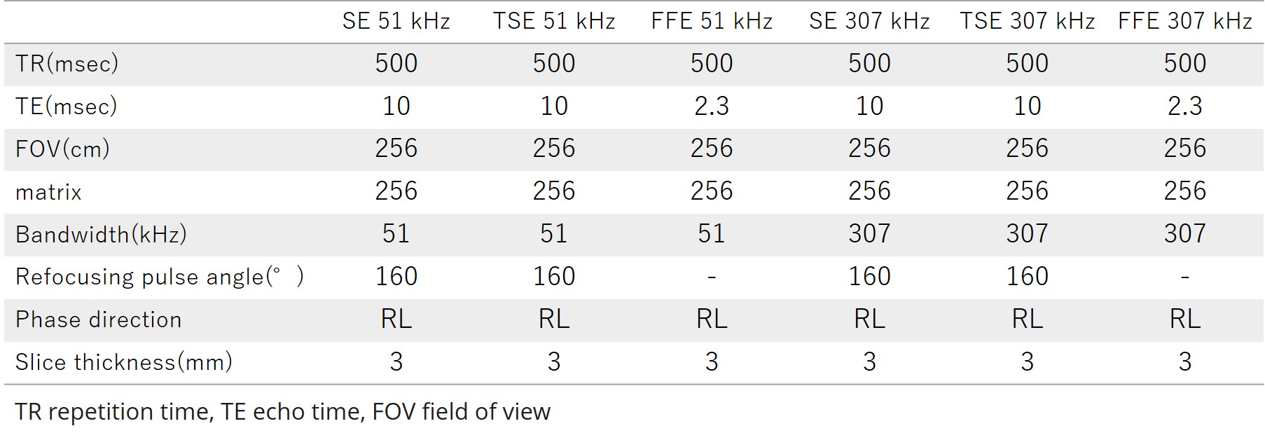
Figure 1. MRI pulse sequence protocol
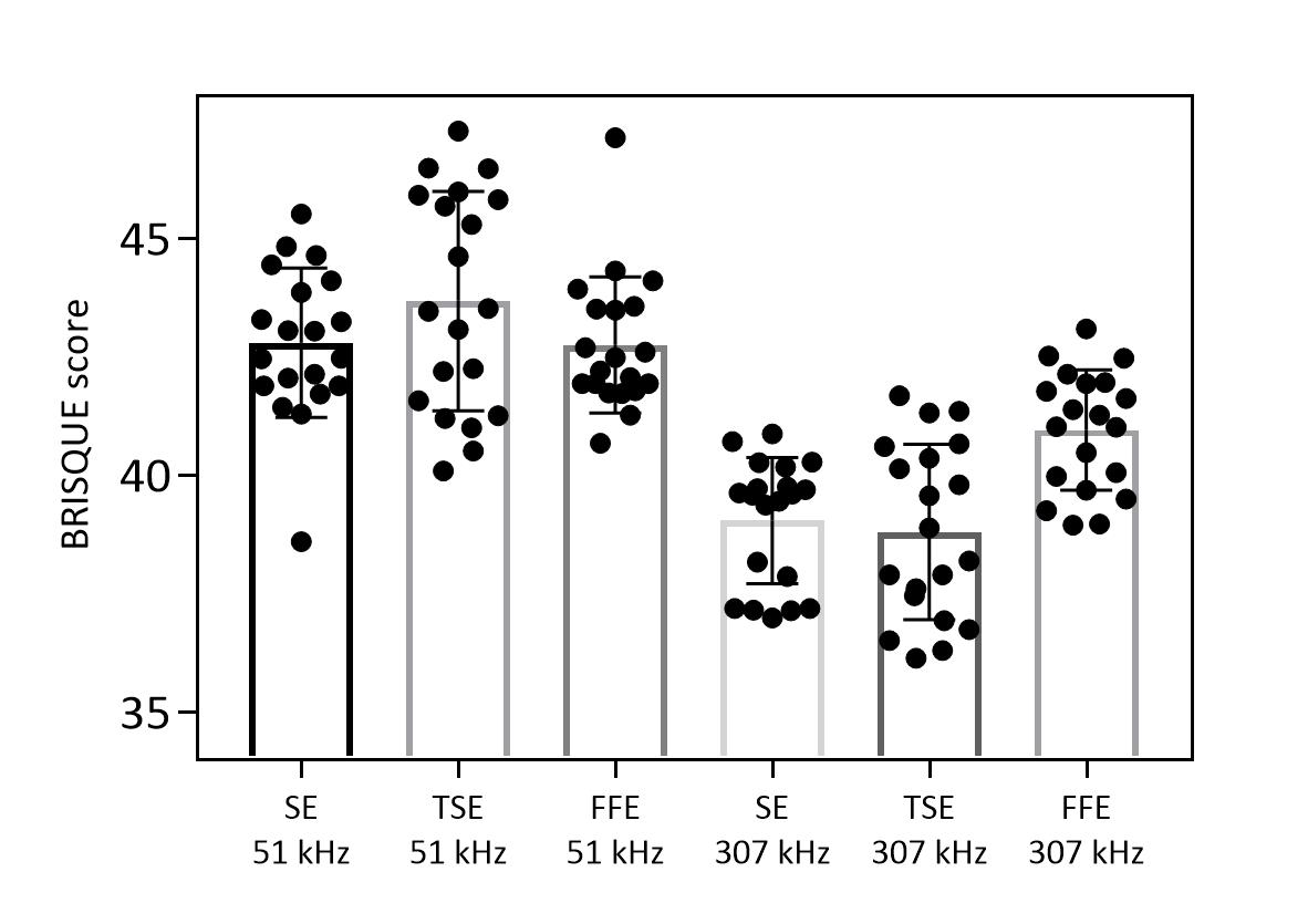
Figure 2. Quantitative evaluation of metal artifacts by BRISQUE score
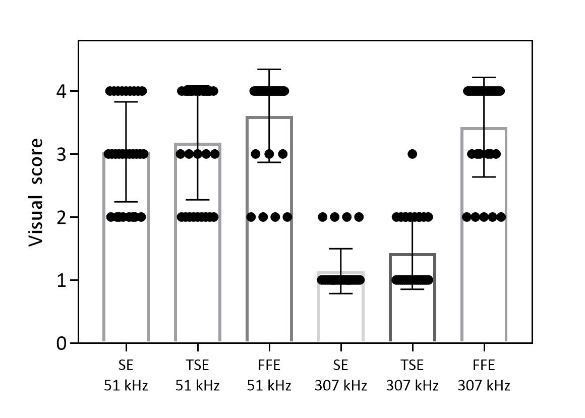
Figure 3. Qualitative evaluation of metal artifacts by visual score
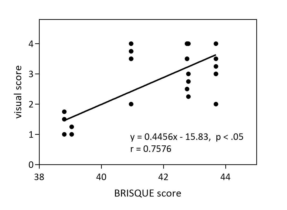
Figure 4. Correlations Between BRISQUE score and visual score
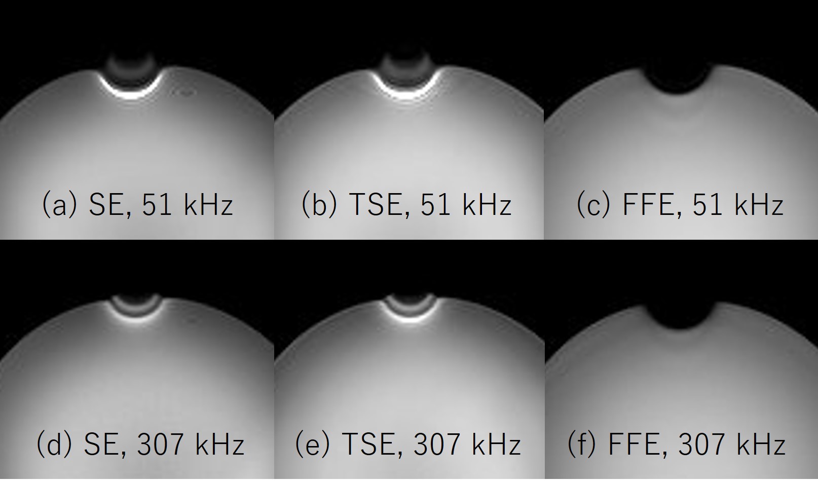
Figure 5. Axial MRI images of the phantom with a lingual sheath (a)SE 51 kHz (b)TSE 51 kHz (c)FFE 51 kHz (d)SE 307 kHz (e)TSE 307 kHz (f)FFE 307 kHz