S12
The Effect of Enlarging Shim Volume on B0 homogeneity of MRS Voxel on 7T1Center for Clinical Spectroscopy, Brigham and Women's Hospital, Boston, MA, United States
Synopsis
B0 homogeneity is important for clinical spectroscopy but is highly region dependent and time consuming. Enlarging shim volume could be a quick and easy way to improve B0 homogeneity in the clinical practice of spectroscopy and the goal of this study is test if this method is beneficial. We used different methods to measure the potential improvement in B0 homogeneity in different brain regions. While our results showed that increasing shim volumes did not improve spectral linewidths or data quality, we demonstrate a robust method of measuring B0 homogeneity in clinical spectroscopy.
Background and Purpose:
A good B0 shim for brain MR spectroscopy with narrow spectral lines will provide more reliable separation and quantification of metabolites which in turn improves diagnostic accuracy1,2. However, from a clinical perspective, shimming is time consuming and highly user dependent. Studies have shown that increasing the shim volume can improve spectral linewidth2 and therefore may provide a simple method that can be readily employed in the clinical setting. To test this method, we examined the efficacy of increasing shim volumes as a way of improving B0 homogeneity on a clinical 7T scanner. In particular, we wanted to test this in challenging but clinically important regions such as the hippocampus3,4,5 and thalamus4. The primary measures of improvement were linewidths expressed in full width half maximum (FWHM) and the fitting of the major brain metabolites expressed in Cramer-Rao Lower Bounds (CRLB).Teaching Points:
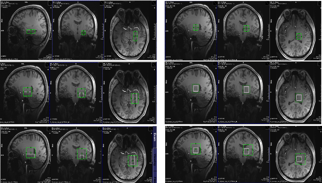
Equipment and protocol:MR exams were performed after informed consent on a Siemens Magnetom Terra 7 Tesla MRI scanner (Syngo MR VE13) using a Nova medical head coil (1TX/32RX). Two calcium titanate dielectric pads6 were placed on each of the head, one in the back and the other on the left side of the head (pad on the right side cannot be placed due to large head size). The scan protocol includes: 3-plane localizer, T1 MPRAGE (TR/TE 2290/2.95ms, flip angle 7°), single-voxel spectroscopy with semi-LASER localization (sLASER-SVS; TR/TE 4s/35ms, 48 averages, water reference) in two locations:left hippocampus (1.5x3.5x1.5cm3 voxel ) and left thalamus (2x2x2cm3 voxel) with three sizes of shim volumes:8, 32 and 64cc (Figure 2).
Left Hippocampus:
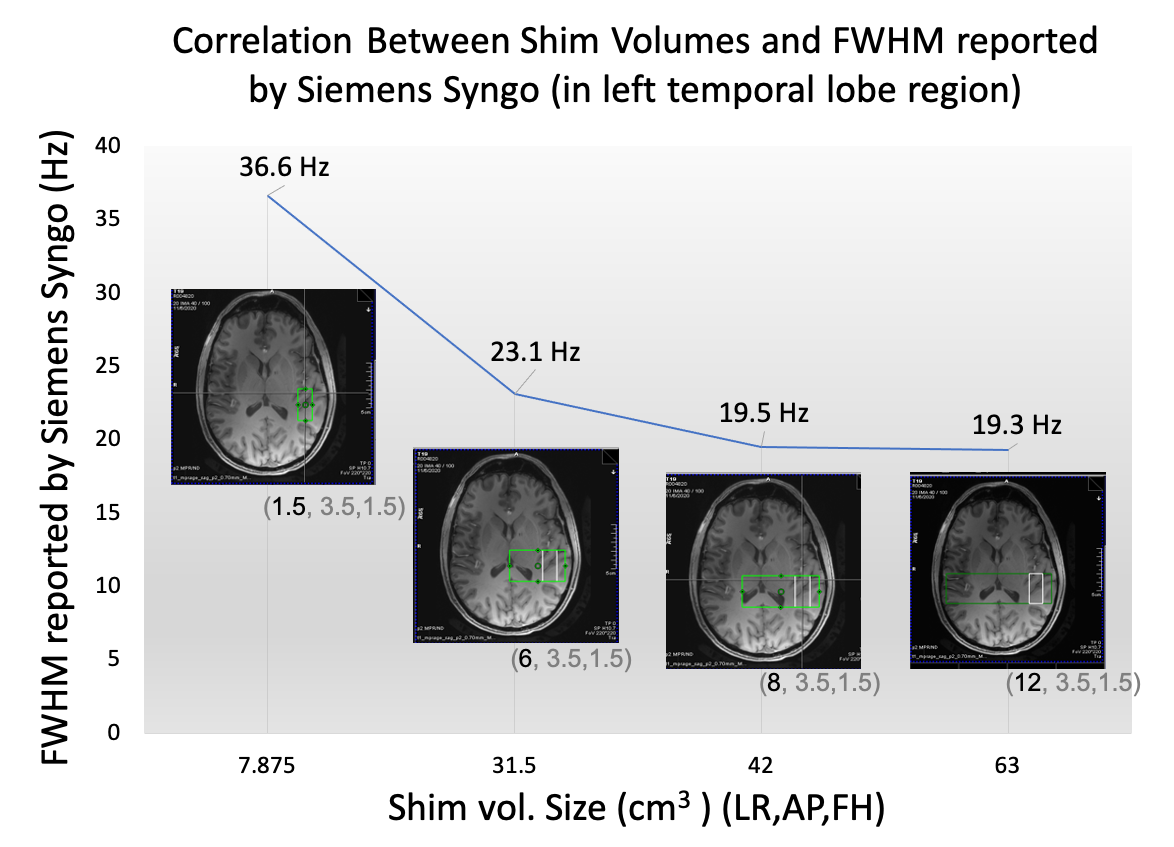
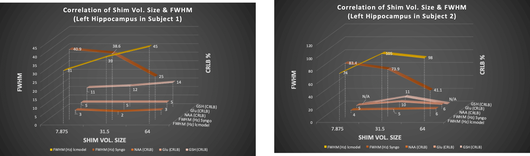
FWHM- The linewidths (FWHM) reported by Siemens Syngo software in left temporal lobe showed a trend of decreased linewidth with increased shim VOI (Figure 1). We tested again in the left hippocampus in another subject (Figure 2) and this time the linewidths were measured using LCModel which provides a more accurate linewidth measure. The results are shown in yellow in Figure 3 showed a different trend: linewidths increased instead of decreasing with increased shim VOI sizes. A similar result is shown in subject 2.
Metabolites – Since the linewidths showed inconsistent trends between the values reported by Siemens Syngo and LCModel, the cramer rao lower bound (CRLB or SD%) of metabolites were obtained to evaluate the trends further. The CRLBs for N-Acetylaspartic acid (NAA), Glutamate (Glu), and Glutathione (GSH) were measured. NAA, as a prominent peak with high concentration, its CRLB values were small and stable in both subjects (even in subject 2 who had large linewidth). Glu which has a lower concentration, also showed small and stable CRLB. Both NAA and Glutamate’s CRLBs (10%) were not affected much by enlarged shim VOI. However, metabolite with small concentration like GSH was highly affected by the shim values. With good shim values shown in subject 1, GSH’s CRLBs displayed a similar trend as the linewidth estimated by LCModel, which the CRLBs were worsen when the Shim VOI sizes increased.
Left Thalamus:
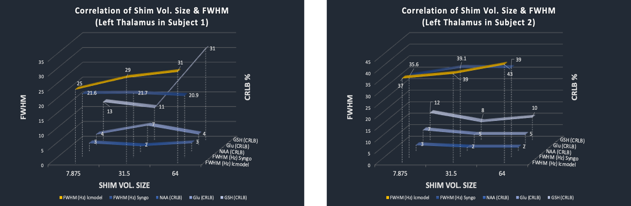
FWHM- The overall linewidths in left thalamus were better than left hippocampus (Figure 4). Different from the left hippocampus, the FWHM values reported by Siemens Syngo had not changed much when shim VOI increased. However, the linewidths estimated by LCModel showed linewidth increased with shim VOI.
Metabolites- Similar to what we observed in left hippocampus, the CRLBs of NAA and Glu were relatively stable as shim VOI increased. With all linewidths reported by either Siemens Syngo or LCModel less than 45Hz, the CRLBs of GSH of both subjects were below 20% except for the 64 cc shim VOI in subject 1 which gave 31%.
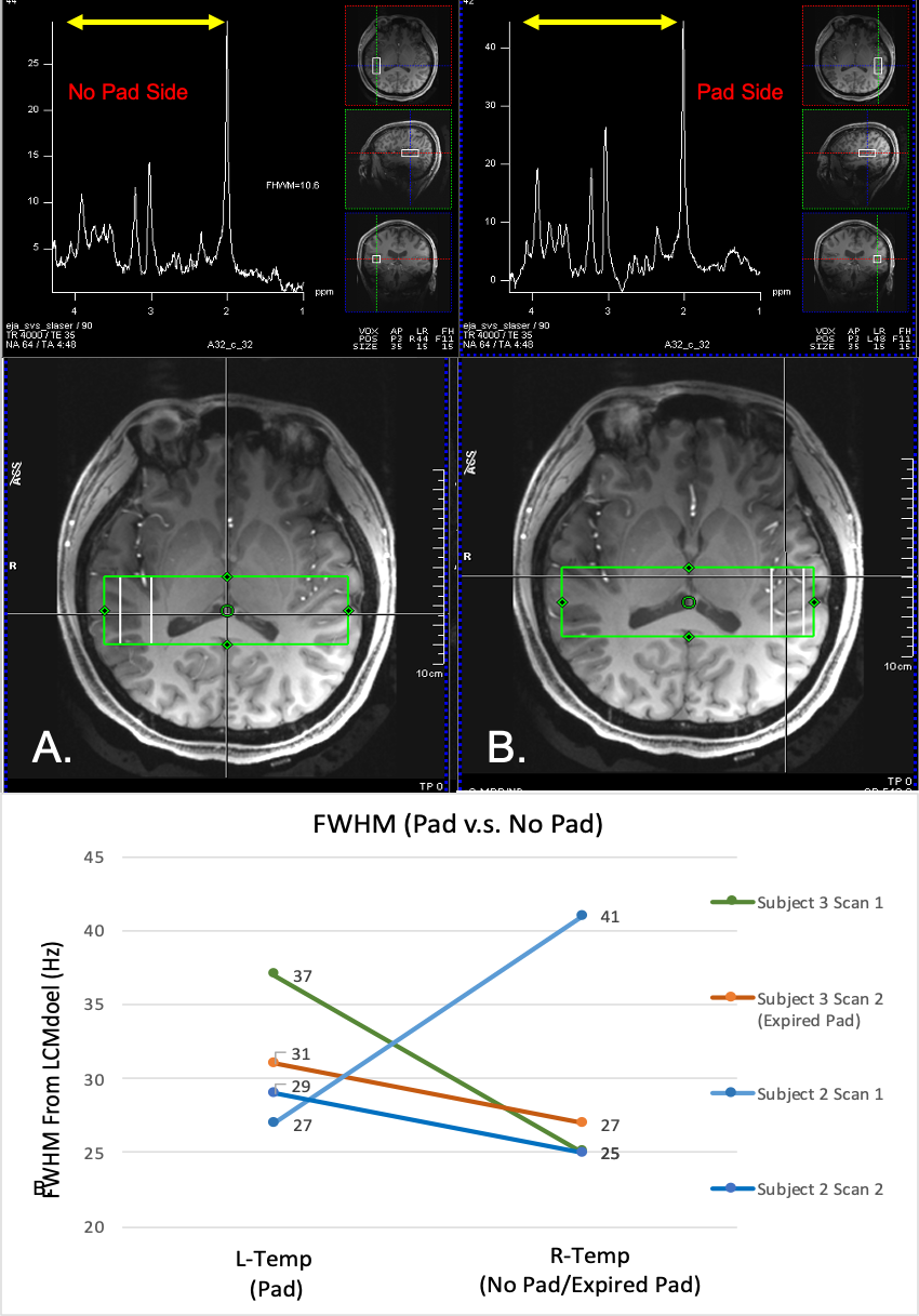
Dielectric pads effect on shim: Since enlarging shim VOI by four and eight times did not seemed to improve linewidths of the acquisition voxel, we also examined the effect of dielectric pads to see if that could improve the linewidths since the dielectric pads have been shown to improved SNR6 (Figure 5). The same equipment and scan protocol as described above was used, but with an elongated shim VOI to cover both temporal lobe area (12x 3.5x1.5cm3) and acquired sLASER-SVS in both left (with pads) and right temporal lobes (without pad or with expired pad). However, FWHM reported by LCModel did not show improvement when pad was added, which was similar to the previous finding6.
Summary and Conclusion:
B0 homogeneity is highly subject and region dependent. Enlarging shim VOI by four or eight folds in inhomogeneous regions like left hippocampus and left thalamus did not improve the B0 homogeneity of the MRS voxel. As suggested by experts’ consensus recommendations, “a moderate extension of the shim VOI beyond the voxel is useful...(e.g. using 25x25x25mm3 shim VOI for a 20x20x20mm3 voxel)” 7 which is about 2 times of the voxel size, so our results showed B0 homogeneity might not be improved if the shim VOI size is larger than this. Also, unlike NAA and Glu, we found that low concentration metabolites as GSH are particularly sensitive to B0 homogeneity, which the inappropriate enlargement of the shim VOI could worsen its measurement. Despite these negative results, this study shows a robust method of measuring B0 homogeneity and its effects on spectroscopy data.Acknowledgements
No acknowledgement found.References
1. Lin A, Tran T, Bluml S, et al. Guidelines for Acquiring and Reporting Clinical Neurospectroscopy. Semin Neurol. 2012;32:432–453.
2. Juchem C and Graaf R A, B0 Magnetic Field Homogeneity and Shimming for In Vivo MR Spectroscopy. Anal Biochem. 2017 July 15; 529: 17–29.
3. Singh S, Khushu S, Kumar P, et al. Evidence for regional hippocampal damage in patients with schizophrenia. Neuroradiology. 2018 Feb;60(2):199-205.
4. Akbaş S, Nahir M, Pirzirenli ME, et al. Quantitative analysis of the amygdala, thalamus and hippocampus on MRI in pediatric bipolar disorders and compared with the children of bipolar parents and healthy control. Psychiatry Res Neuroimaging. 2017 Dec 30;270:61-67. doi: 10.1016/j.pscychresns.2017.08.007.
5. Gigante AD, Lafer B, Yatham LN. (1)H-MRS of hippocampus in patients after first manic episode. World J Biol Psychiatry. 2014 Feb;15(2):145-54.
6. Liao H et al. The Effects of Dielectric Pads on Brain MR Images and Spectroscopy at 7T. Proc Soc Magn Rad Tech. SMRT 28th Annual Meeting, Montreal, 2019.
7. Juchem C, Cudalbu C, de Graaf R A, et al. B0 shimming for in vivo magnetic resonance spectroscopy: Experts' consensus recommendations. Spec. iss. rev. art. NMR in Biomedicine. 28 June 2020.
Figures