Imaging Biomarkers of Cerebral Amyloid Angiopathy
1Massachusetts General Hospital, Boston, MA, United States
Synopsis
Cerebral amyloid angiopathy (CAA), deposition of ß-amyloid in small cortical and leptomeningeal vessels, is a common age-related pathology associated with lobar hemorrhage and cognitive impairment. Among MRI-based markers of CAA are lobar hemorrhages and microbleeds, convexity subarachnoid hemorrhage and cortical superficial siderosis, microinfarcts, altered structural connectivity on diffusion-tensor imaging, dilated perivascular spaces in centrum semiovale, multi-spot pattern white matter hyperintensities, and impaired vascular reactivity to visual stimulation. Current diagnosis of CAA per the Boston Criteria depends on detection of multiple strictly lobar hemorrhagic lesions. Ongoing studies suggest that incorporation of emerging white matter markers may improve sensitivity without compromising specificity.
 Slide #1
 Slide #2
 Slide #3
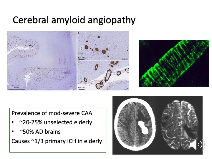 Slide #4
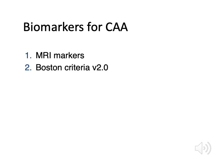 Slide #5
 Slide #6
 Slide #7
 Slide #8
 Slide #9
 Slide #10
 Slide #11
 Slide #12
 Slide #13
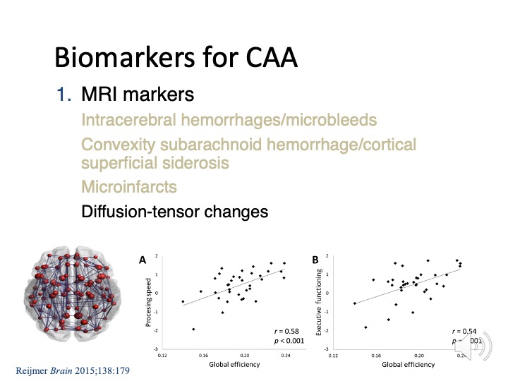 Slide #14
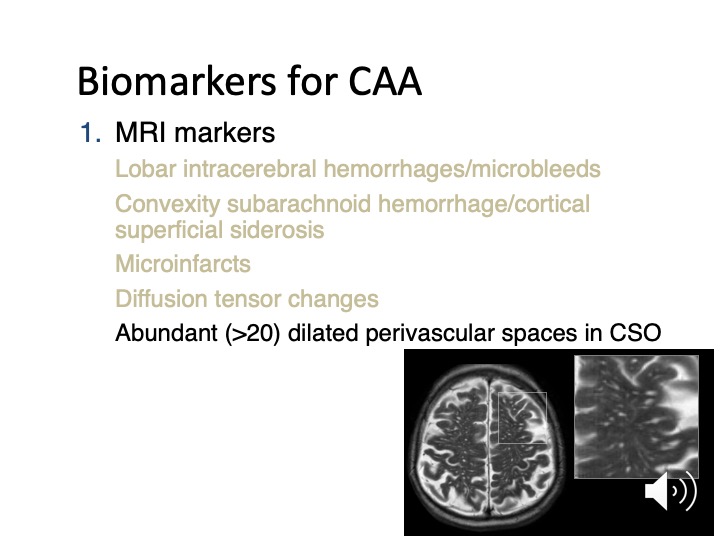 Slide #15
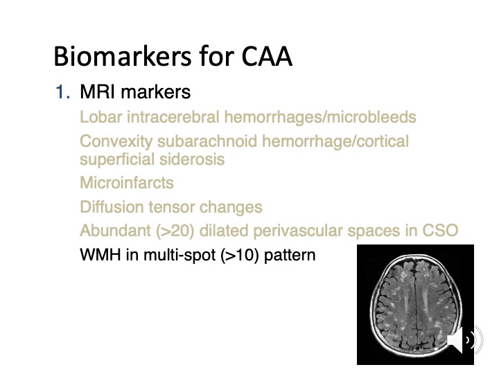 Slide #16
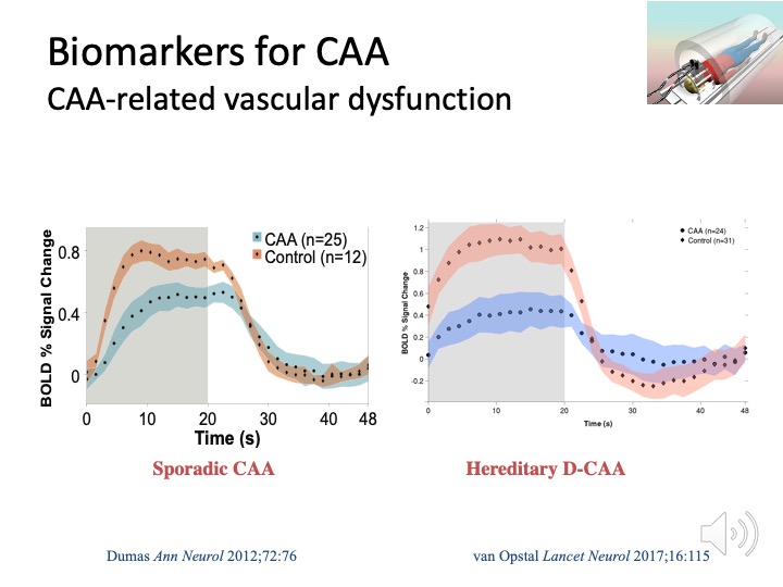 Slide #17
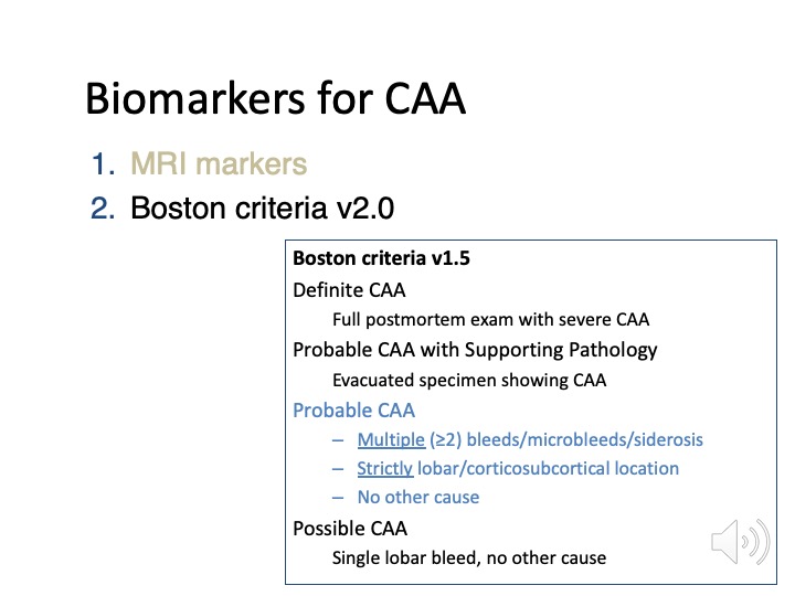 Slide #18
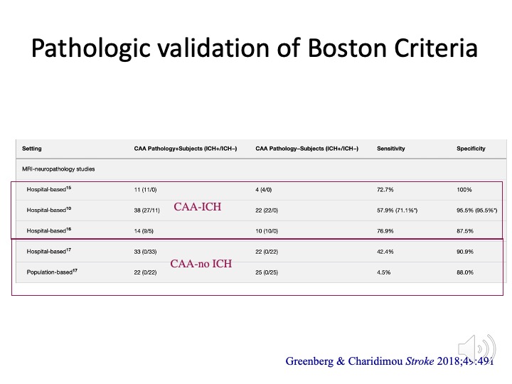 Slide #19
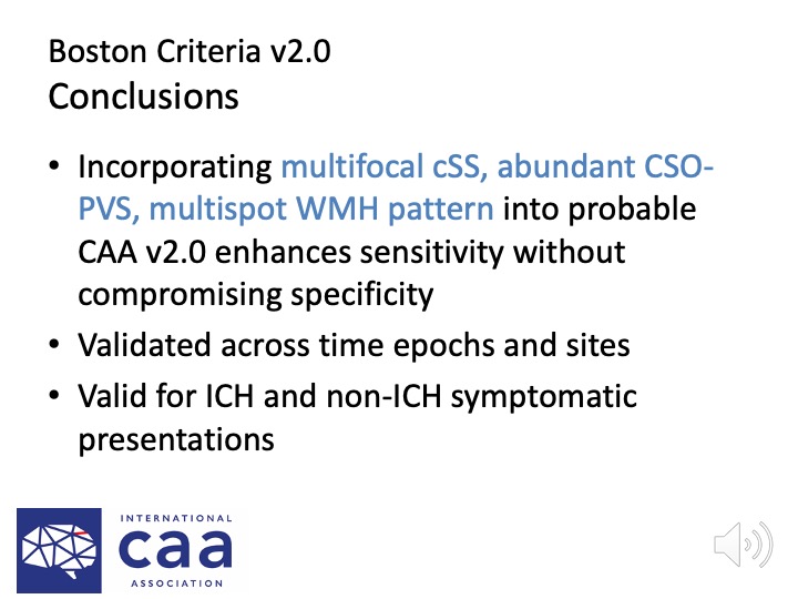 Slide #20