The AI-Augmented MSK Radiologist
Hollis G. Potter1
1Radiology and Imaging, Hospital for Special Surgery, New York, NY, United States
1Radiology and Imaging, Hospital for Special Surgery, New York, NY, United States
Synopsis
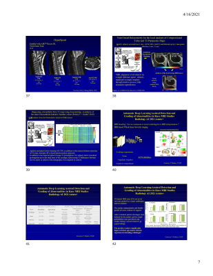
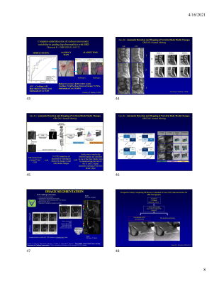
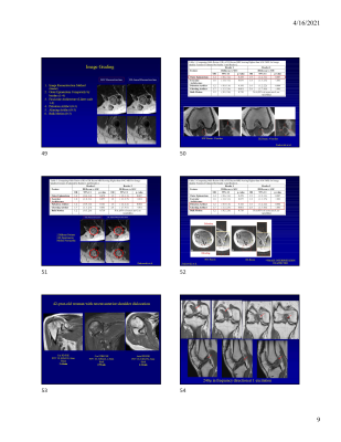
AI is rapidly being adopted into orthopaedic imaging, improving accuracy and speed of acquisition, improving image presentation and reproducibility, and aiding in image interpretation including fracture detection, osteoarthritis metrics and degenerative spine changes. Other applications include cartilage and meniscal segmentation, more efficient interpretation of large quantities of quantitative MR data, and the ability to increase spatial resolution and disease detection. Examples of pitfalls in study design are presented, as well as current applications of AI into both conventional and advanced imaging.

