Myocardial T1, T2, T2* & ECV Mapping: The Clinical Promise
Wiphada Patricia Bandettini1
1NIH/NHLBI, Bethesda, MD, United States
1NIH/NHLBI, Bethesda, MD, United States
Synopsis
• Parametric mapping tissue characterization has moved beyond the research realm and is now applicable within the clinical setting. Parametric mapping adds value to CMR, taking imaging beyond that of anatomy. Native T1, T2, and T2* offer non-contrast options to better characterize the myocardium. Extracellular volume fraction (ECV) helps to identify edema diffuse fibrosis. Edema, inflammation, fibrosis, and infiltration may be identified noninvasively by CMR. The tools of T1, T2, T2*, and ECV are ready for daily clinical use…if the appropriate QA and standards are followed. Parametric mapping offers improved diagnosis, management, and prognosis in the patient with cardiovascular disease.
 Slide #1
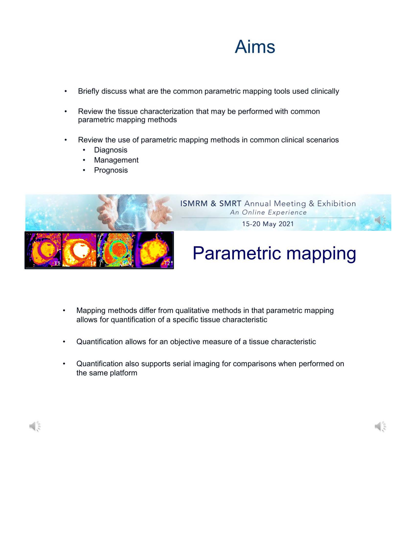 Slide #2
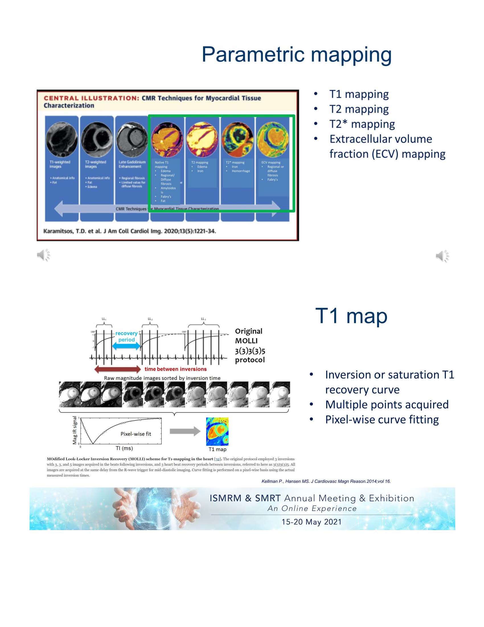 Slide #3
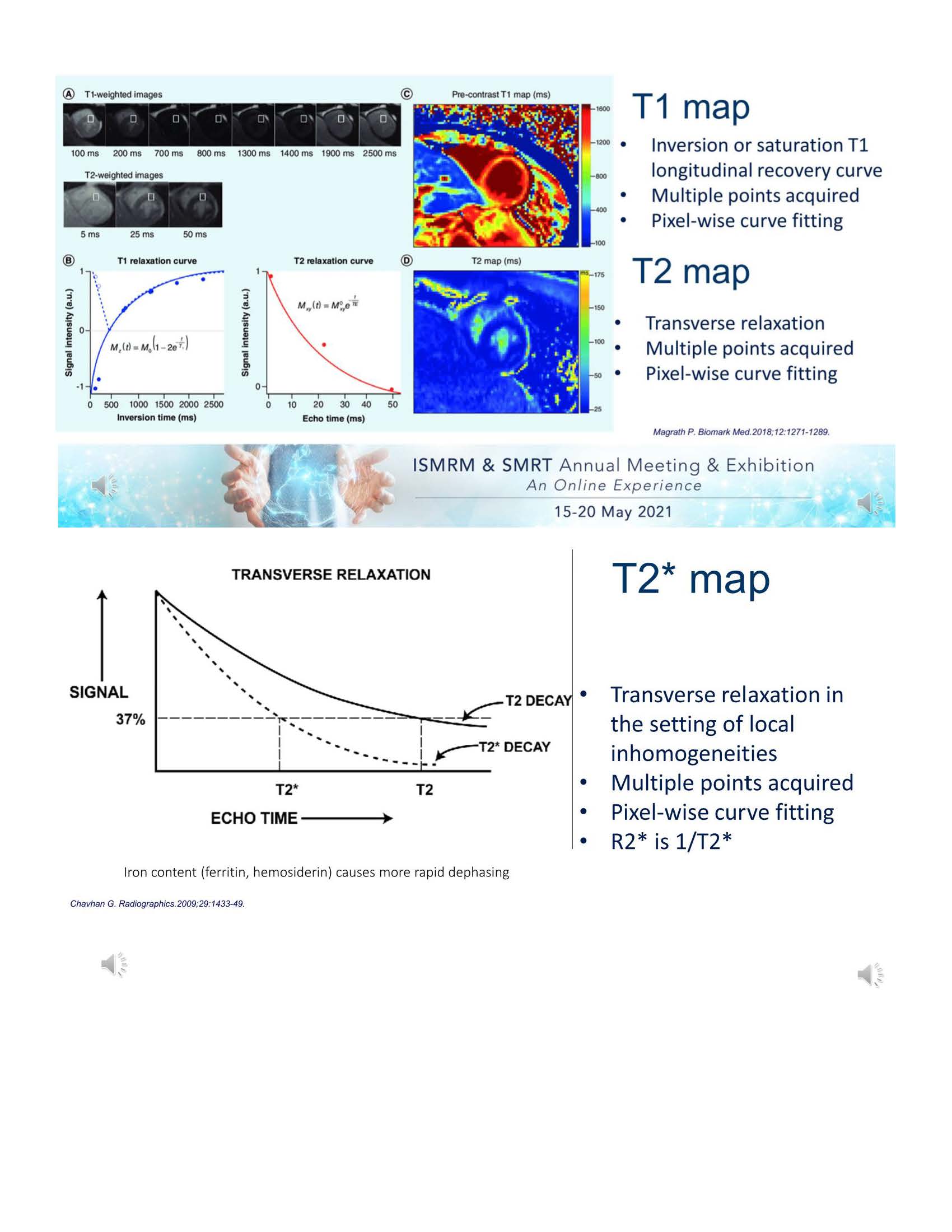 Slide #4
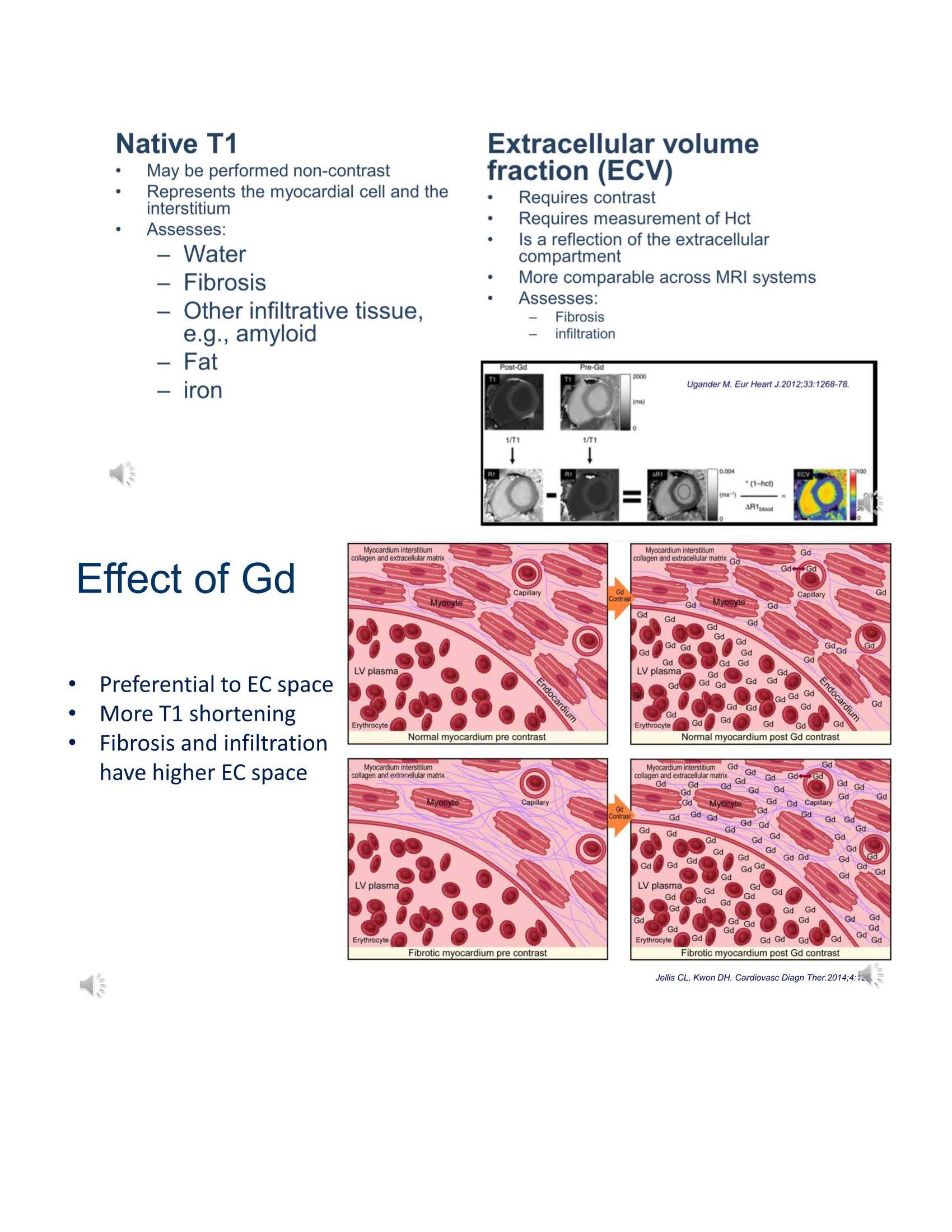 Slide #5
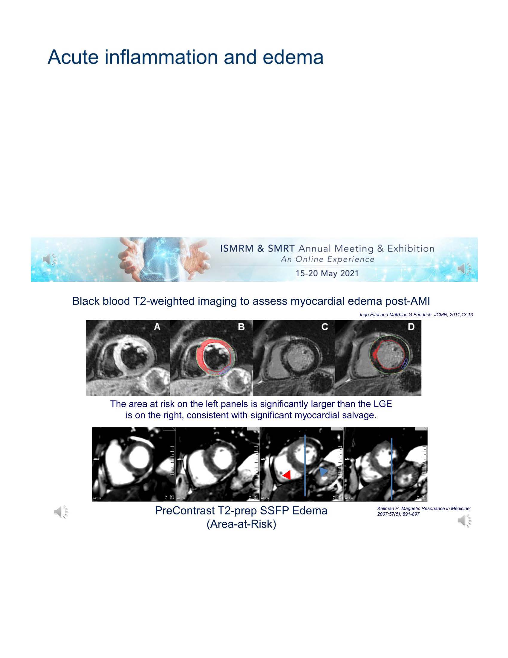 Slide #6
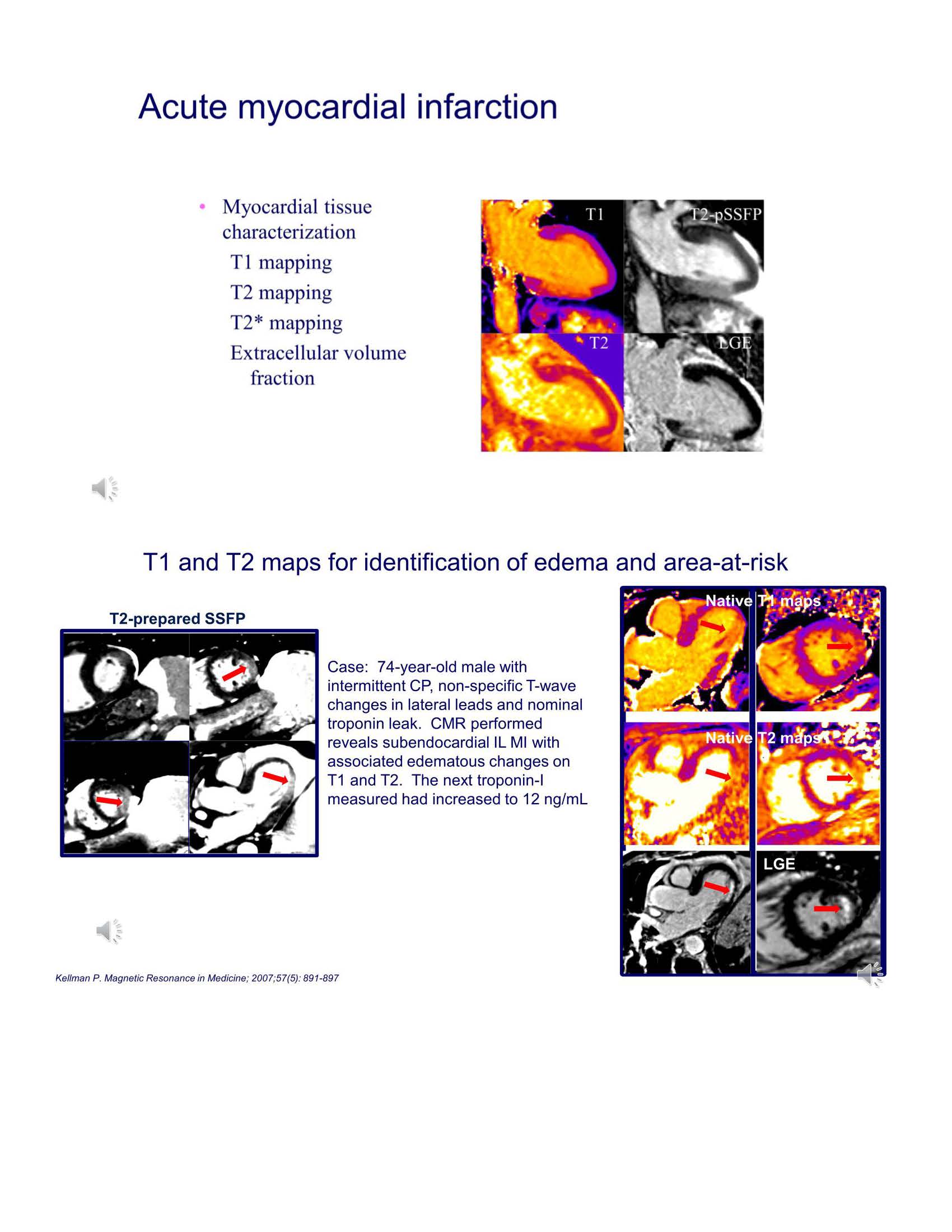 Slide #7
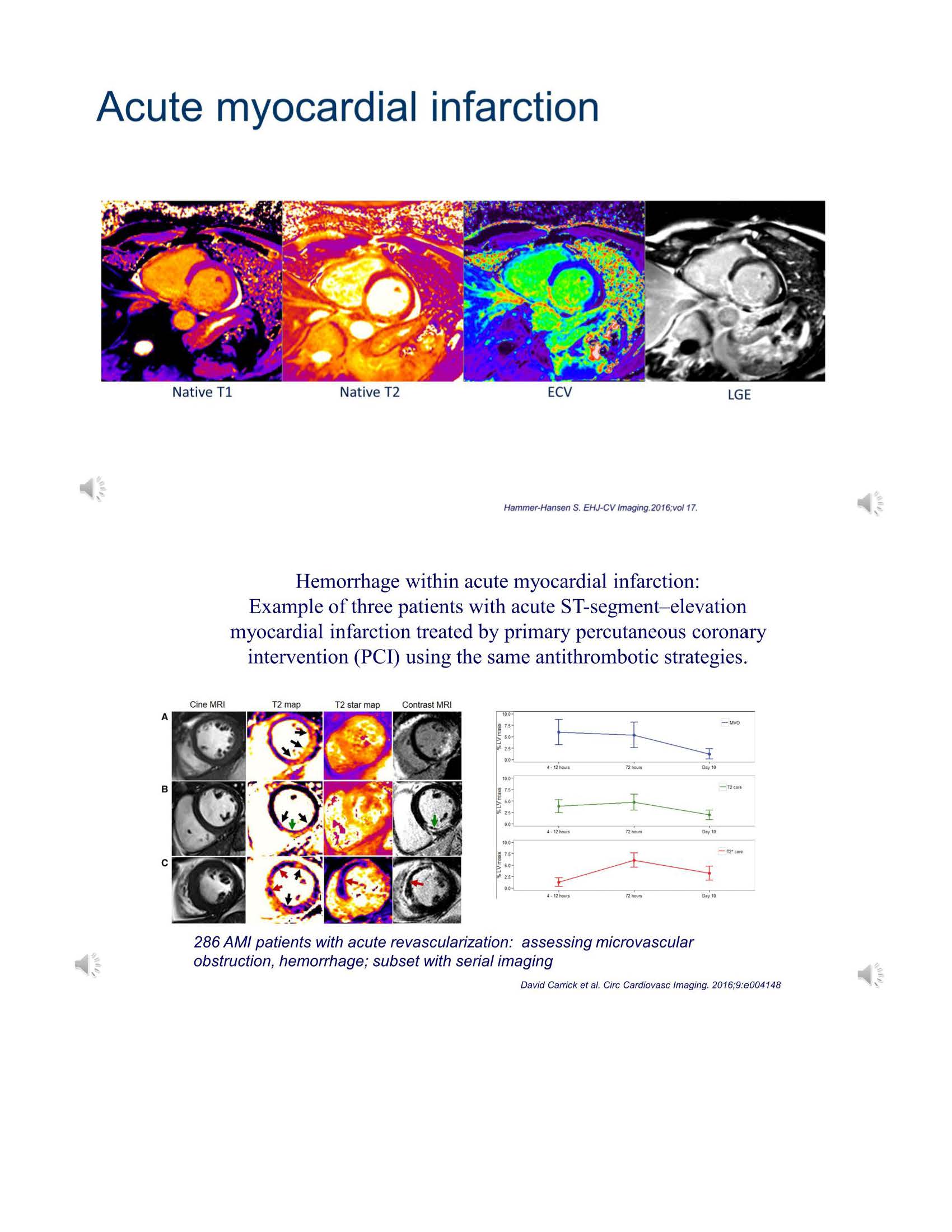 Slide #8
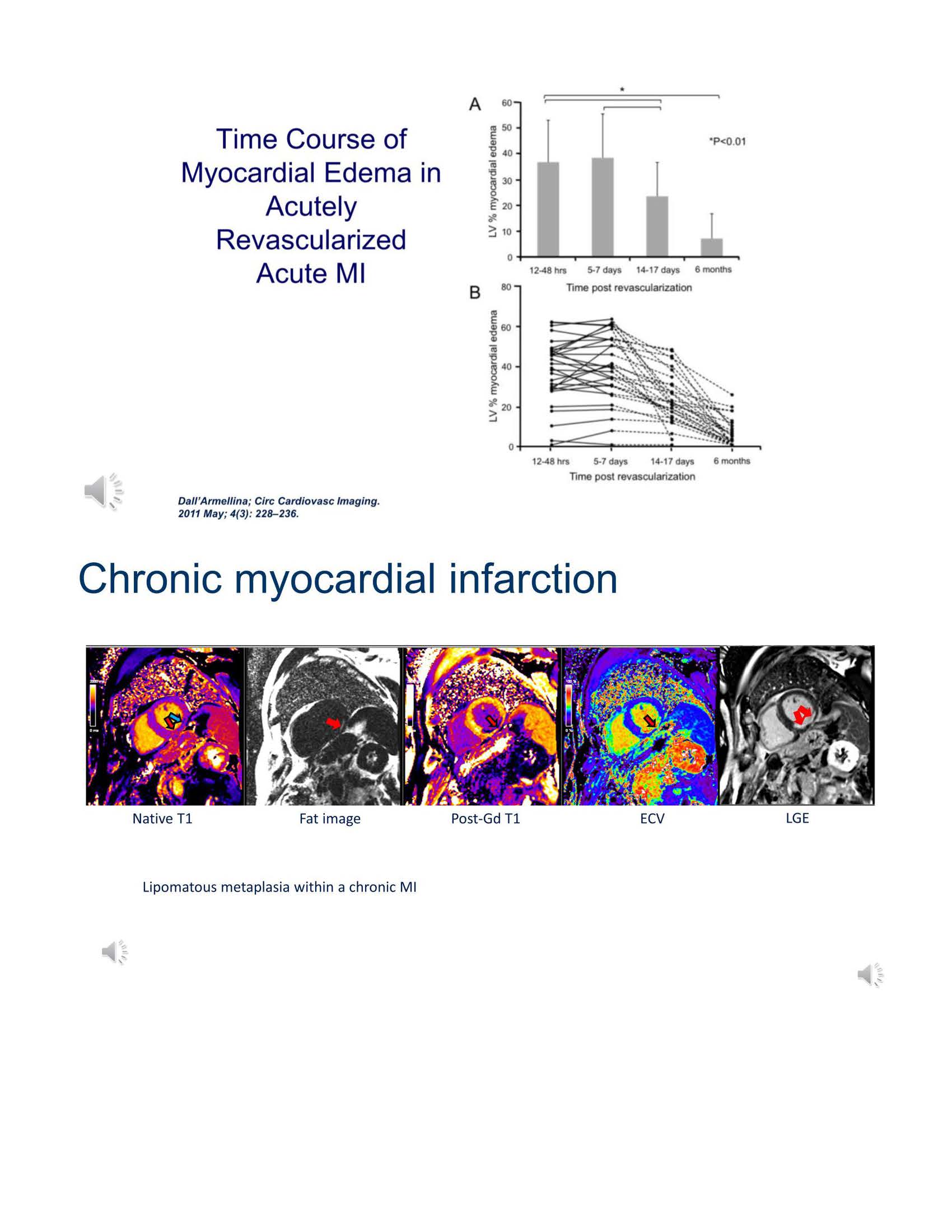 Slide #9
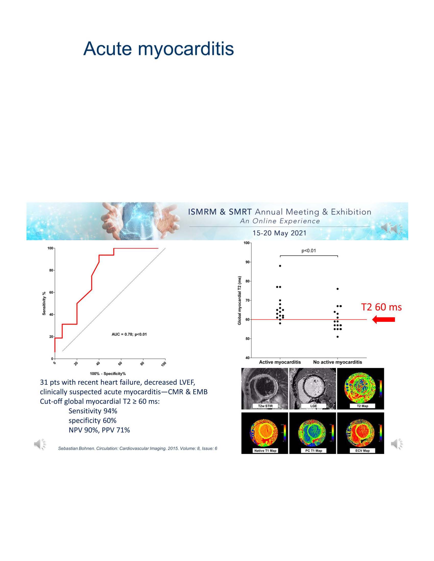 Slide #10
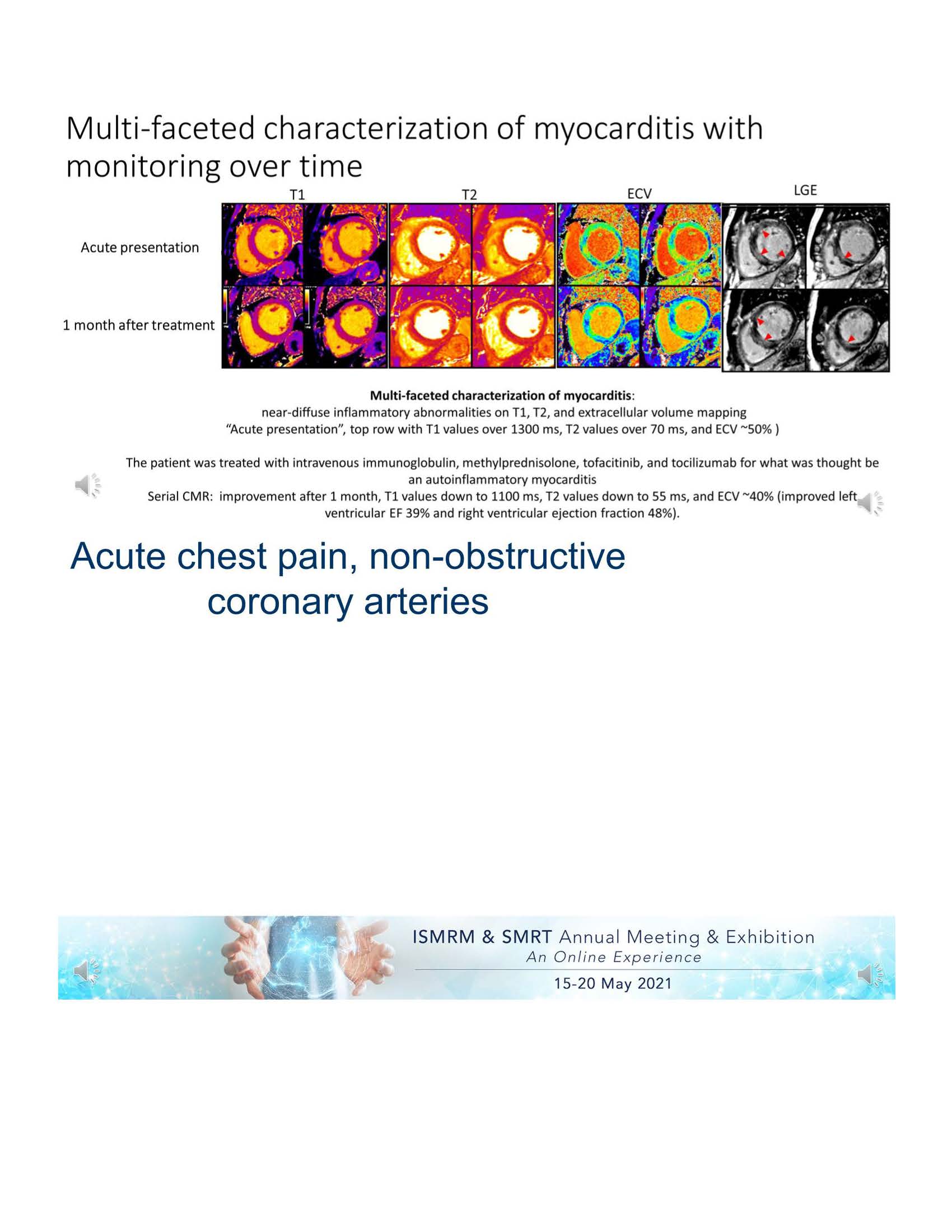 Slide #11
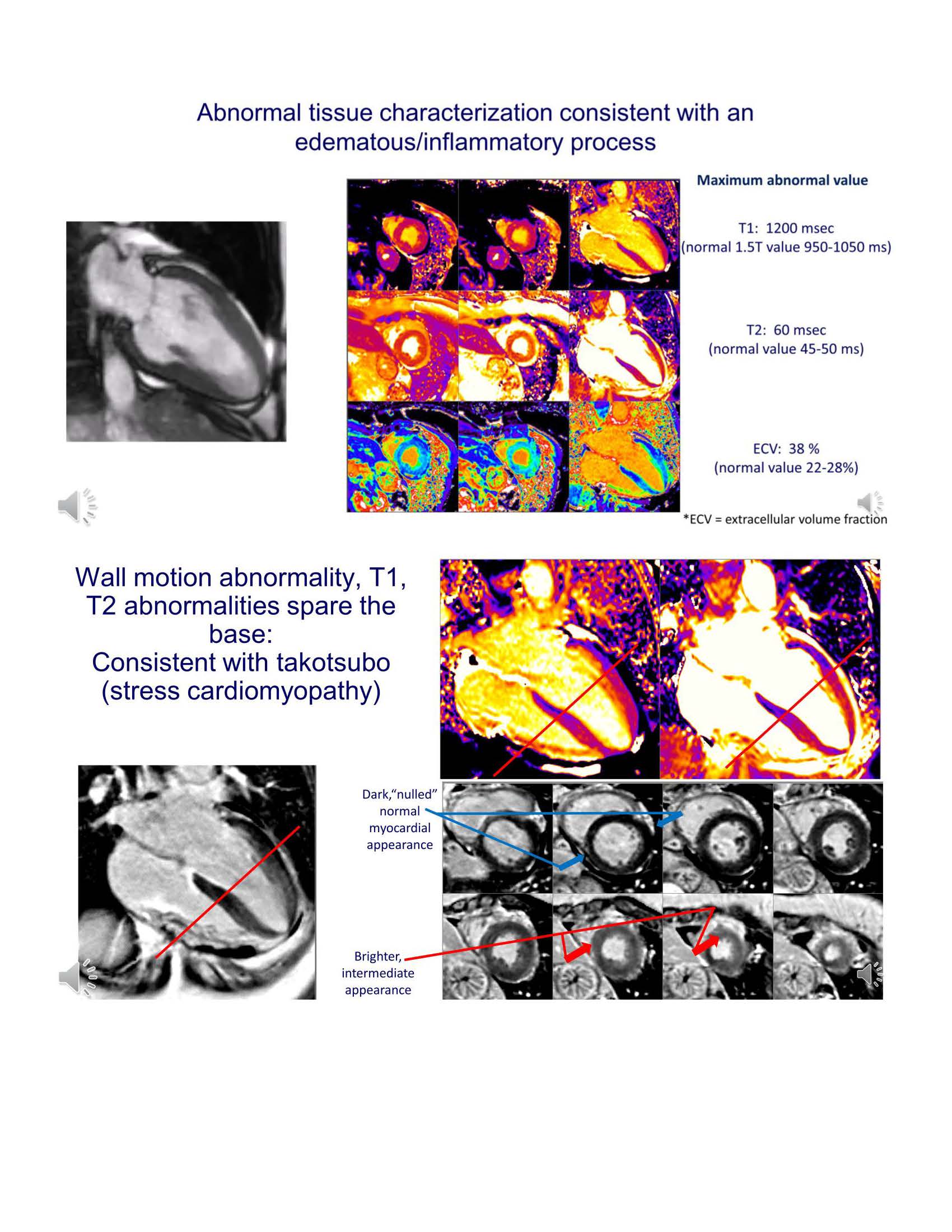 Slide #12
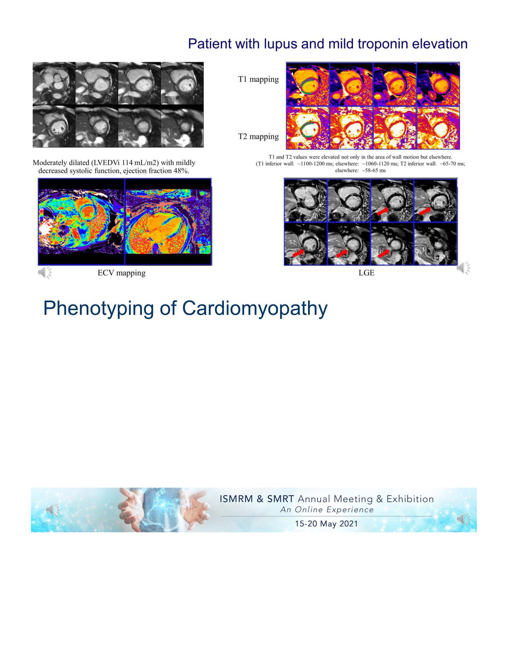 Slide #13
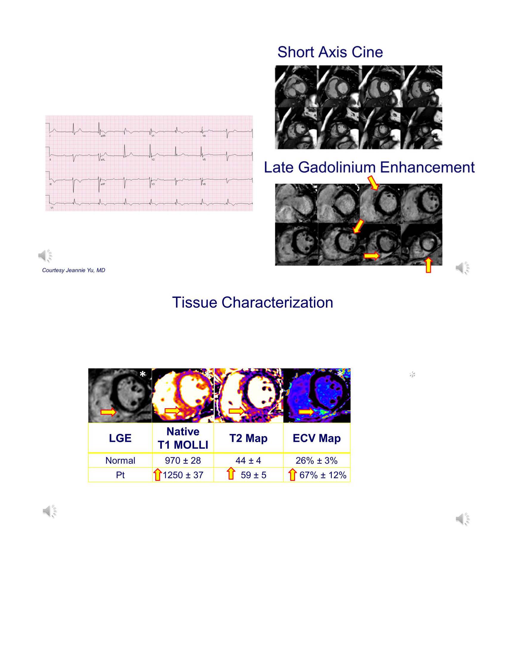 Slide #14
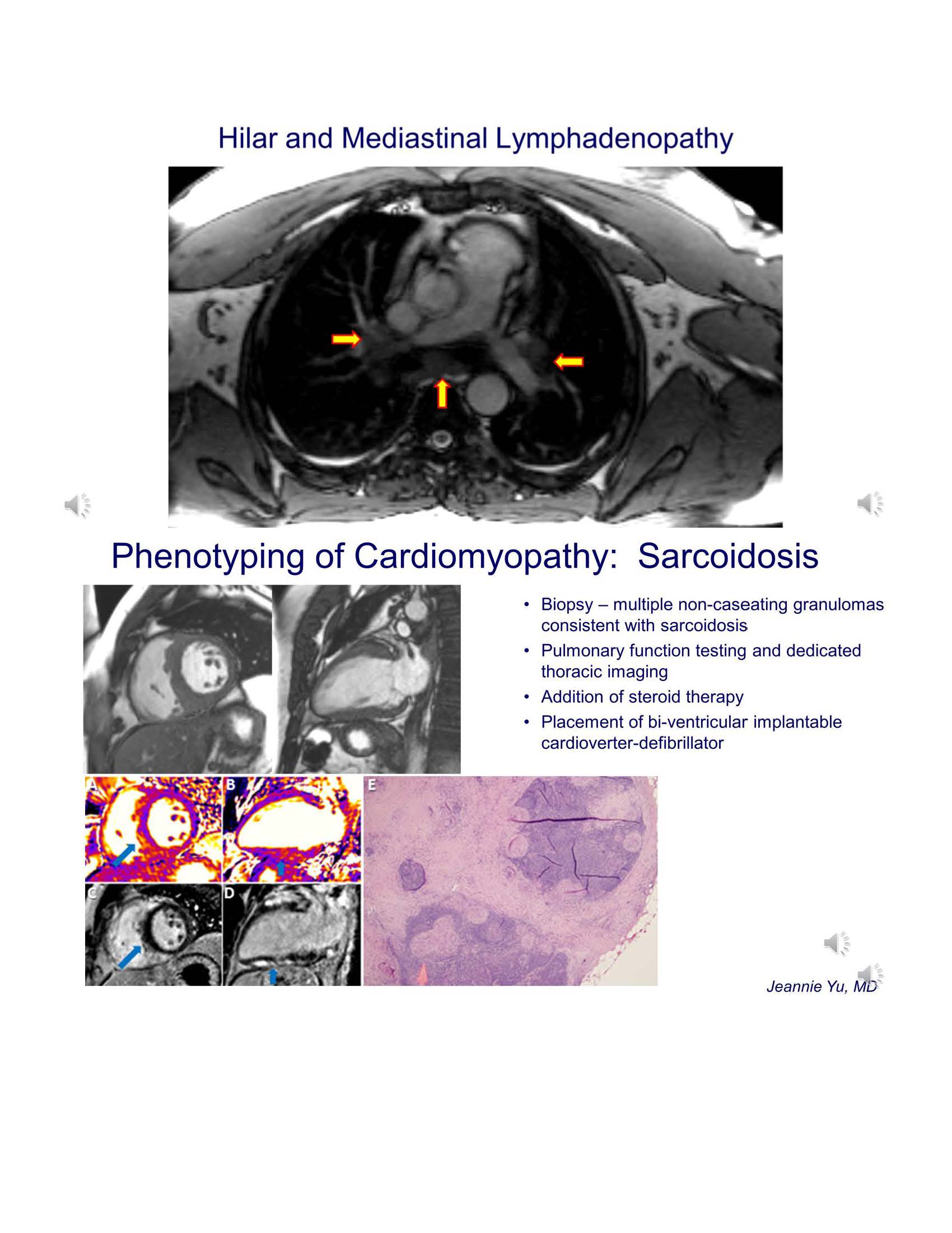 Slide #15
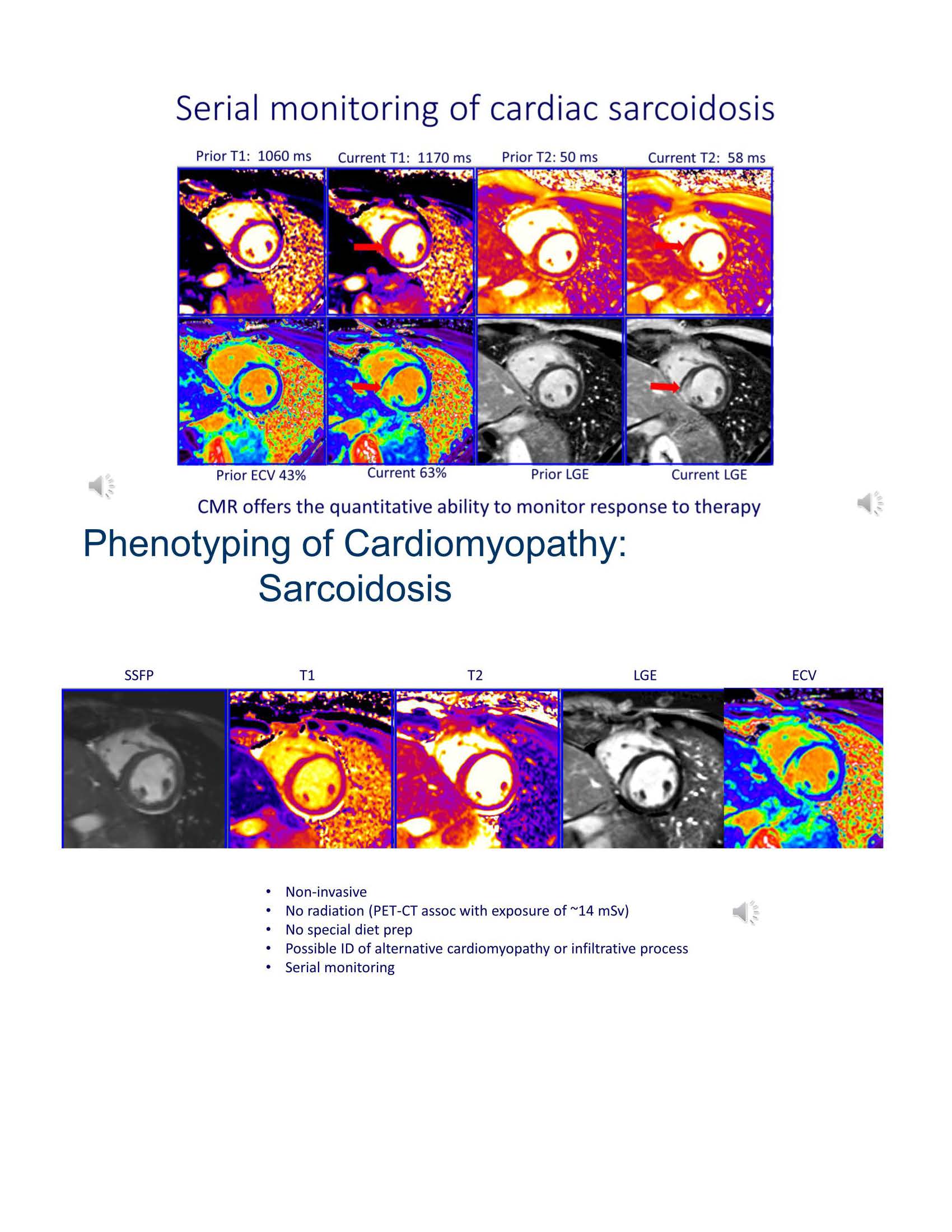 Slide #16
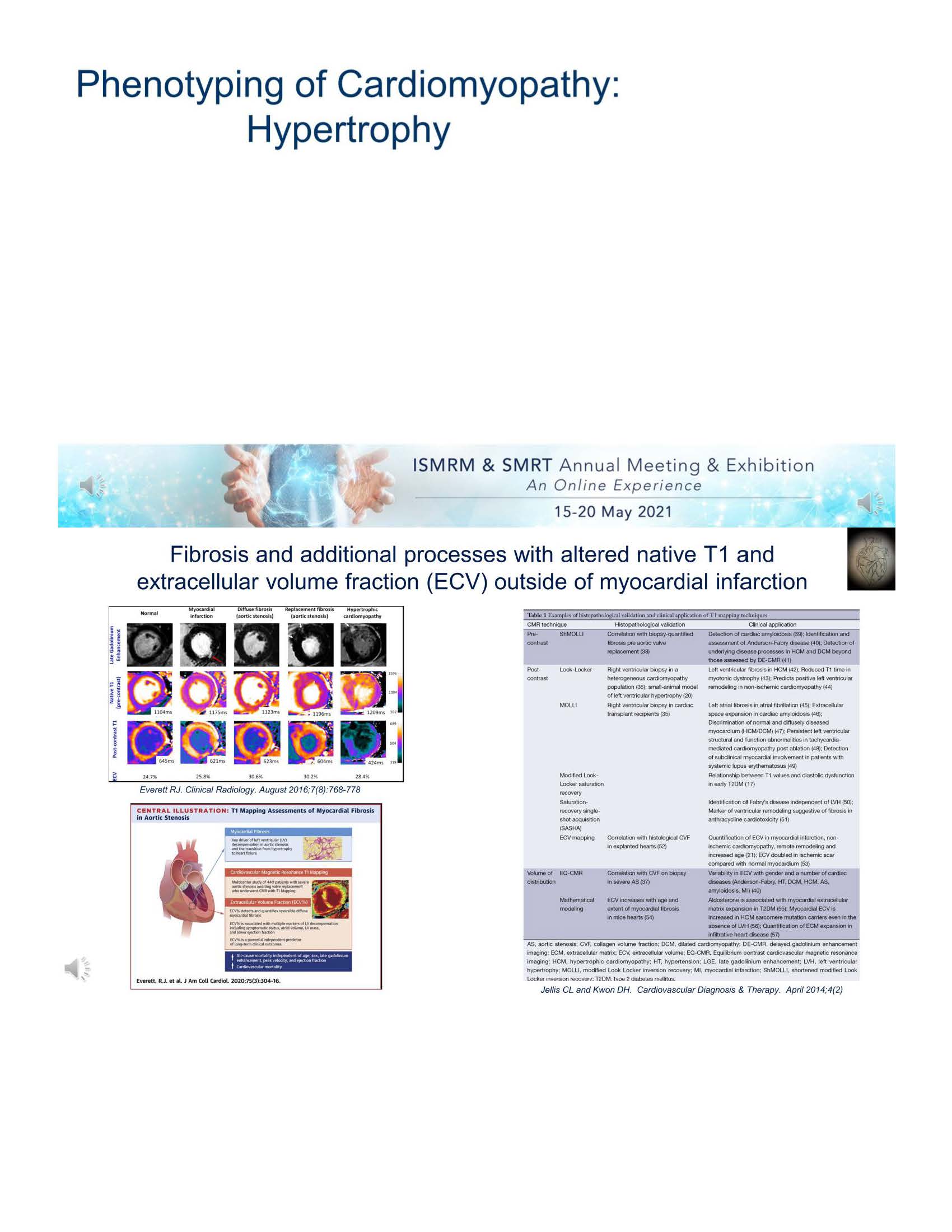 Slide #17
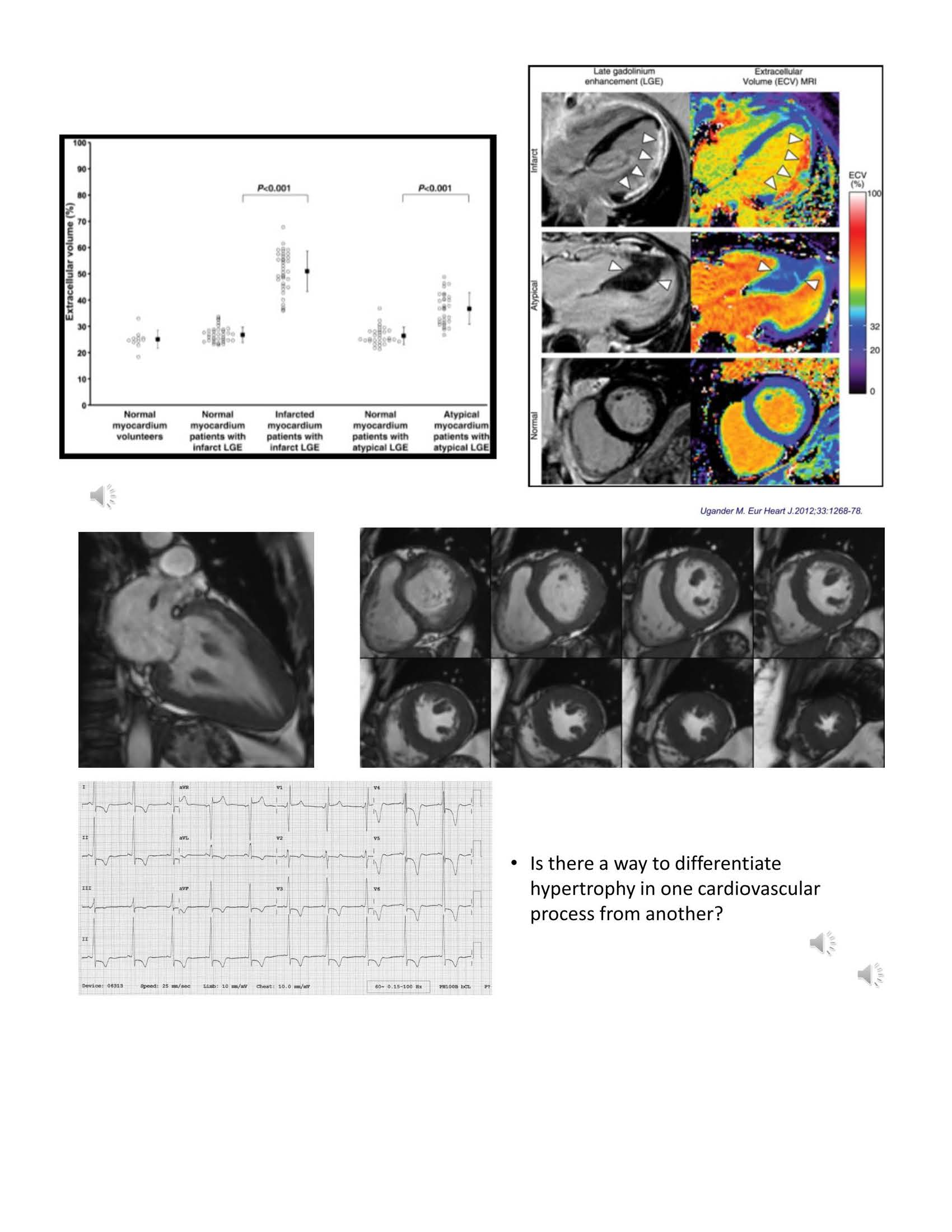 Slide #18
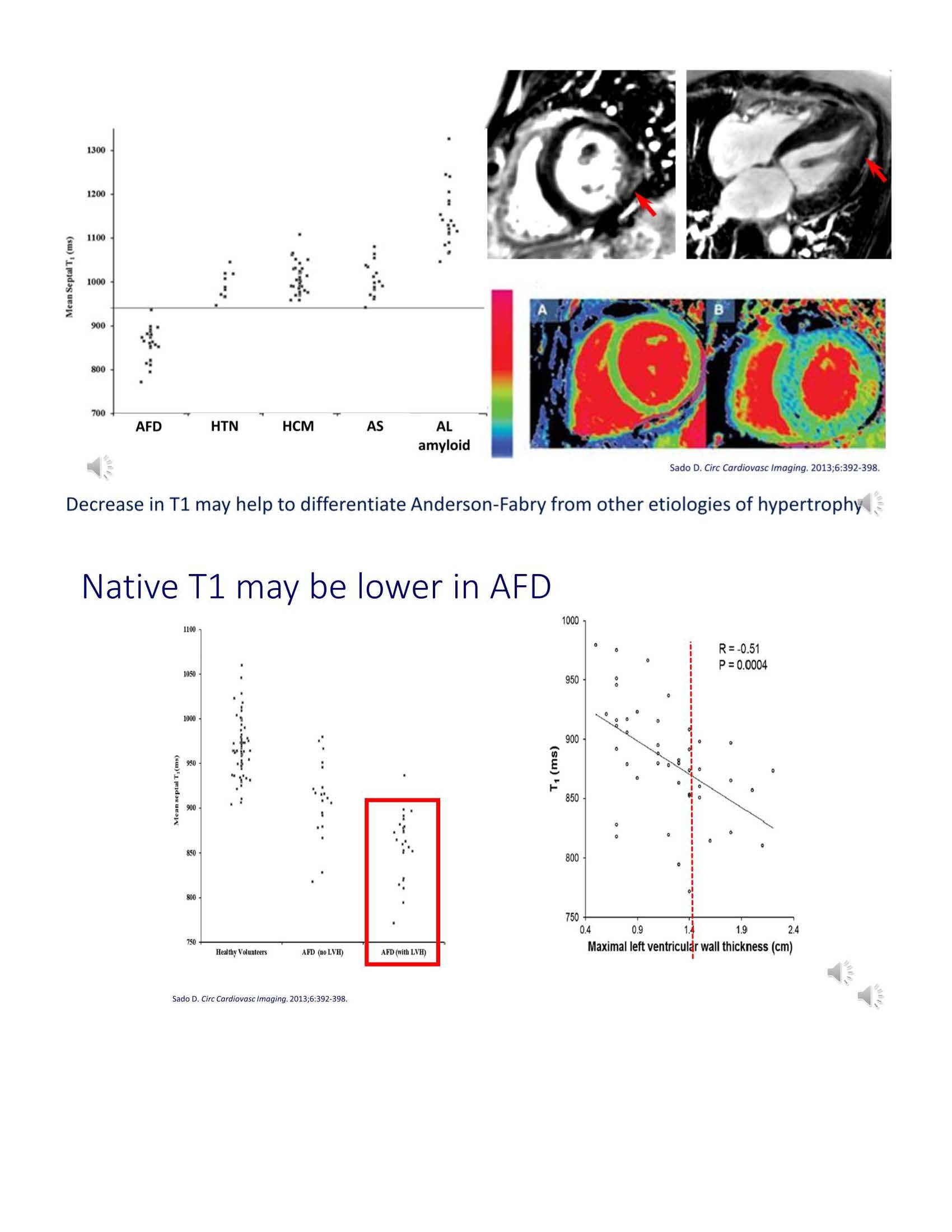 Slide #19
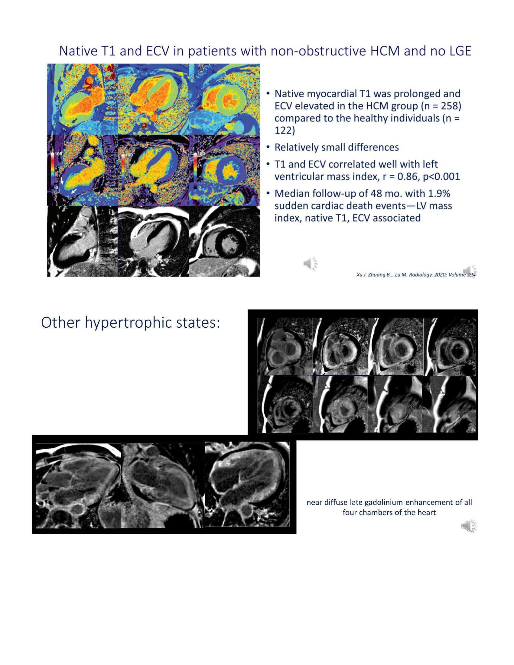 Slide #20
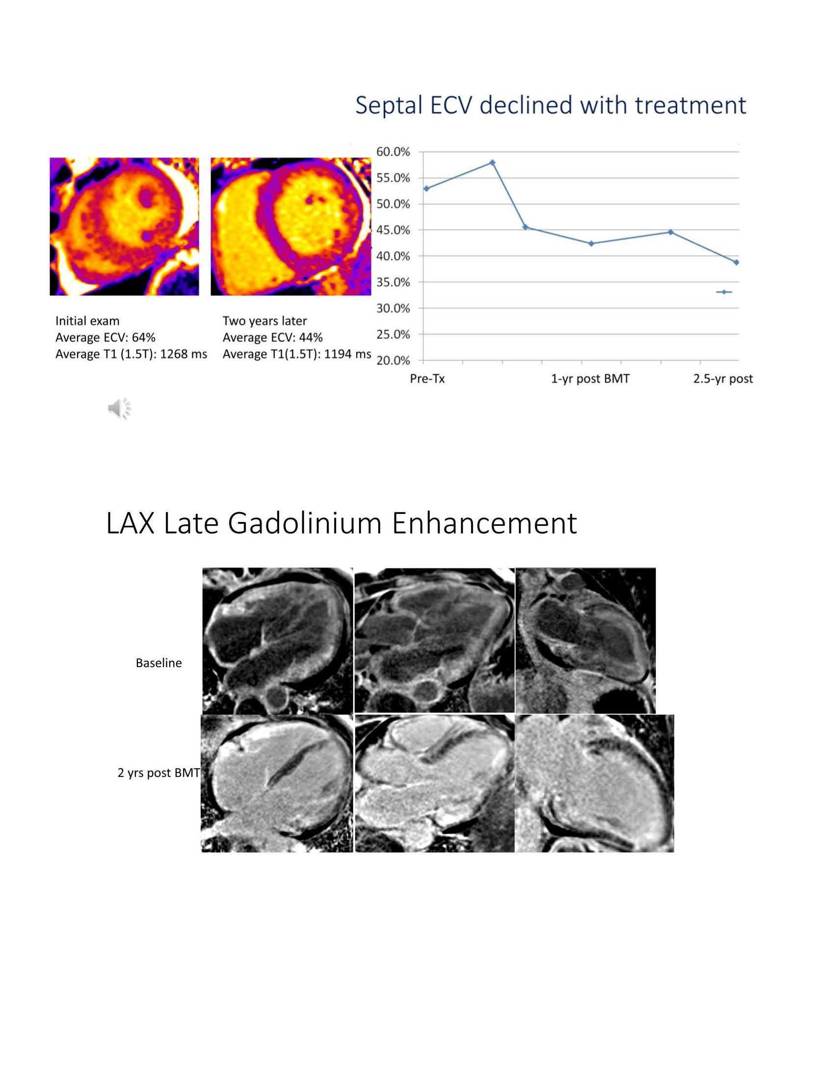 Slide #21
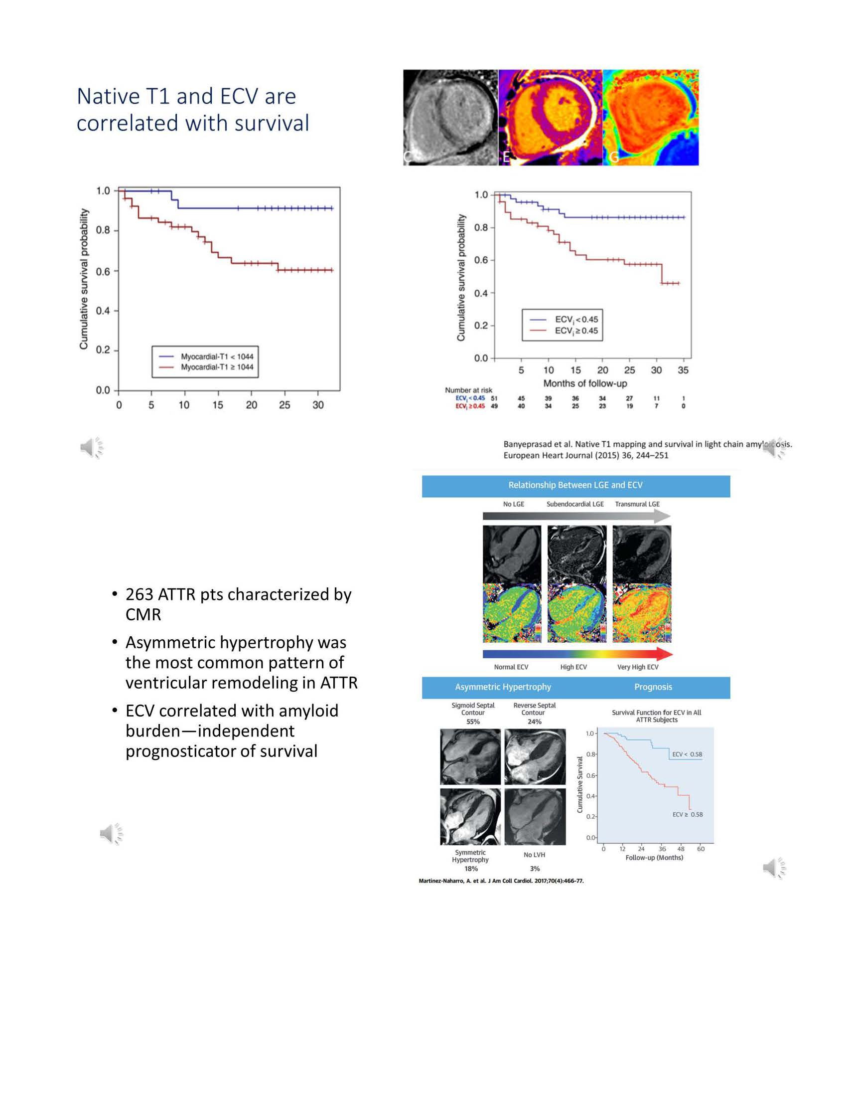 Slide #22
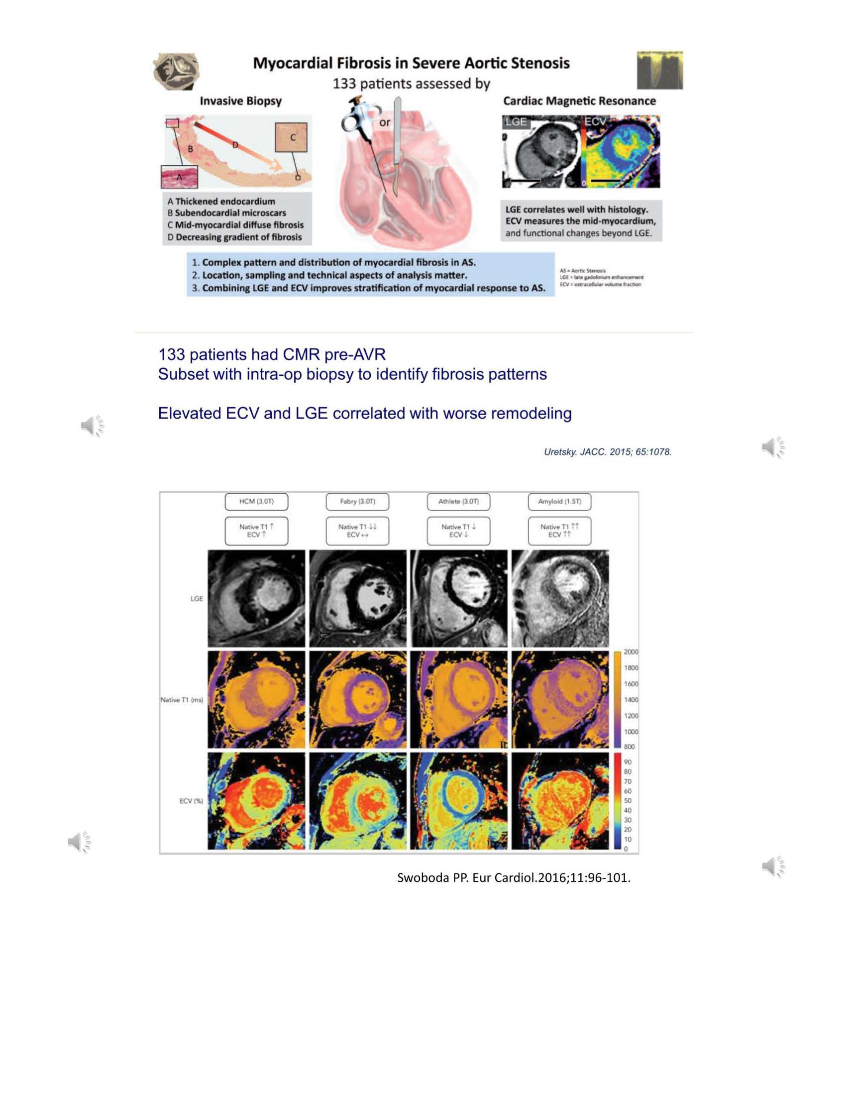 Slide #23
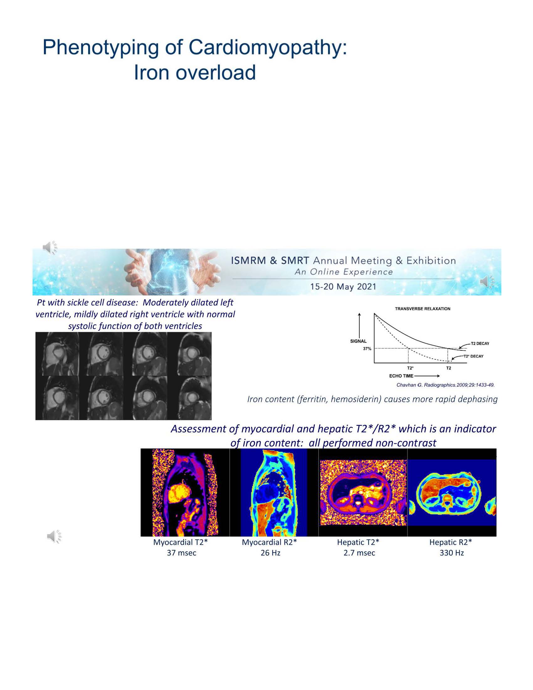 Slide #24
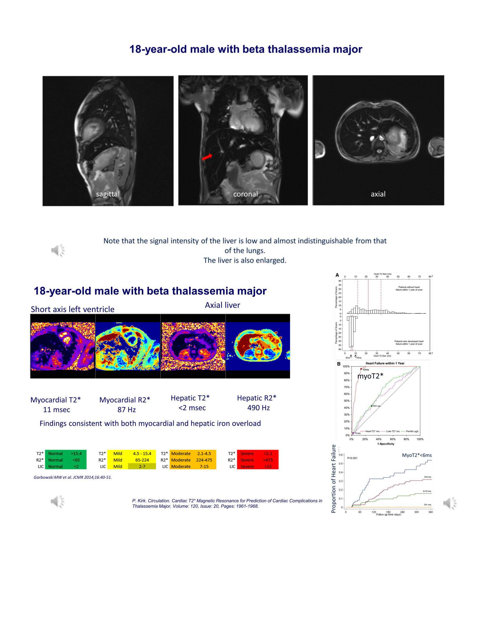 Slide #25
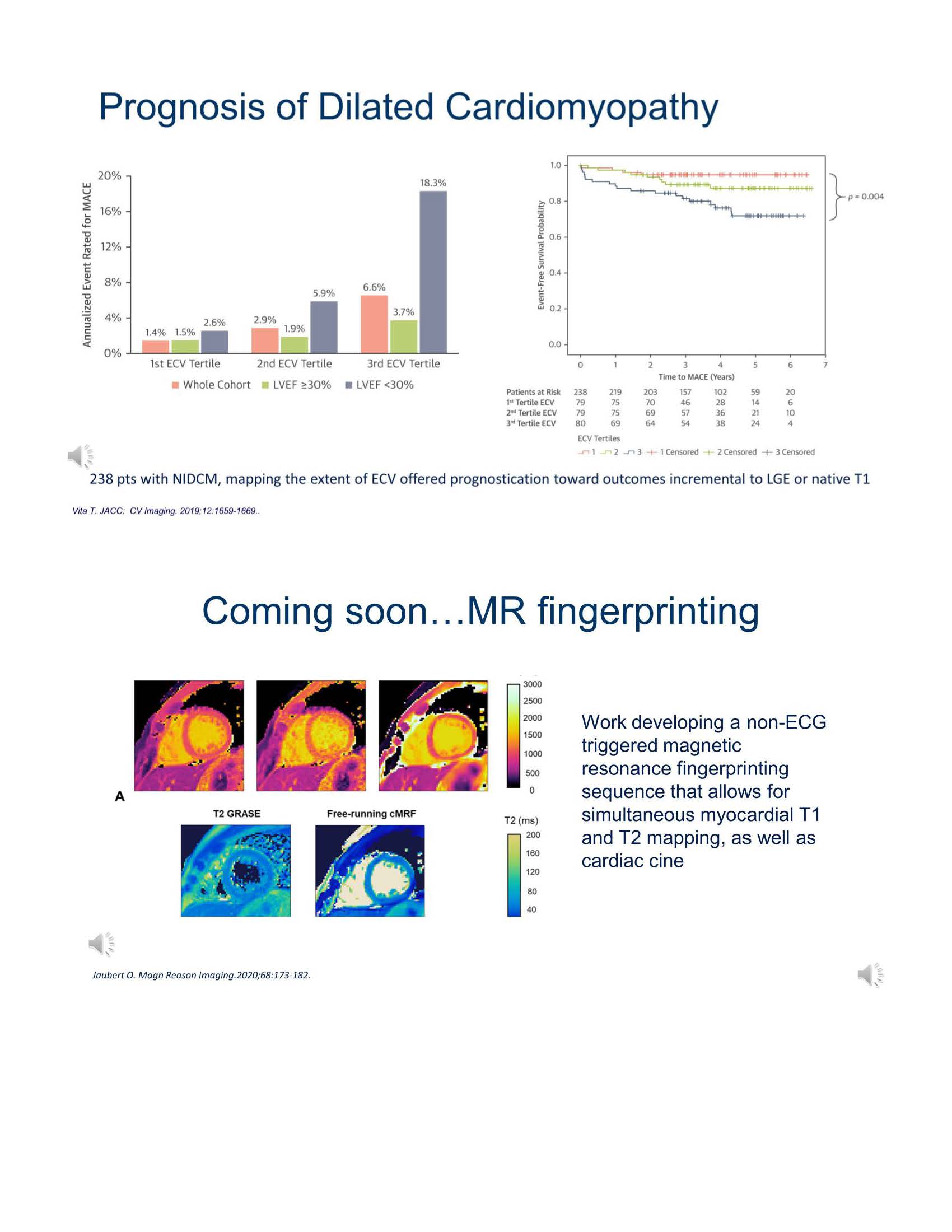 Slide #26
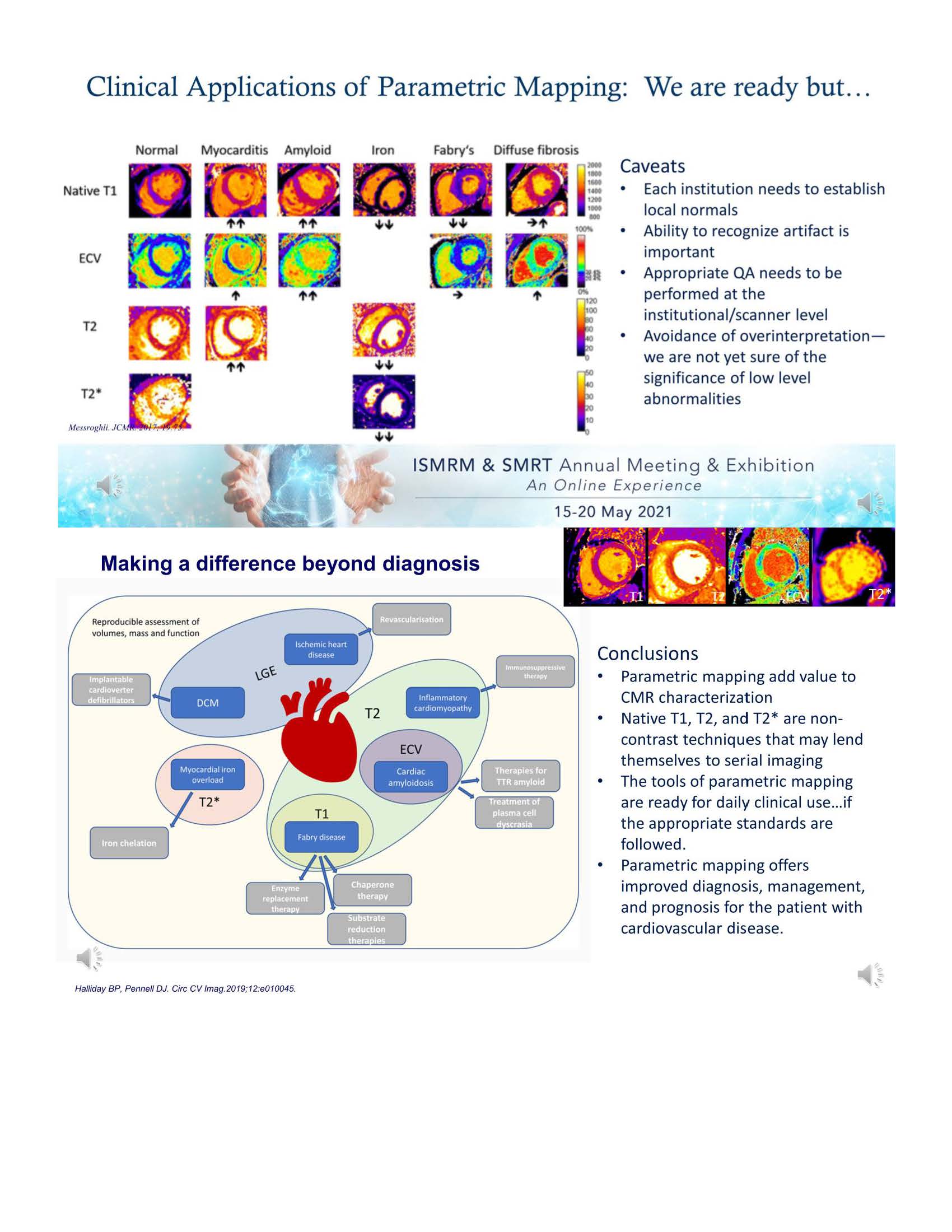 Slide #27
 Slide #28