Brain MR & SARS-CoV-2: Imaging Findings, Complications & Outcomes
Simonetta Gerevini1
1ASST Papa Giovanni XXIII, Bergamo, Italy
1ASST Papa Giovanni XXIII, Bergamo, Italy
Synopsis
A relevant number of COVID-19 patients may present with neurological symptoms in the acute/subacute phase of the disease. Neuroimaging can reveal a wide spectrum of CNS abnormalities, from ischemic lesions to microhemorrhages as well as meningoencephalitis and extensive white matters lesions. Advanced imaging techniques (DWI, DTI, f-MRI) may reveal underlining “inflammation of the brain” in case of persistent neurological symptoms and an unremarkable MRI examination.Long term neurological and neuropsychological sequelae are reported up to 30–40% in COVID-19 survivors, includingfatigue, myalgia, headache, dysautonomia and cognitive impairment (“brain fog”). A complete understanding of these manifestations is mandatory.
 Slide #1
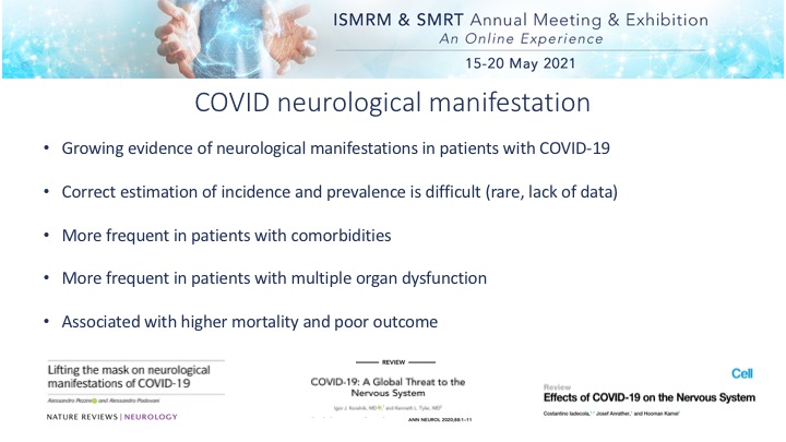 Slide #2
 Slide #3
 Slide #4
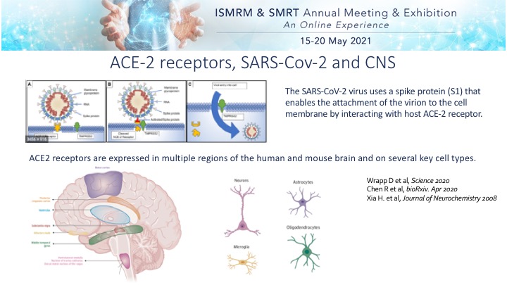 Slide #5
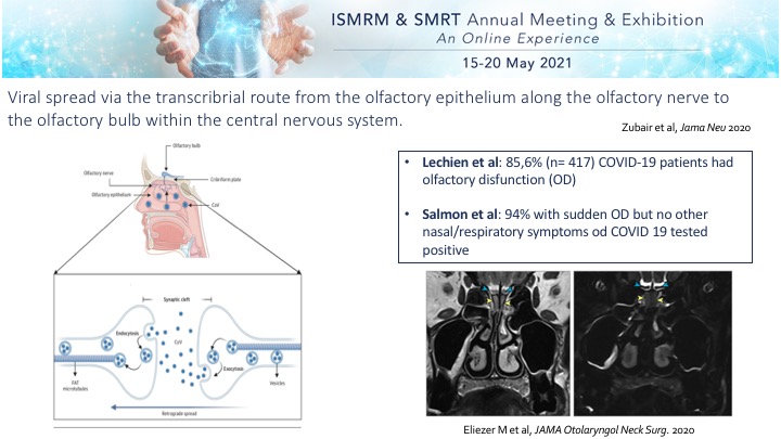 Slide #6
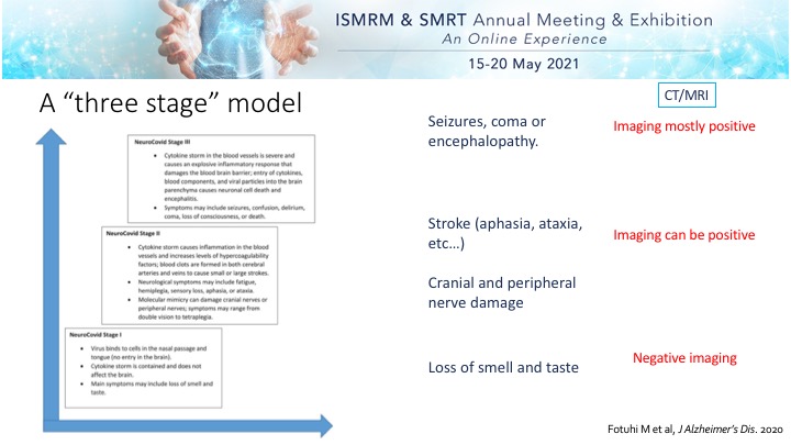 Slide #7
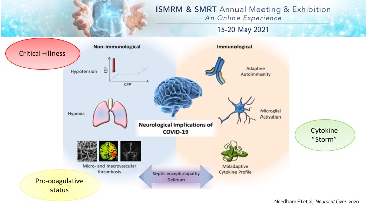 Slide #8
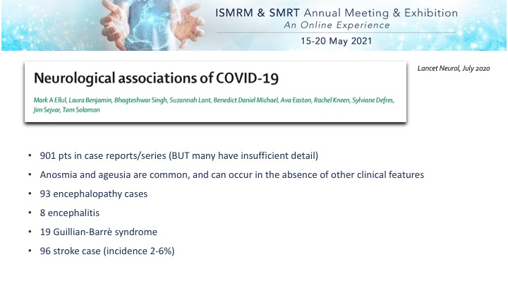 Slide #9
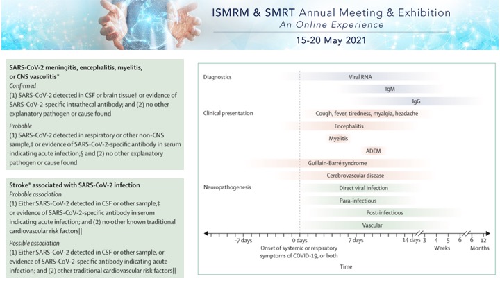 Slide #10
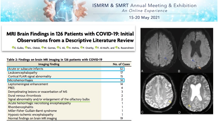 Slide #11
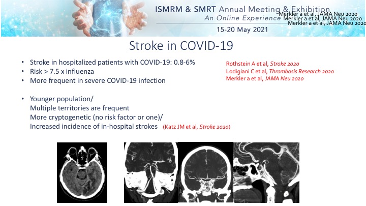 Slide #12
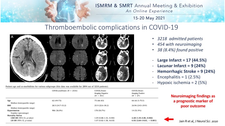 Slide #13
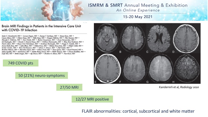 Slide #14
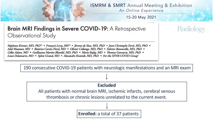 Slide #15
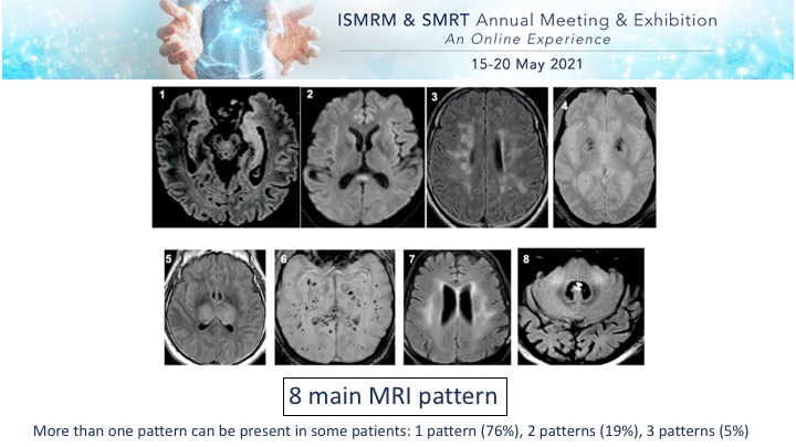 Slide #16
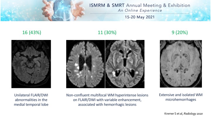 Slide #17
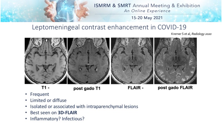 Slide #18
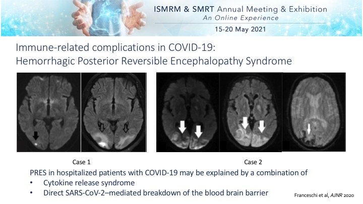 Slide #19
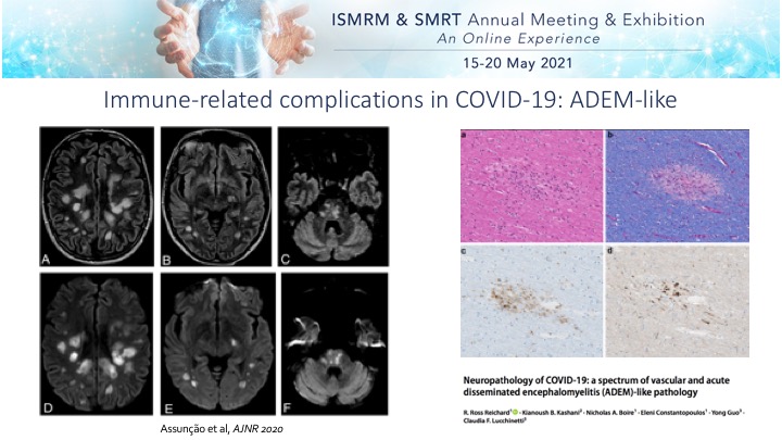 Slide #20
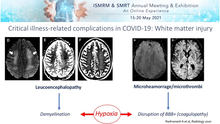 Slide #21
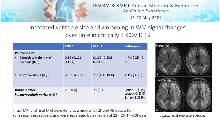 Slide #22
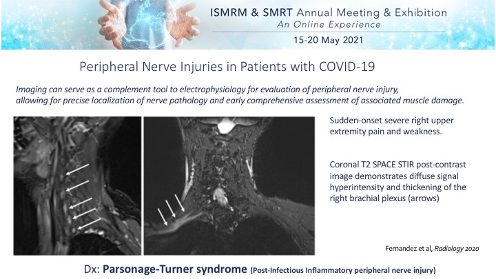 Slide #23
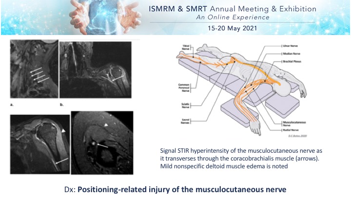 Slide #24
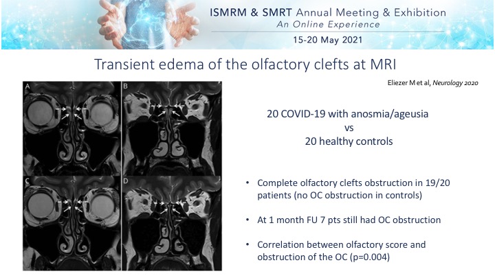 Slide #25
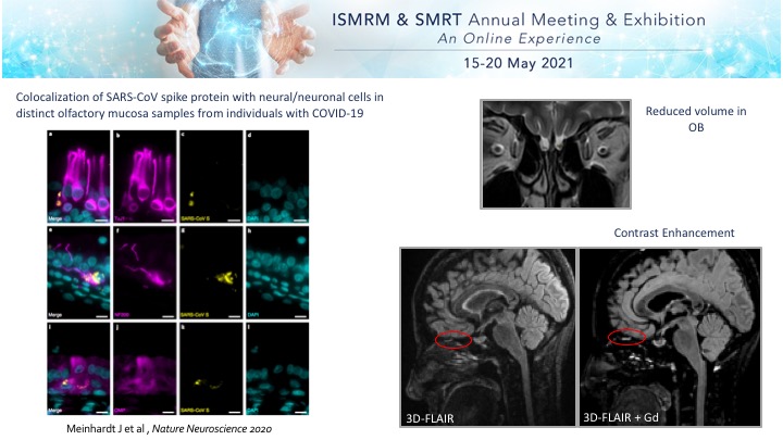 Slide #26
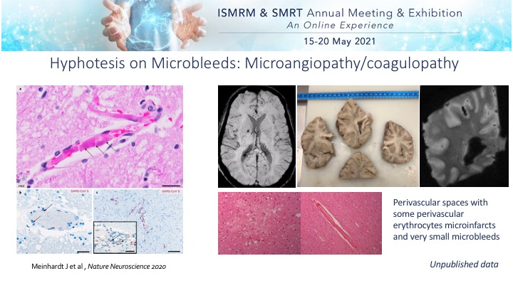 Slide #27
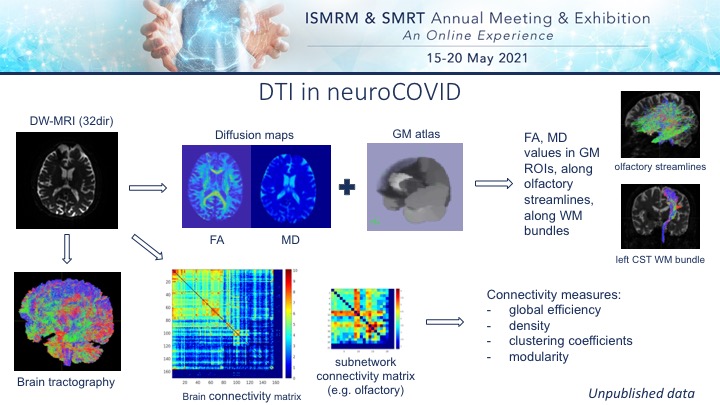 Slide #28
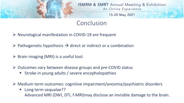 Slide #29
 Slide #30