PC-MRI: State-of-the-Art Techniques
Olivier Baledent1
1University hospital of Picardie Jules Verne, Amiens, France
1University hospital of Picardie Jules Verne, Amiens, France
Synopsis
Cerebral hydrodynamics knowledge has benefited considerably from the introduction of phase-contrast magnetic resonance imaging (PCMRI), the unique technique to investigate CSF and cerebral blood flows oscillations. Using post-processing software, key parameters of flow can be easily calculated. These flows data can be functional information’s complementary to the morphological imaging to better investigate the cranio-spinal system. The objective of this presentation is to describe the power and the limit of such clinical 2D PCMRI protocol concerning CSF and blood flow investigations in different healthy and pathological populations.
The rapid amplitude change of the cerebral systolic arterial input flow increasesthe brain volume. Then CSF is quickly displaced out of the cranium toward the spinal canal; ICP increase is therefore limited. Nevertheless, this first CSF response is also limited and has to be supplemented with the cerebral bloodvenous outflow. The venous contribution is slower than the CSF but at the end drains from the cranium all the blood input volume. Finally, due to the narrowaqueduct of Sylvius, a small CSF ventricular flows out of the fourth ventricle. Cerebral hydrodynamics knowledge has benefited considerably from the introduction of phase-contrast magnetic resonance imaging (PCMRI), the unique technique to investigate the small but rapid CSF oscillations. Using post-processing software, key parameters of flow can be easily calculated. In tenminutes CSF flow is quantified in the spinal subarachnoid spaces, the pontine cistern, the foramens of Magendi and the aqueduct of Sylvius. Blood flow isquantified in the internal carotid and the vertebral arteries, straight and sagittal sinus, jugular and epidural veins. These flows data can be functionalinformation’s complementary to the morphological imaging to better investigatethe cranio-spinal system in case of patients presented hydrocephalus, Chiari malformation, syringomyelia, cerebral hemorrhage, intracranial hyper or hypo tension. The objective of this presentation is to describe the power and the limitof such clinical 2D PCMRI protocol concerning CSF and blood flow investigations in different healthy and pathological populations.
Acknowledgements
No acknowledgement found.References
No reference found.Figures
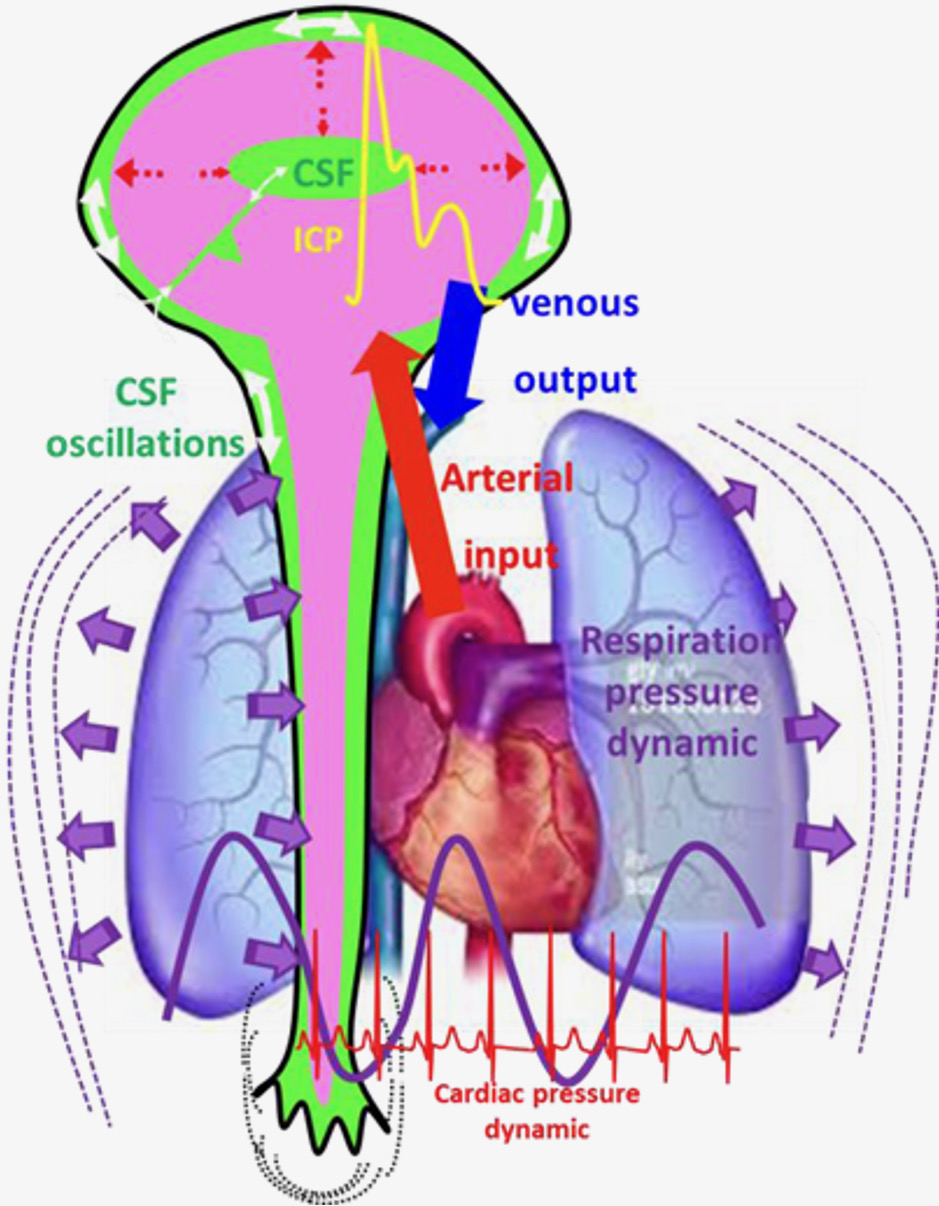
cerebral dynamics