Methods for T2 & T2* Mapping
Richard Dortch1
1Barrow Neurological Institute, Phoenix, AZ, United States
1Barrow Neurological Institute, Phoenix, AZ, United States
Synopsis
Researchers have developed an array of methods for mapping T2 and T2* values in tissue. The choice of the method depends on numerous factors, including the signal model (single versus multi-compartment), scan time, SNR, and the tissue of interest. In all cases, care must be taken to optimize sequence parameters and minimize the impact of confounding features (B0 and B1 variations). Moving forward, it is likely that more efficient and robust methods for estimating T2 and T2* in tissue will continue to be developed, especially as multi-compartment models continue to illustrate their ability to quantify microstructural features (e.g., myelin content).
 Slide #1
 Slide #2
 Slide #3
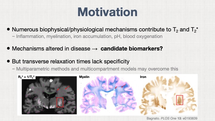 Slide #4
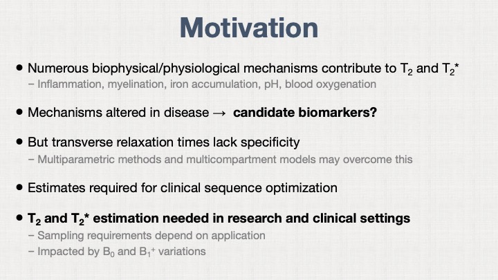 Slide #5
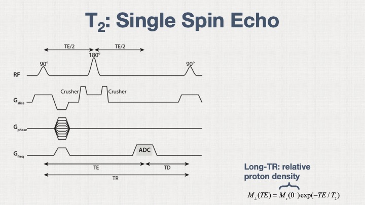 Slide #6
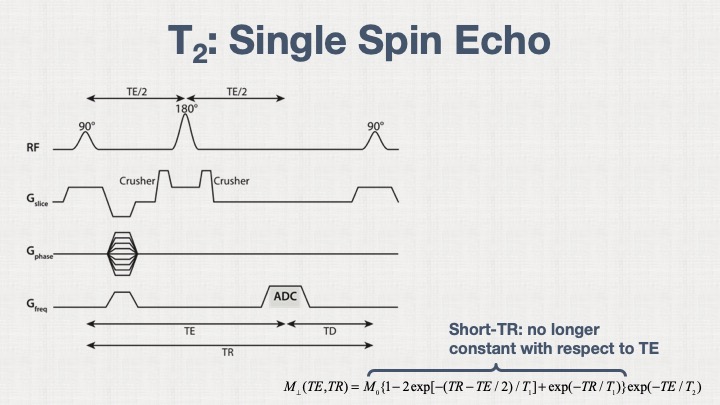 Slide #7
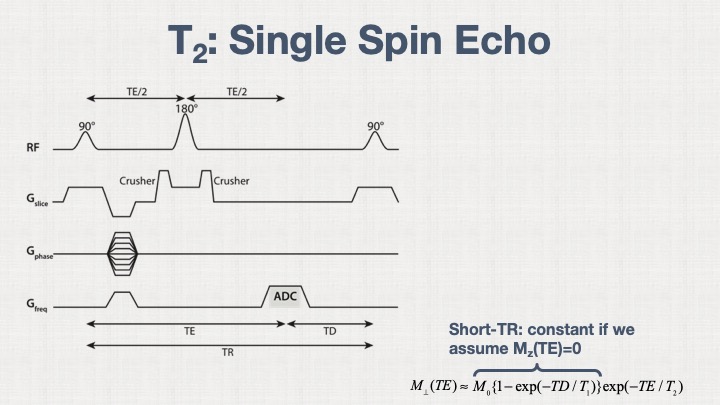 Slide #8
 Slide #9
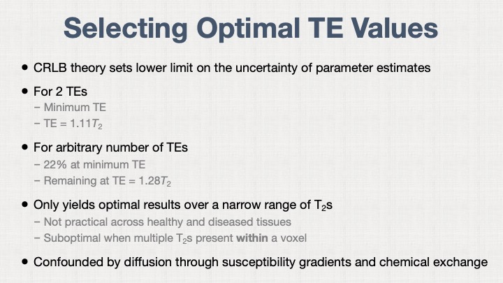 Slide #10
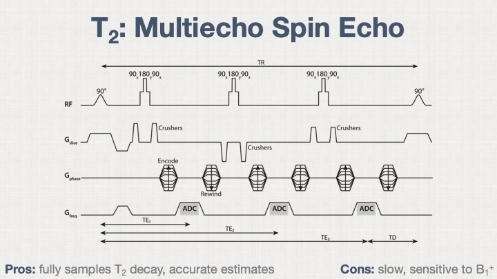 Slide #11
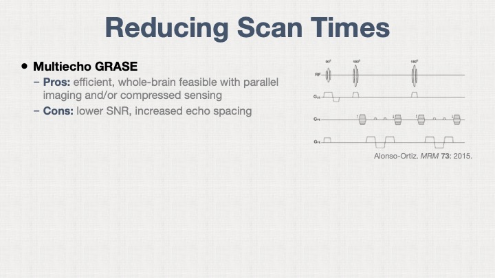 Slide #12
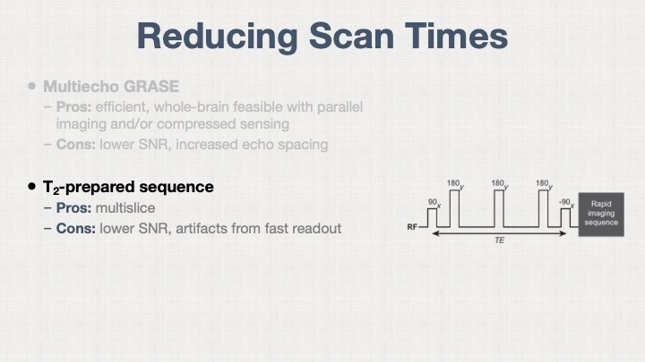 Slide #13
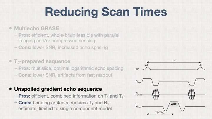 Slide #14
 Slide #15
 Slide #16
 Slide #17
 Slide #18
 Slide #19
 Slide #20
 Slide #21
 Slide #22
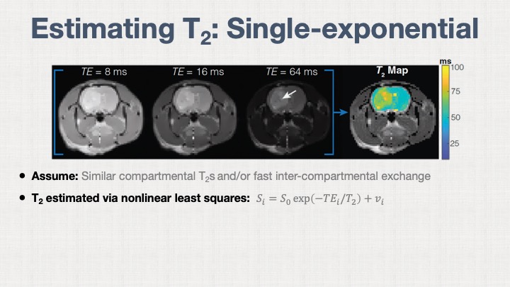 Slide #23
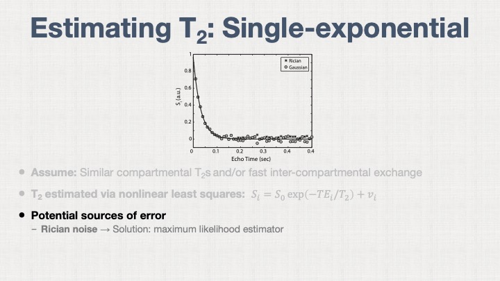 Slide #24
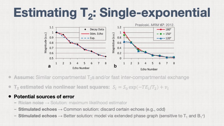 Slide #25
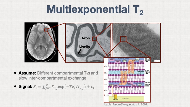 Slide #26
 Slide #27
 Slide #28
 Slide #29
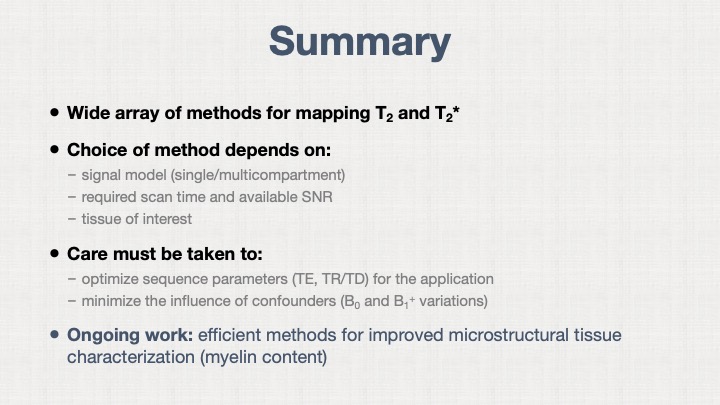 Slide #30