resting state fMRI: Neuronal Components
Silvina G Horovitz1
1National Institute of Neurological Disorders and Stroke, Bethesda, MD, United States
1National Institute of Neurological Disorders and Stroke, Bethesda, MD, United States
Synopsis
This lecture explores the neuronal contributions to the BOLD signal fluctuations and fMRI functional connectivity. We then provide an overview on electrophysiological and behavioral changes that alter fMRI functional connectivity. Finally, we discuss the implications in the understanding of resting state studies.
Since fist measure in 1995 [1], correlation of BOLD signal fluctuations at rest resting state fMRI became a powerful tool to study brain “functional connectivity”. However, the origin of the BOLD signal fluctuation and their propagation remain elusive. This lecture provides an overview of the construct that resting state fMRI signal have a neuronal component. In animal models, BOLD signal is correlated to the local field potentials (LFP) in response to stimuli [2], and also follows the on-going neuronal activity [3]. LFP [4] and BOLD signals [5,6] fluctuate, propagate in space, and follow the power law. Meta-stable states can help understand and model networks' behavior[7,8]. BOLD fluctuations correlations display spatial structure which is consistent across time and subjects[ 5]. Supporting the neuronal component, correlations are modulated by state [9,10], diseases[11], neuromodulation[12,13], and drugs[14]. Careful consideration of the neuronal pathways that could support the signal propagation, together with an understanding of the relationship between physiological, arousal and neuronal components are key for proper interpretation.
Acknowledgements
SGH is supported by the NINDS Intramural Research ProgramReferences
[1] Biswal et al, 1995
[2] Logothetis et al, 2001
[3] Shmuel and Leopold, 2008
[4] Arieli et al, 1995
[5] Fukunaga et al, 2006
[6] He et al, 2010
[7] Tagliazucchi et al, 2015
[8] Rabinovich and Varona, 2011
[9] Horovitz et al, 2009
[10] Bianciardi et al, 2009
[11] Vanhaudenhuyse et al, 2010
[12] Censor et al, 2014
[13] Scheinost et al, 2013
[14] Tagliazucchi et al, 2014
Figures
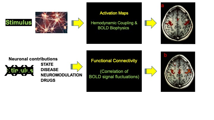
schematic: BOLD fluctuations have a neuronal component