Deuterium Metabolic Imaging (DMI)
Xiao-Hong Zhu1
1University of Minnesota, Minneapolis, MN, United States
1University of Minnesota, Minneapolis, MN, United States
Synopsis
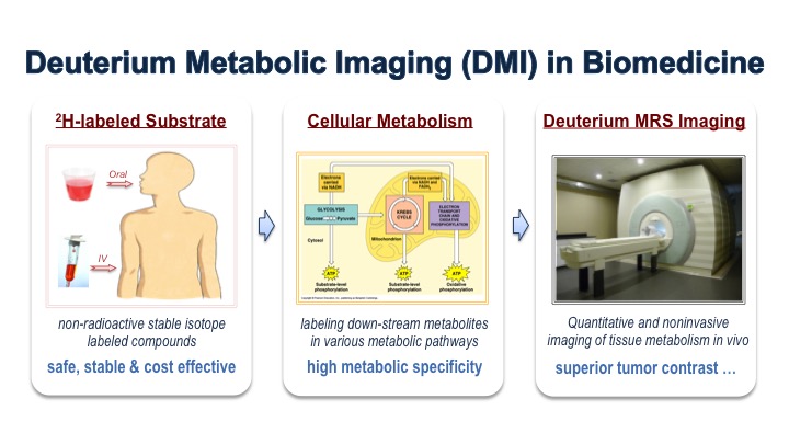
Metabolic imaging provides valuable tools for evaluating cellular metabolism under physiopathological conditions. Deuterium metabolic imaging (DMI) is a newly developed deuterium (2H) MRS imaging technology that can measure the steady-state signal and/or dynamic changes of deuterated metabolites in living organs or tissues after administration of deuterium-labeled substrate. DMI has been used to study various metabolic processes, especially the cerebral glucose metabolism in healthy brain and in brain tumor patients, and has shown its advantages over certain existing methods. This presentation will briefly introduce DMI technology - its past development, current capabilities and future prospects.
 Slide #1