Measuring Ventricular Microstructure & Strain
Daniel B. Ennis1
1Department of Radiology, Stanford University, Stanford, CA, United States
1Department of Radiology, Stanford University, Stanford, CA, United States
Synopsis
Cardiac structure and function are inextricably bound. MRI is an unrivaled technology for exploring and discovering the structure-function mechanisms of cardiac function and dysfunction. This talk reviews the basic microstructural constituents of the hearts (i.e. “myofibers” and “sheets”), two methods for measuring regional cardiac function (i.e. MRI tagging and cine DENSE), and the use of cardiac diffusion tensor MRI to measure microstructural organization in vivo. Methods to jointly integrate measures of structure and function to reveal microstructurally anchored measures of cardiac function (.e.g “myofiber” strain) are also described.
 Slide #1
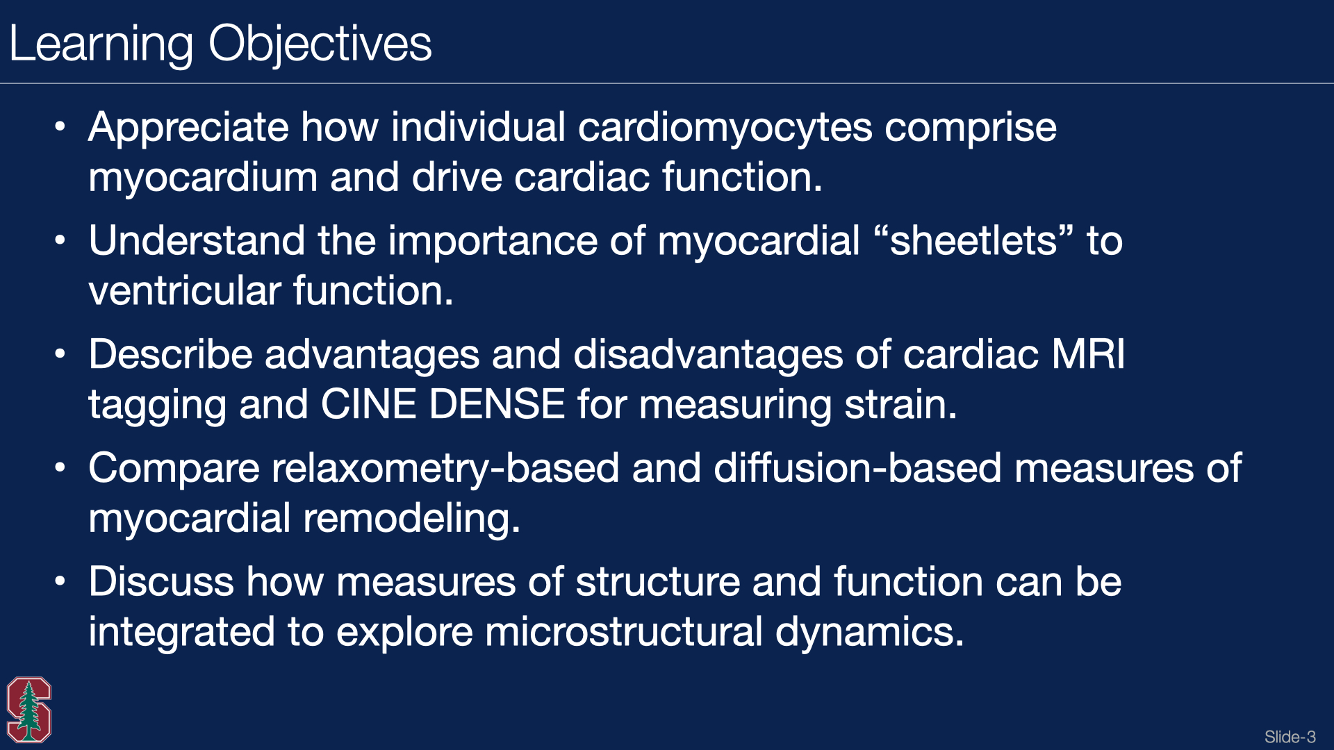 Slide #2
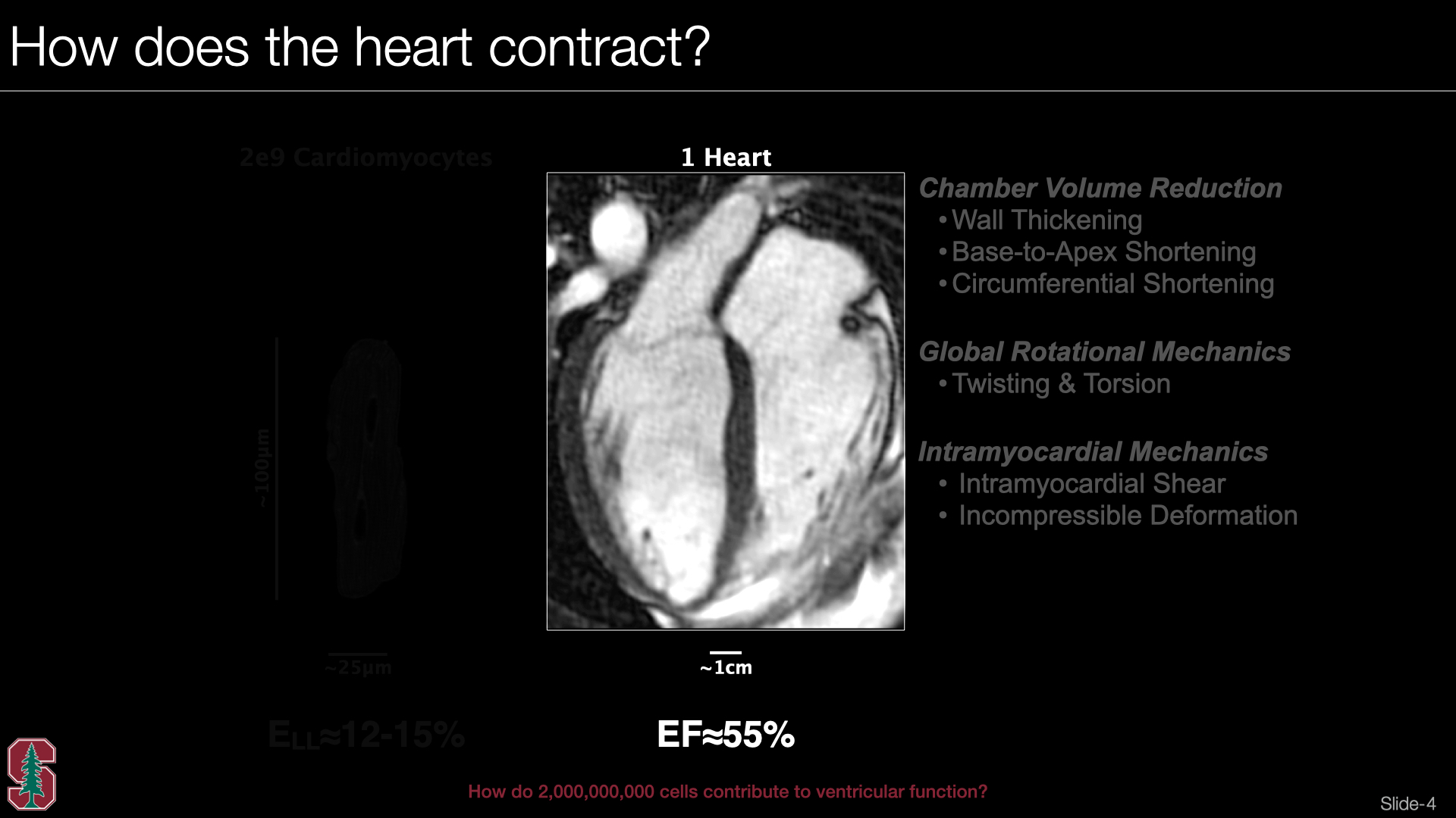 Slide #3
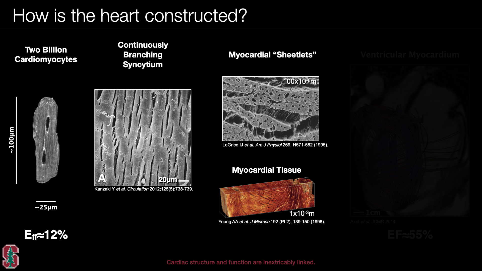 Slide #4
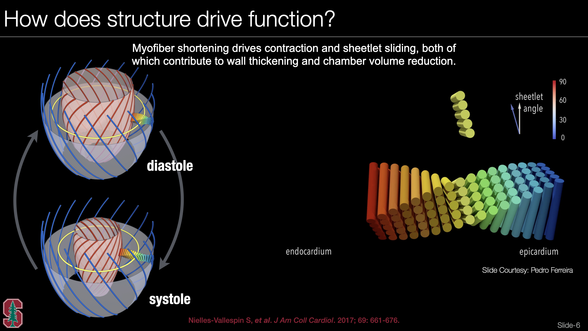 Slide #5
 Slide #6
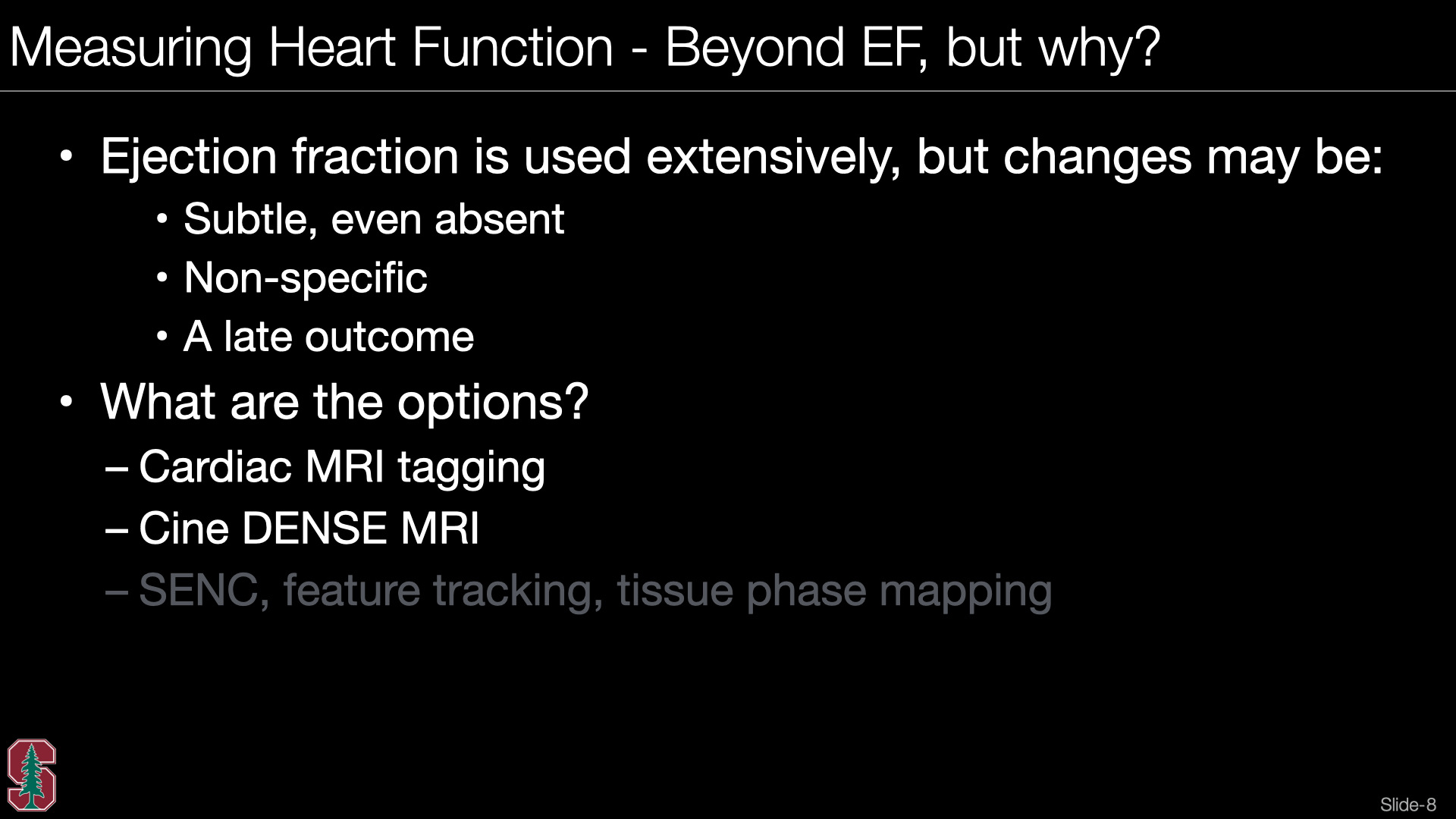 Slide #7
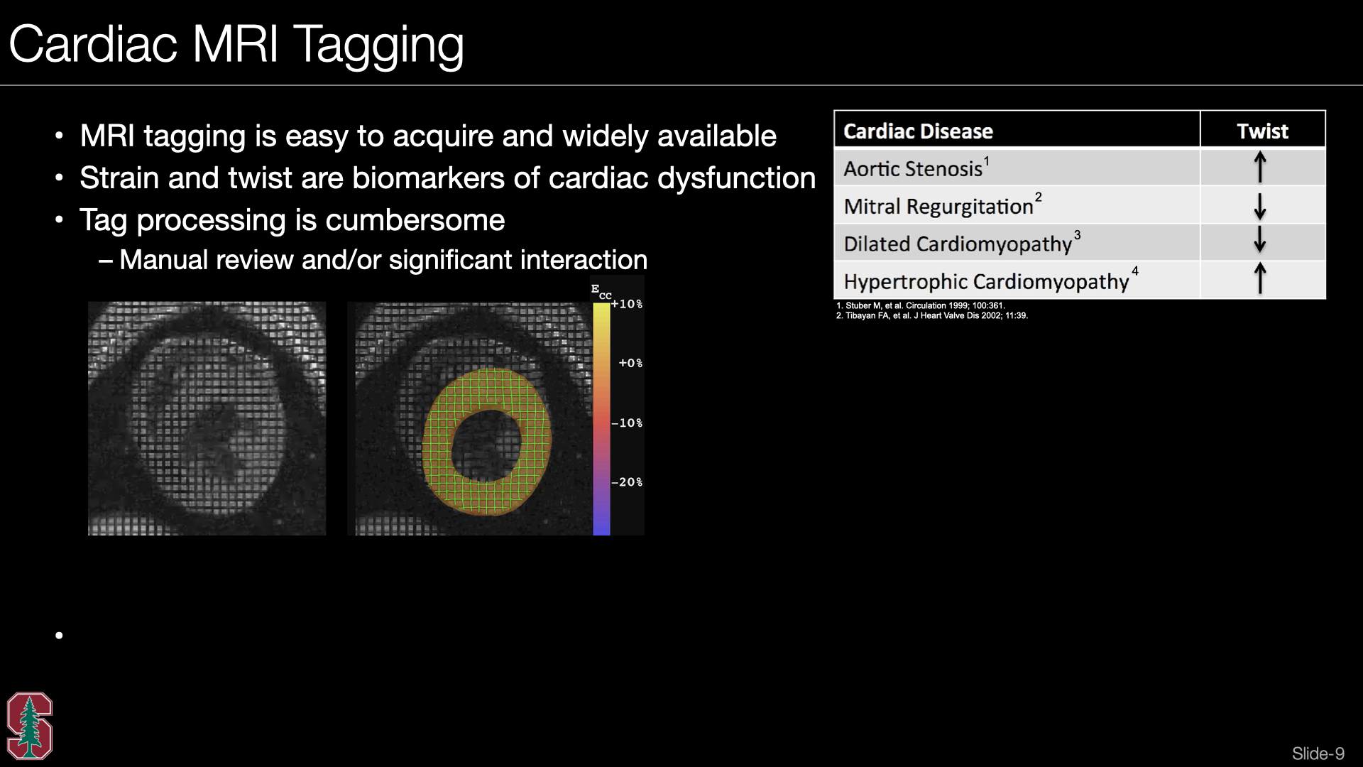 Slide #8
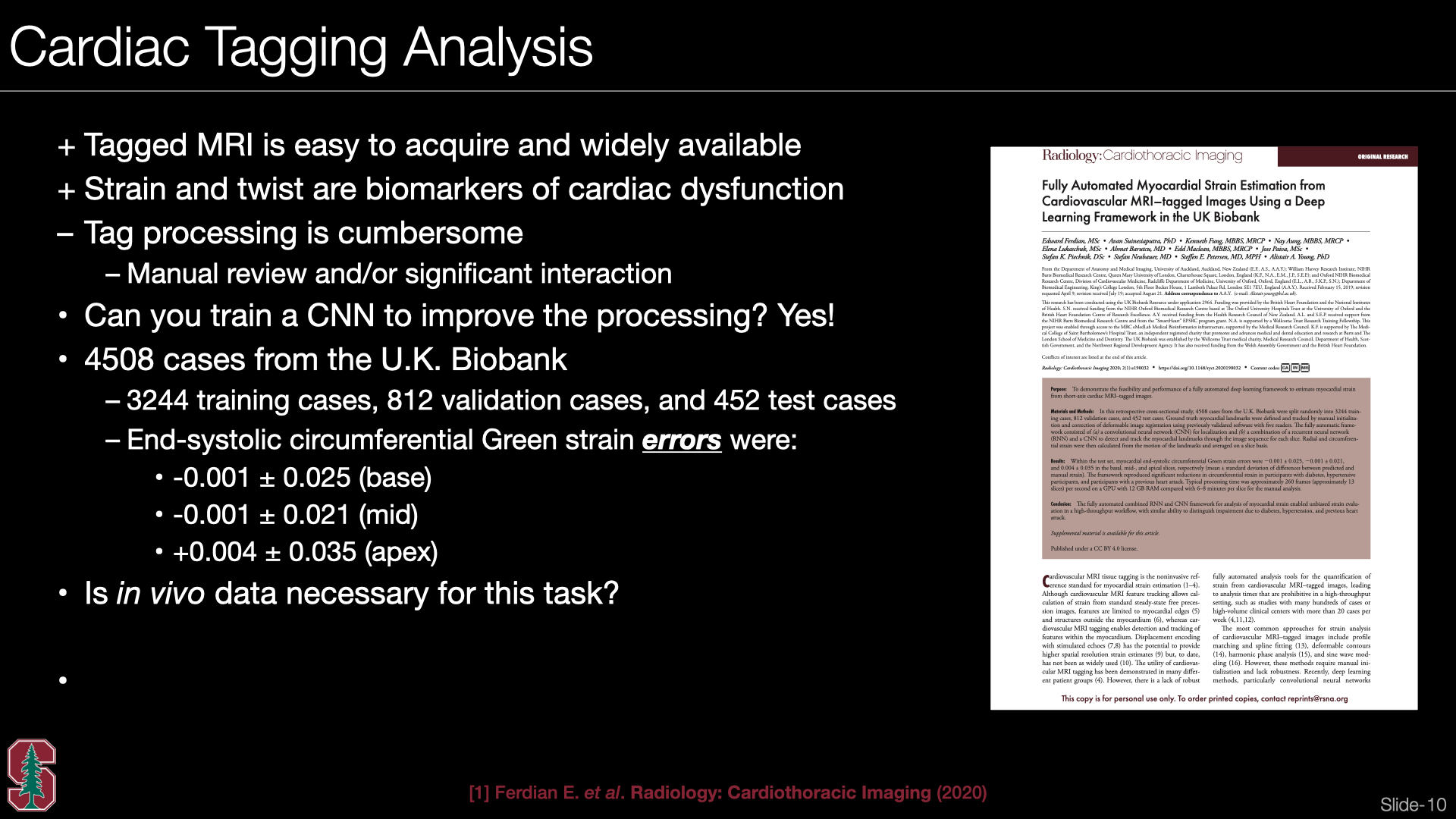 Slide #9
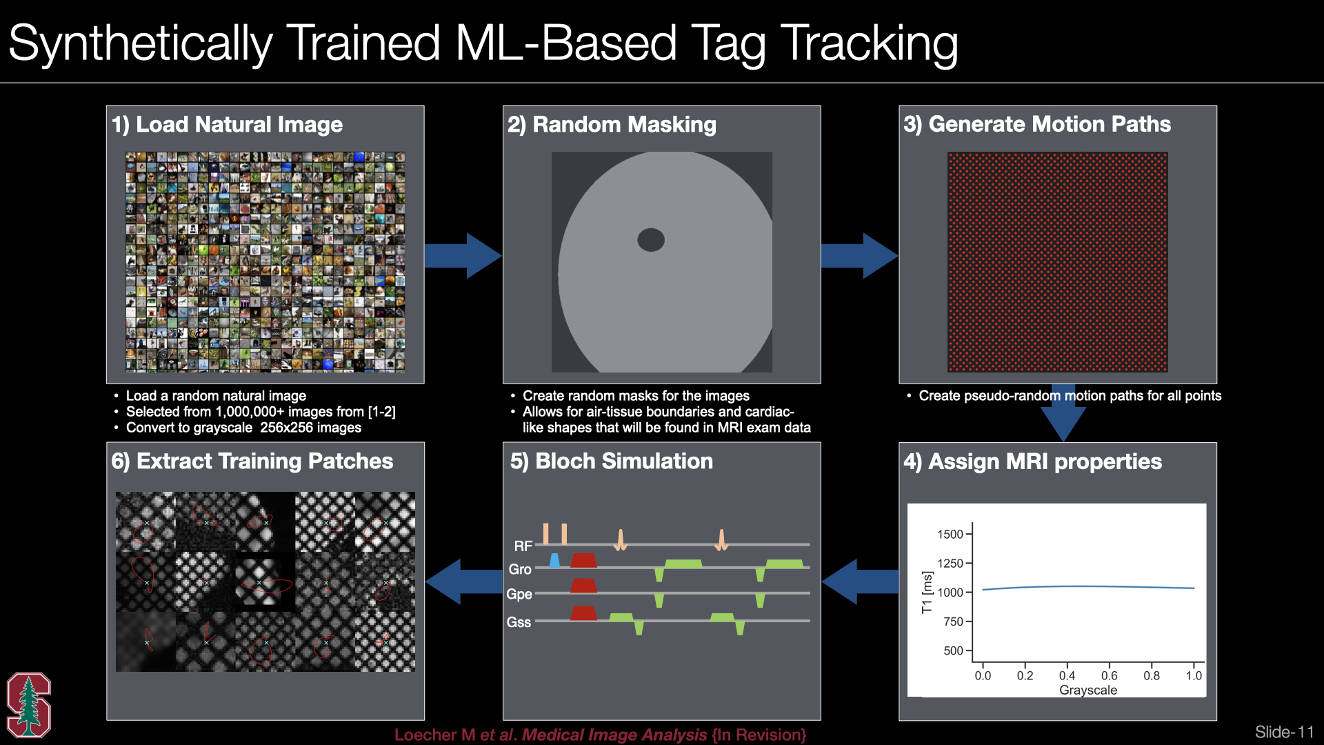 Slide #10
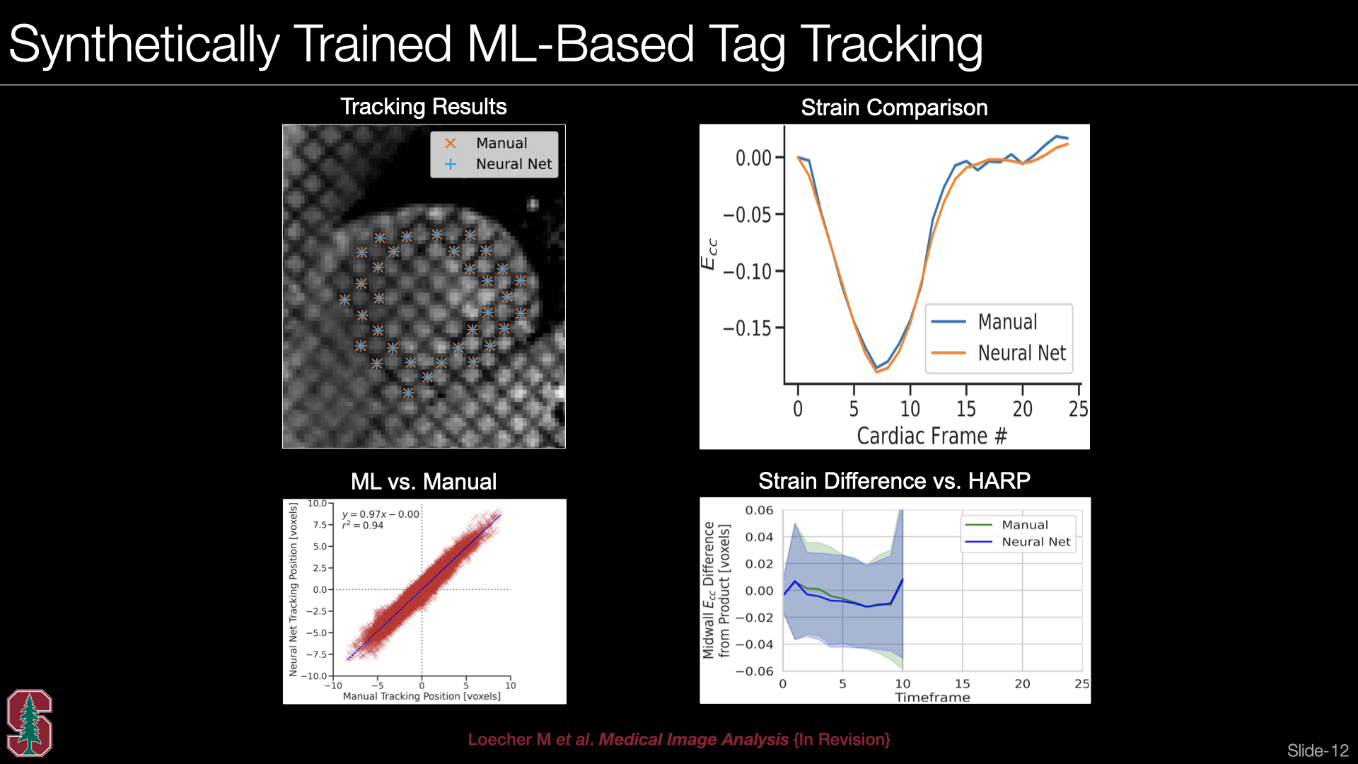 Slide #11
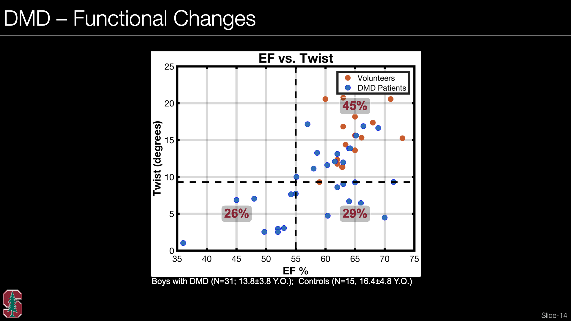 Slide #12
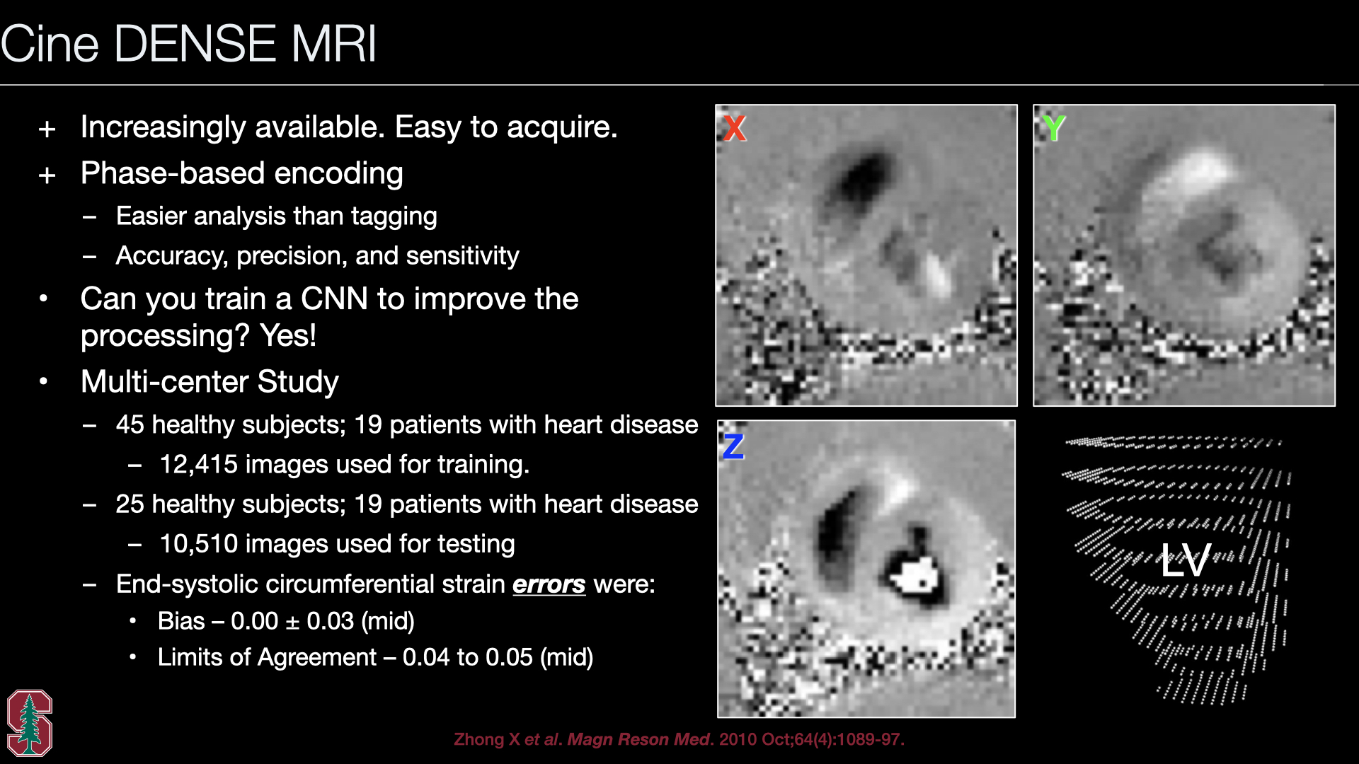 Slide #13
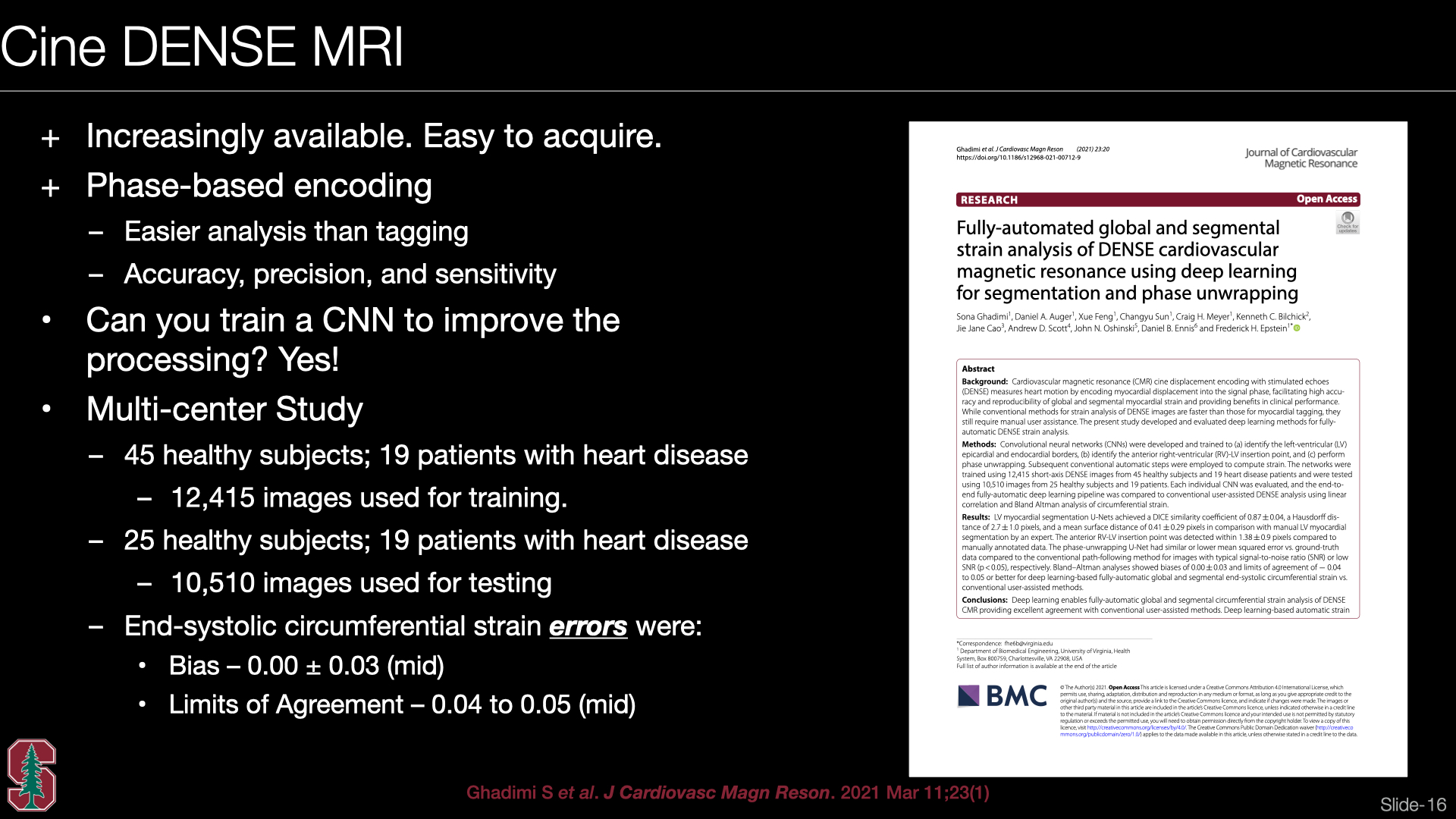 Slide #14
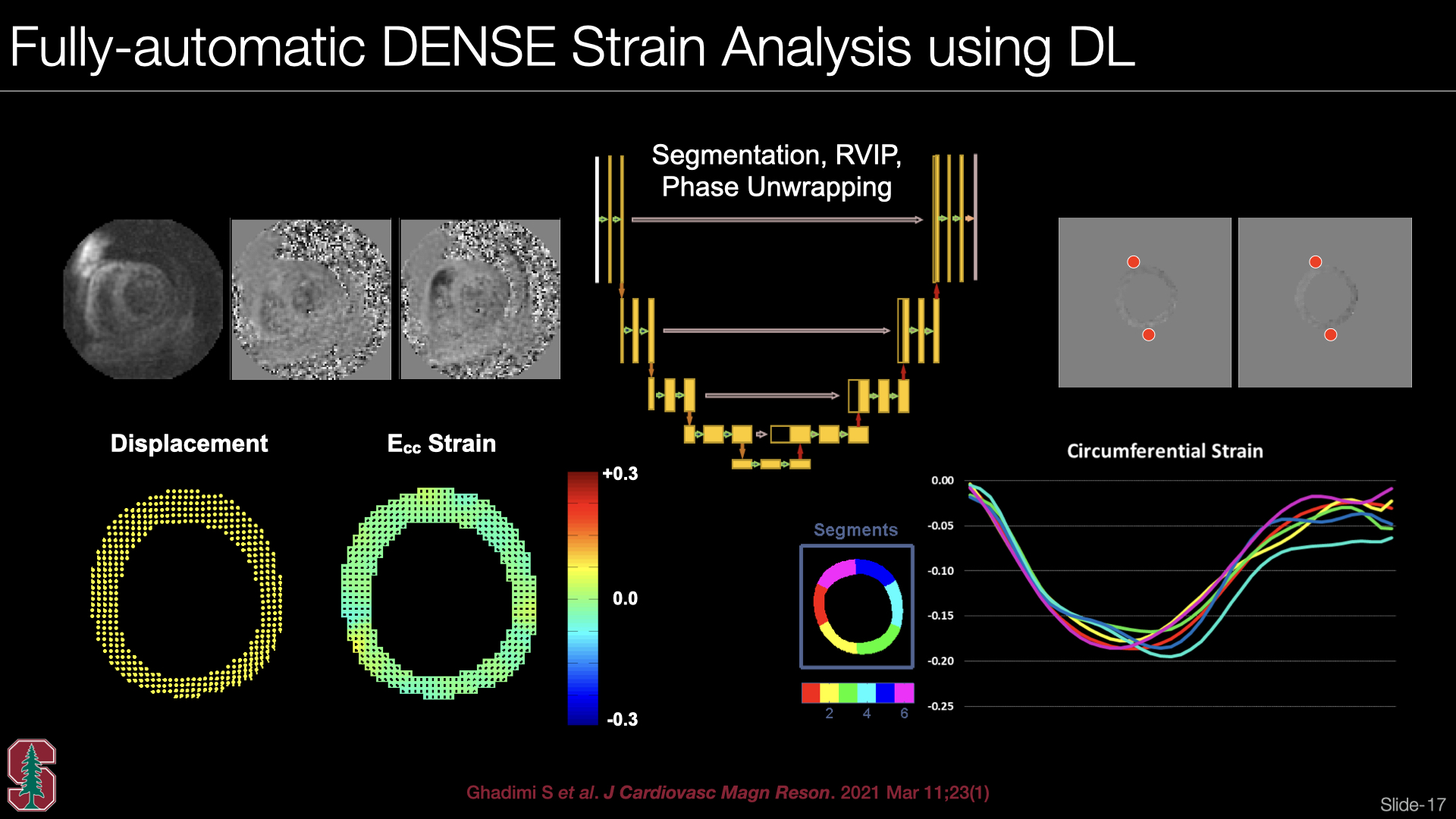 Slide #15
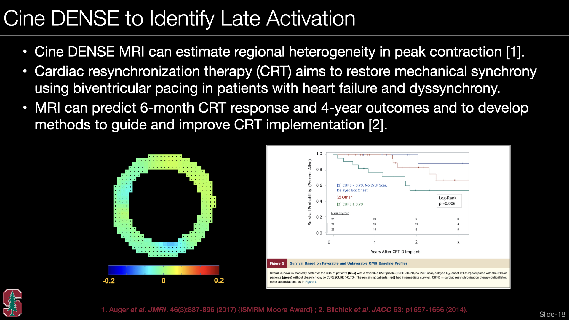 Slide #16
 Slide #17
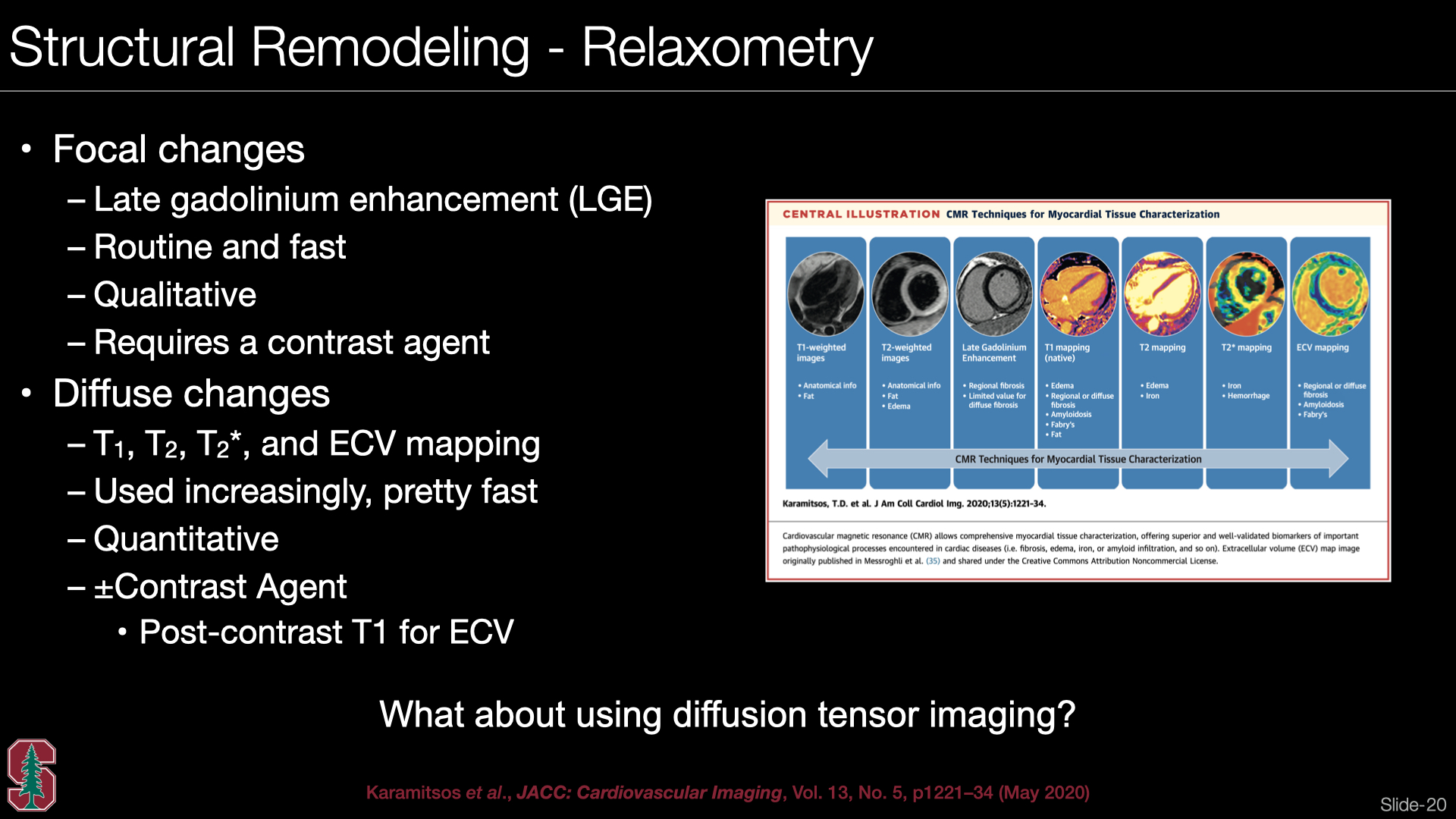 Slide #18
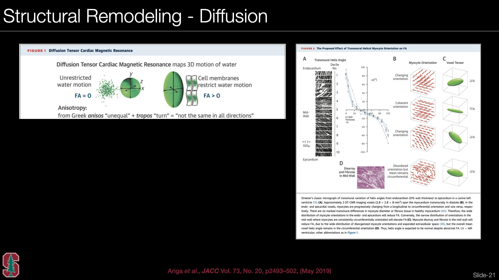 Slide #19
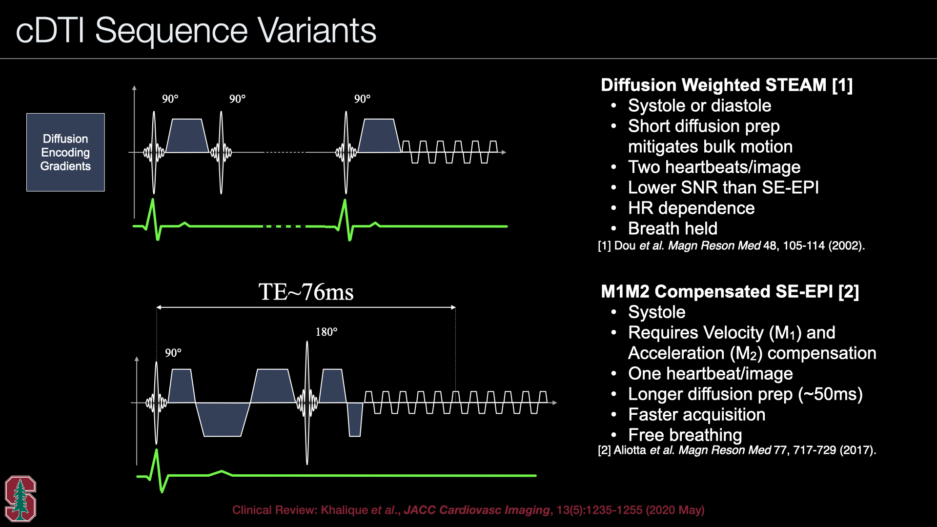 Slide #20
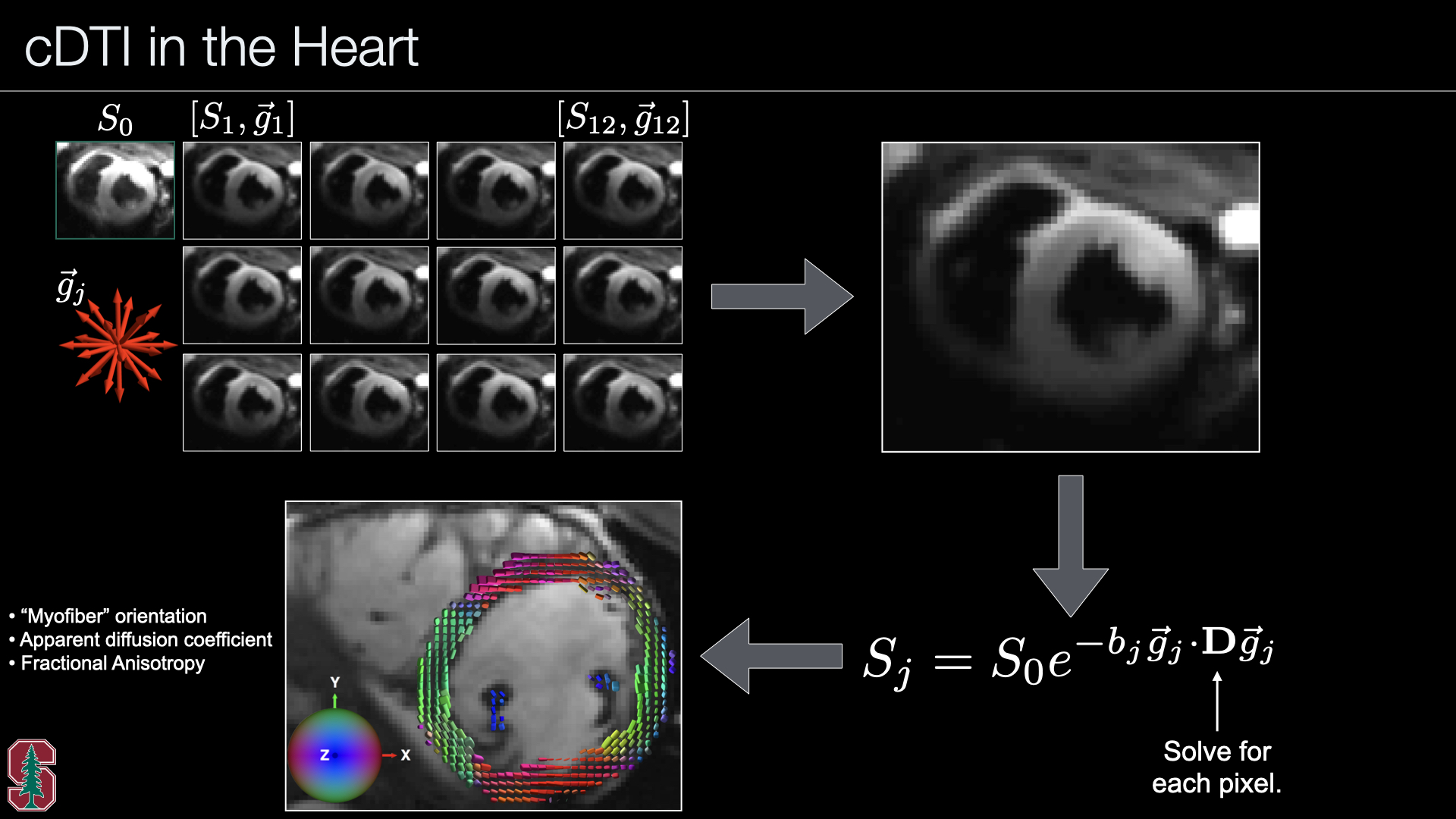 Slide #21
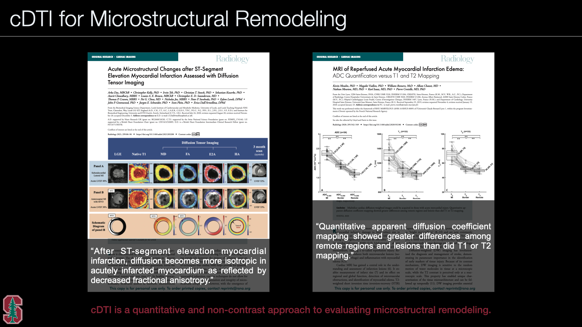 Slide #22
 Slide #23
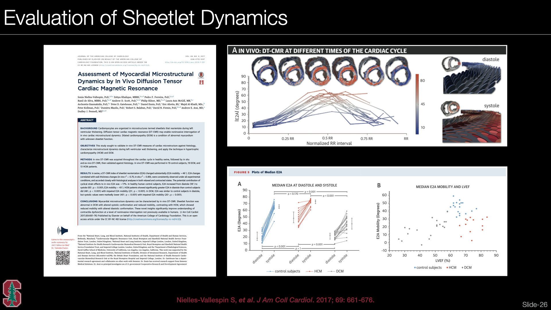 Slide #24
 Slide #25
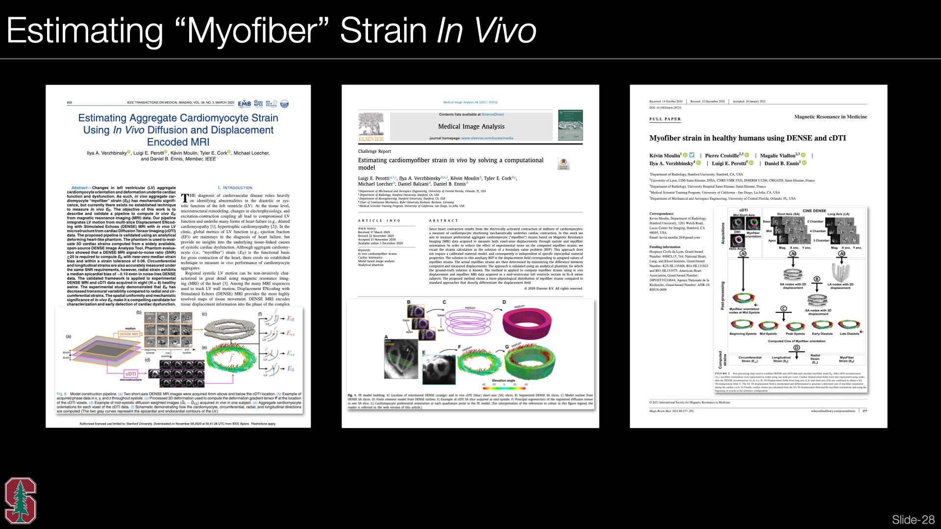 Slide #26
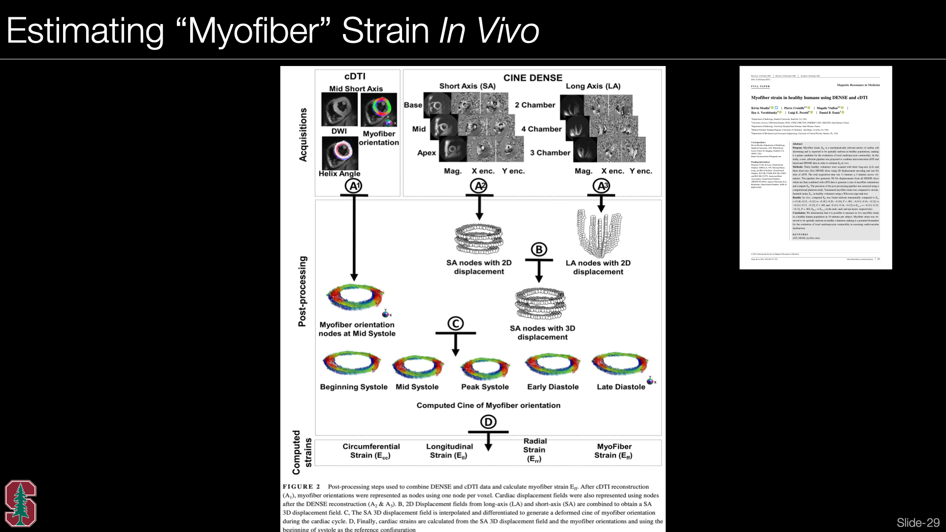 Slide #27
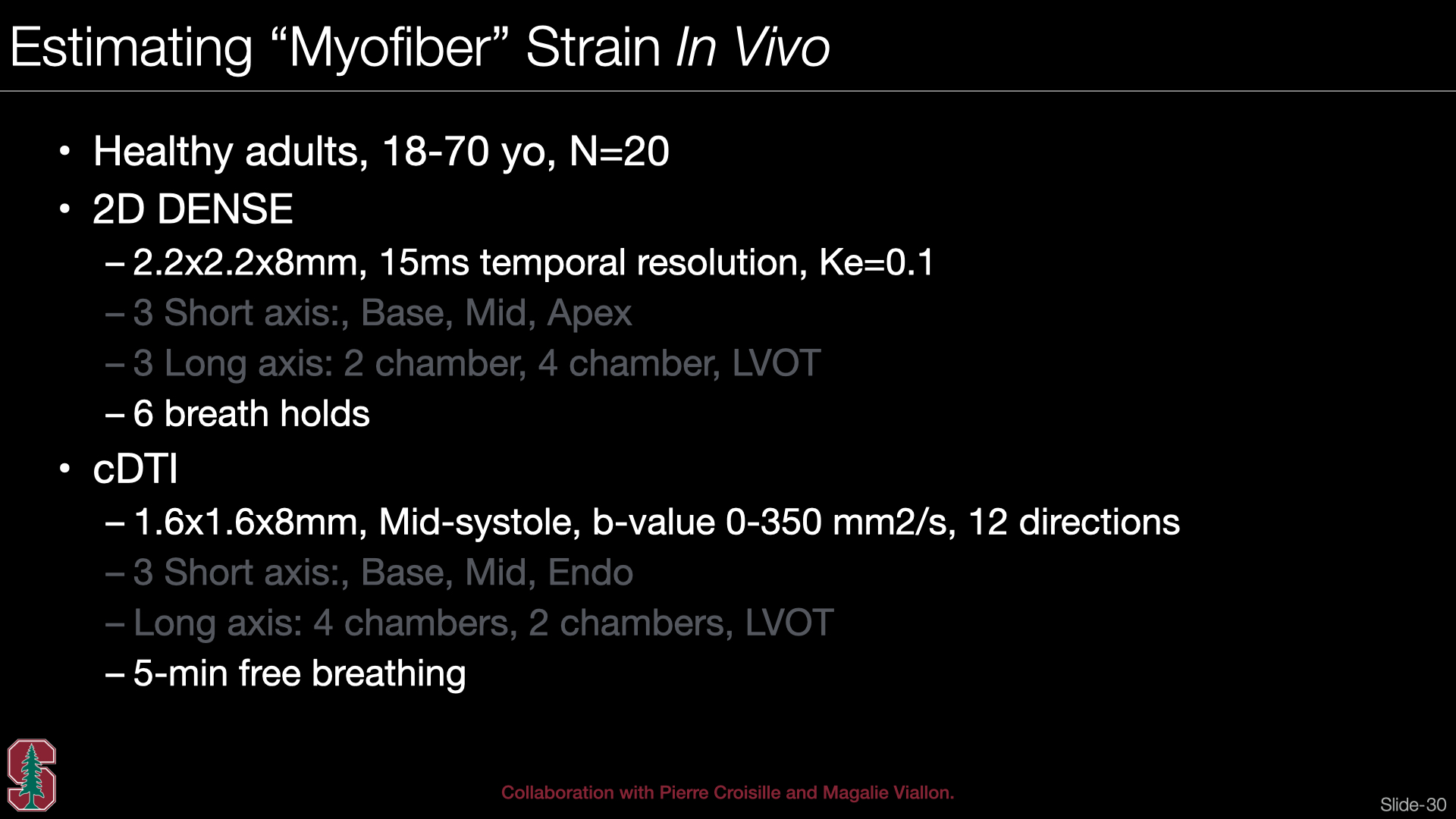 Slide #28
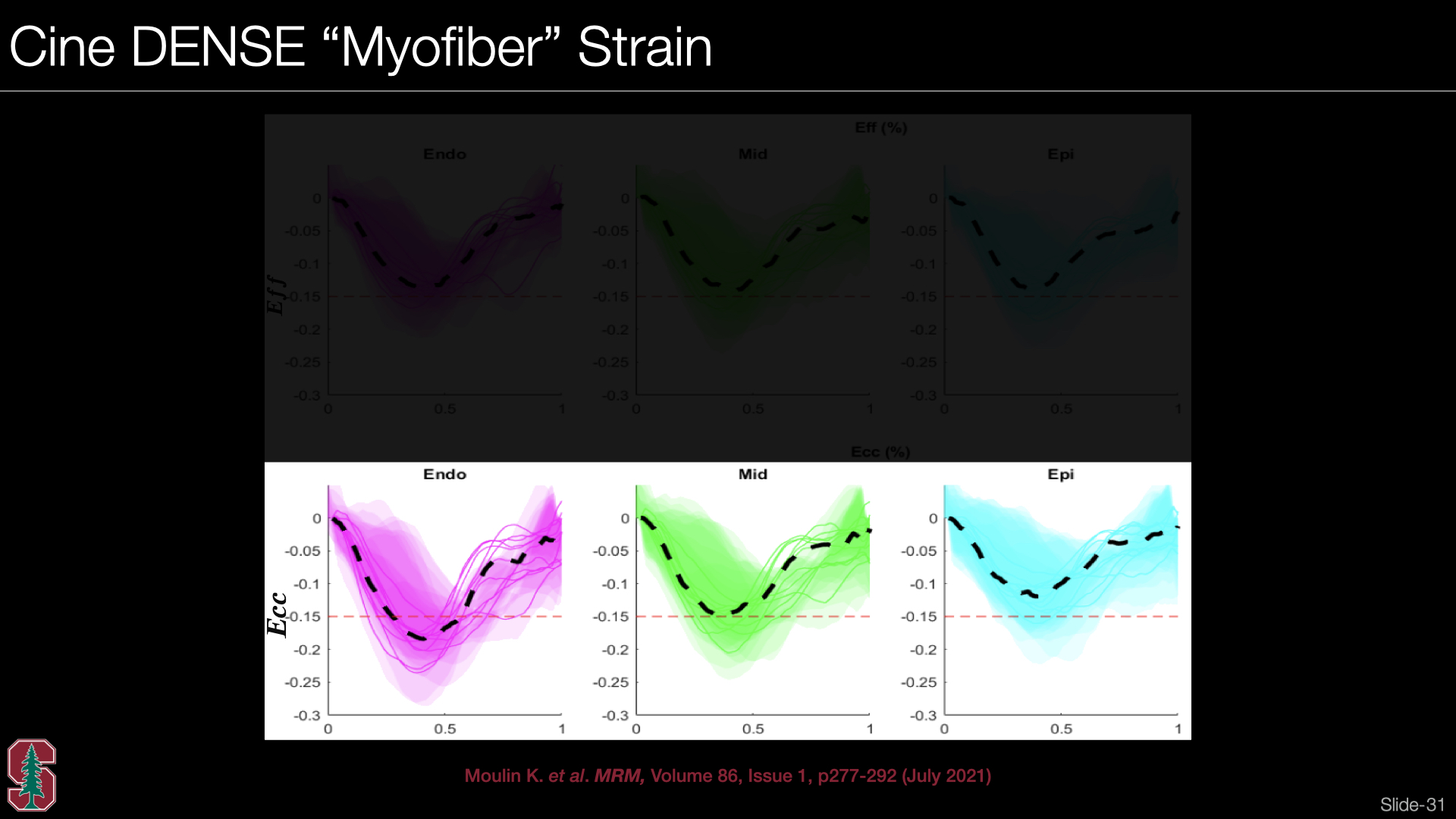 Slide #29
 Slide #30