Cardiac MRI of Myocardial Perfusion & Fibrosis - State of the Art
Graham A. Wright1 and Nilesh Ghugre1
1Sunnybrook Health Sciences Centre, Toronto, ON, Canada
1Sunnybrook Health Sciences Centre, Toronto, ON, Canada
Synopsis
Assessment of perfusion and fibrosis in the heart with MRI is central to a complete cardiac exam. The core clinical methods have focused on dynamic first pass of Gadolinium-based contrast for perfusion and late gadolinium enhancement for fibrosis. Newer methods have targeted improvements in spatial resolution and coverage with reductions in imaging time, and robustness in the presence of implanted devices. There is a growing emphasis on quantitatiion and the introduction of methods that do not require the injection of a contrast agent. Clinical applications include management of ischemic and non-ischemic heart disease as well as complex arrhythmia management.
 Slide #1
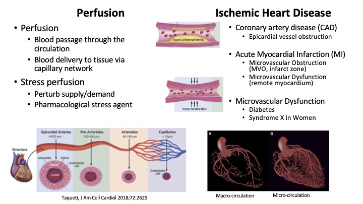 Slide #2
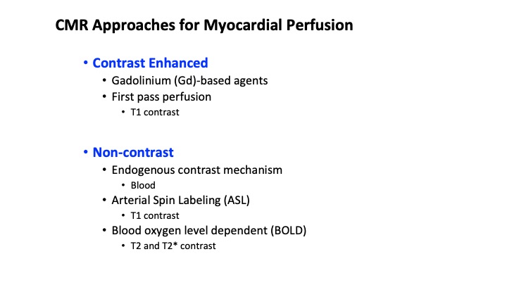 Slide #3
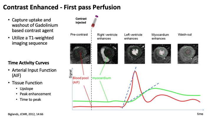 Slide #4
 Slide #5
 Slide #6
 Slide #7
 Slide #8
 Slide #9
 Slide #10
 Slide #11
 Slide #12
 Slide #13
 Slide #14
 Slide #15
 Slide #16
 Slide #17
 Slide #18
 Slide #19
 Slide #20
 Slide #21
 Slide #22
 Slide #23
 Slide #24
 Slide #25
 Slide #26
 Slide #27
 Slide #28
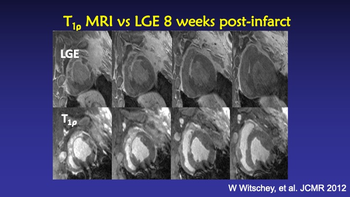 Slide #29
 Slide #30