MR Perfusion in Glioma Management
Kentaro Akazawa1
1Radiology, Kyoto Prefectural University of Medicine, Kyoto, Japan
1Radiology, Kyoto Prefectural University of Medicine, Kyoto, Japan
Synopsis
Magnetic resonance (MR) perfusion identifies tumor angiogenesis or the proliferation of abnormal vessels in tumors. Of the various techniques devised for evaluating cerebral perfusion imaging, the dynamic susceptibility contrast (DSC) method has been employed most widely in clinical practice. DSC perfusion is used to generate hemodynamic parameters such as relative cerebral blood volume (rCBV). CBF maps can be used to assess neovascularity in tumors, which is thought to correlate with tumor grade and malignant histology. This talk will provide knowledge on how DSC perfusion can be useful in the glioma management in a variety of clinical conditions.
 Slide #1
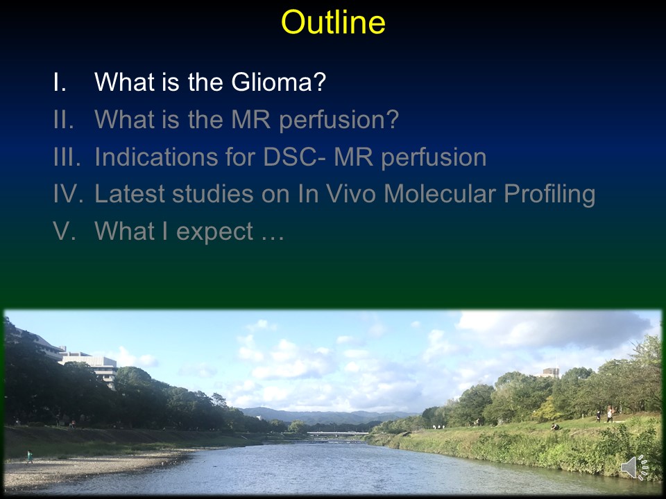 Slide #2
 Slide #3
 Slide #4
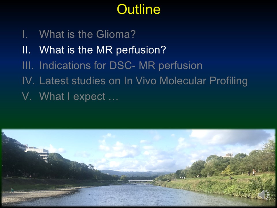 Slide #5
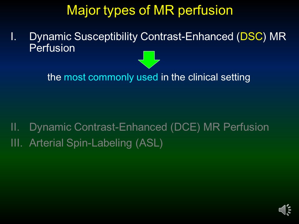 Slide #6
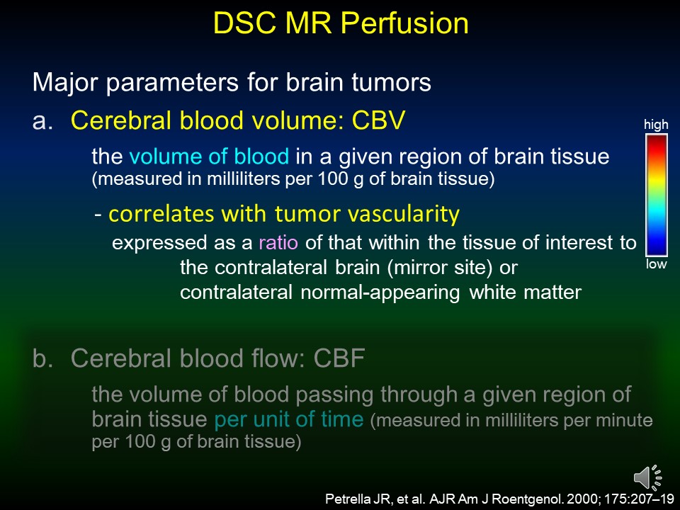 Slide #7
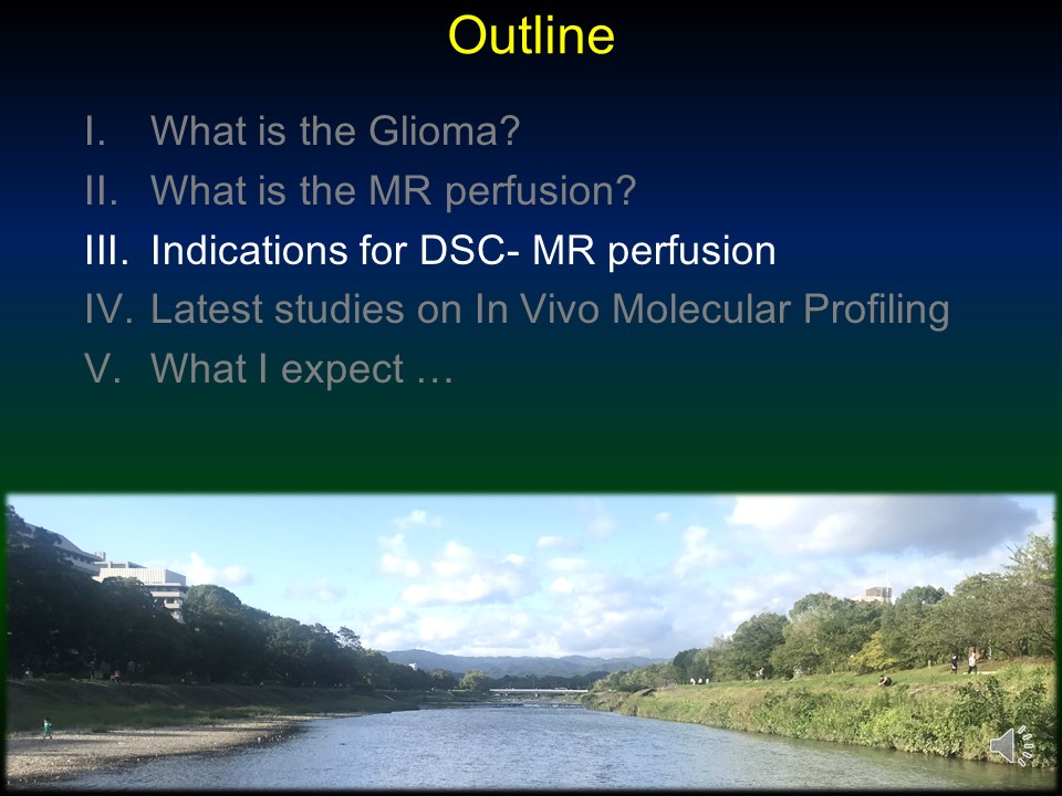 Slide #8
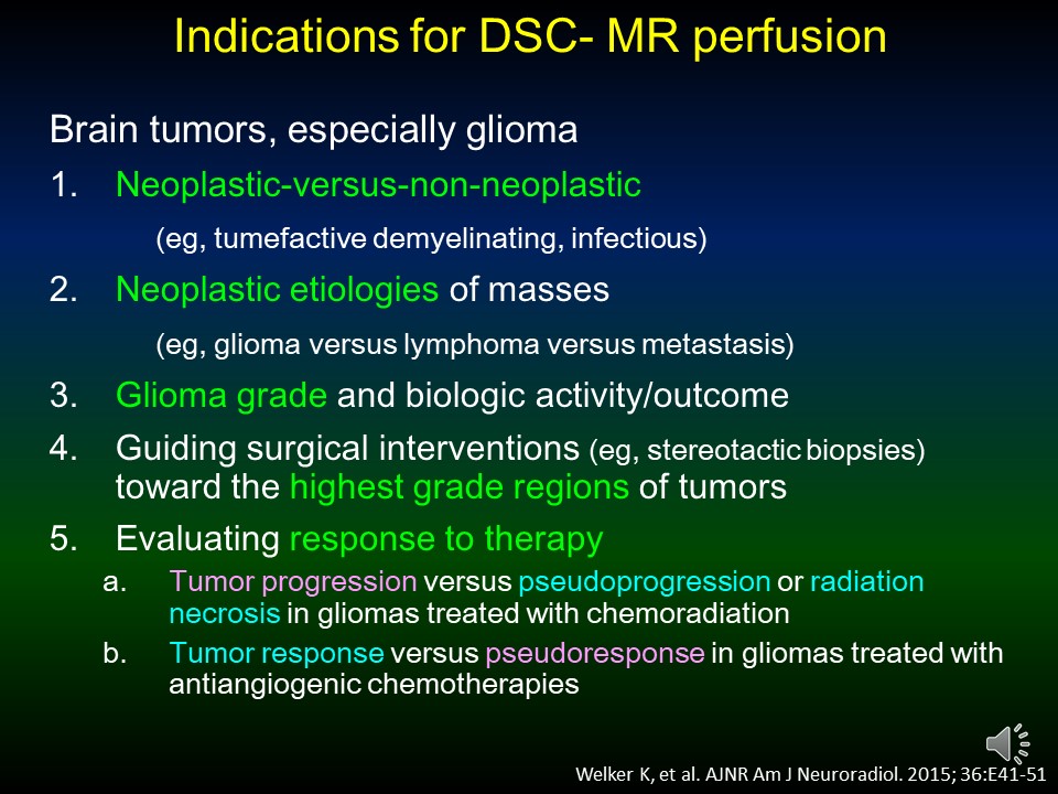 Slide #9
 Slide #10
 Slide #11
 Slide #12
 Slide #13
 Slide #14
 Slide #15
 Slide #16
 Slide #17
 Slide #18
 Slide #19
 Slide #20
 Slide #21
 Slide #22
 Slide #23
 Slide #24
 Slide #25
 Slide #26
 Slide #27
 Slide #28
 Slide #29
 Slide #30