Bowel Imaging: How to Incorporate DWI in My Protocol
Keith Wan-Hang Chiu1
1The University of Hong Kong, Hong Kong, China
1The University of Hong Kong, Hong Kong, China
Synopsis
Magnetic Resonance Enterography (MRE) is routinely used in the diagnosis and assessment of small bowel and colon in patients with inflammatory bowel disease (IBD). Diffusion restricted imaging (DWI) plays an important role in complementing conventional sequences and is very sensitive in detecting bowel wall inflammation. However, it intrinsically lacks anatomical details, can be non-specific and susceptible to artifacts, such as breathing, peristalsis, distortion, aliasing and T2 shine-through. Thus, evaluation of DWI images requires close correlation with T2W and contrast-enhanced sequences. Good understanding of the IBD disease spectrum is also necessary in order to interpret these images accurately.
 Slide #1
 Slide #2
 Slide #3
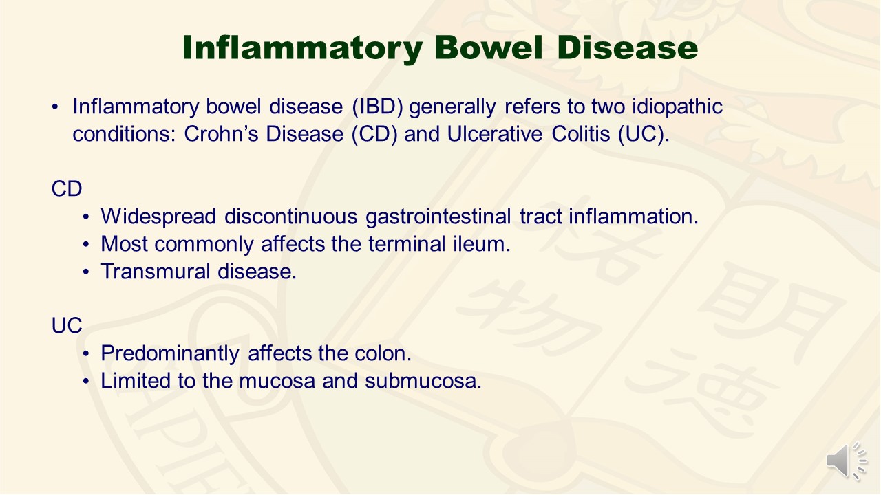 Slide #4
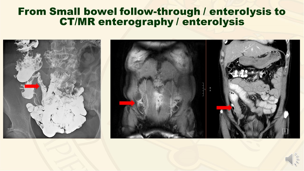 Slide #5
 Slide #6
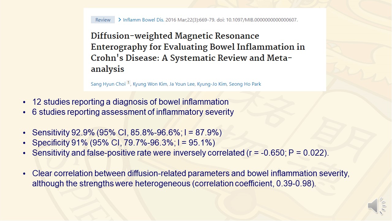 Slide #7
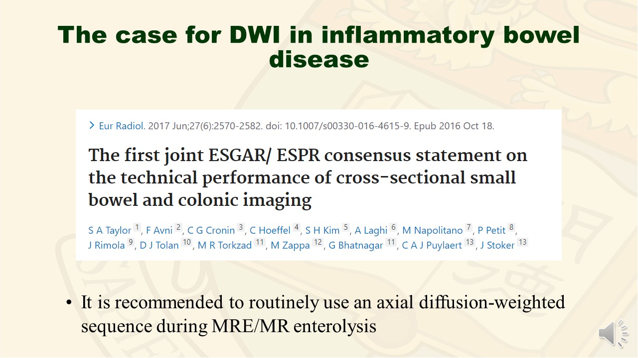 Slide #8
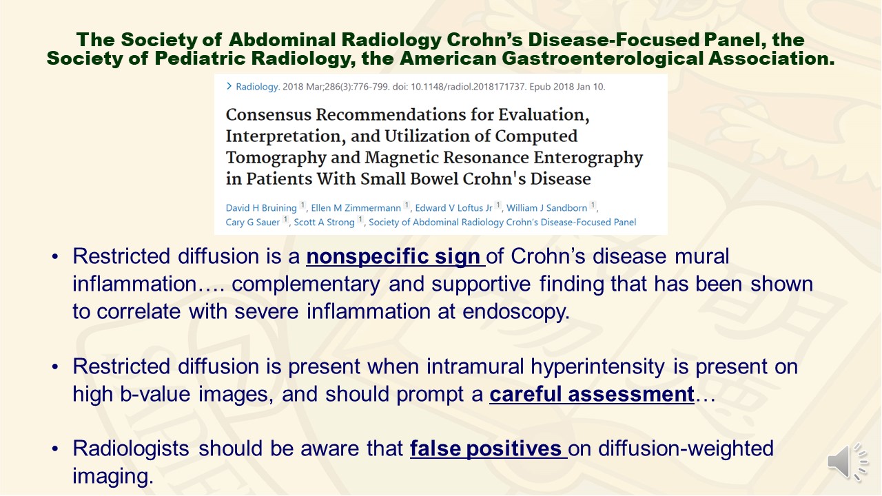 Slide #9
 Slide #10
 Slide #11
 Slide #12
 Slide #13
 Slide #14
 Slide #15
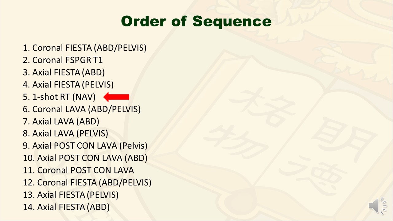 Slide #16
 Slide #17
 Slide #18
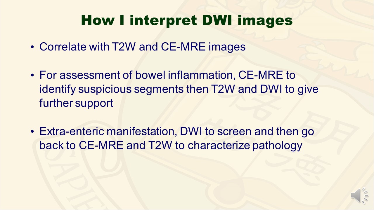 Slide #19
 Slide #20
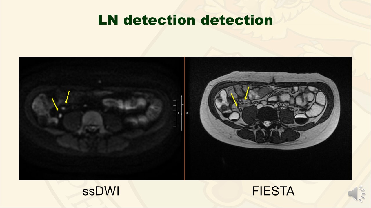 Slide #21
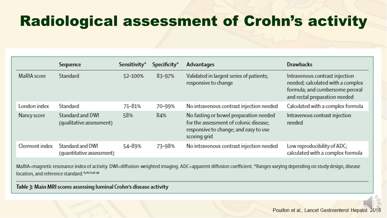 Slide #22
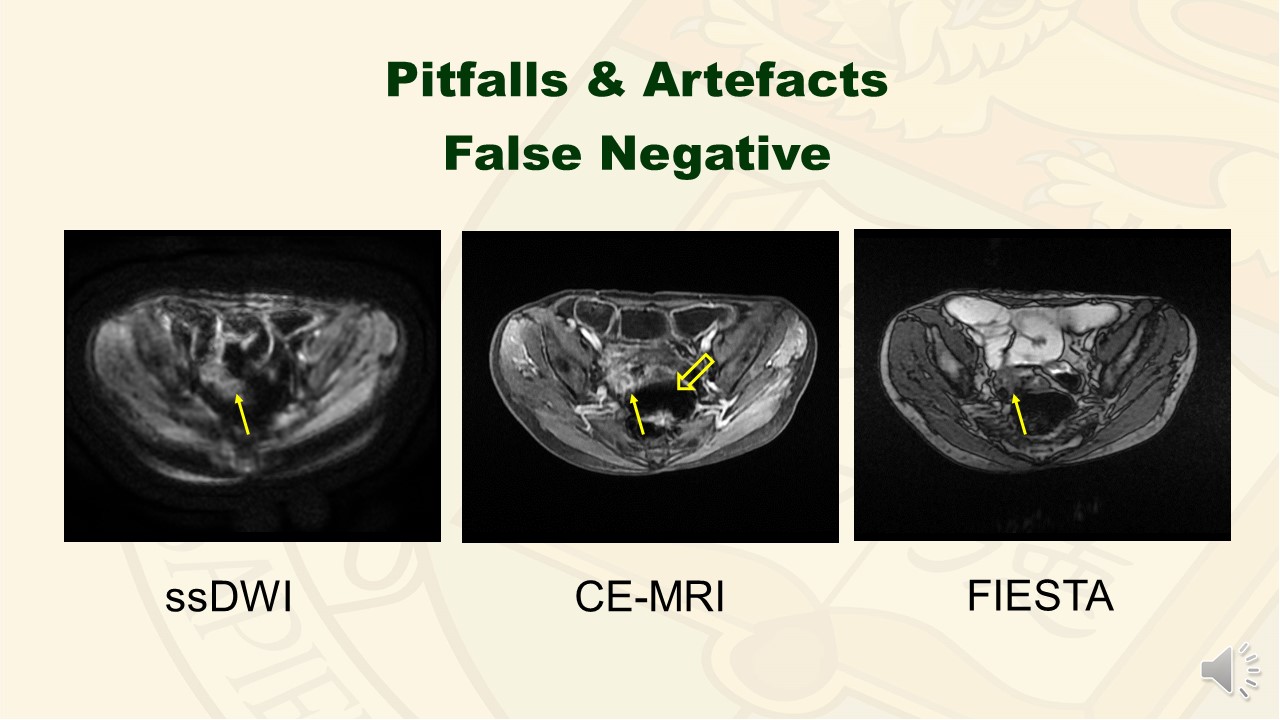 Slide #23
 Slide #24
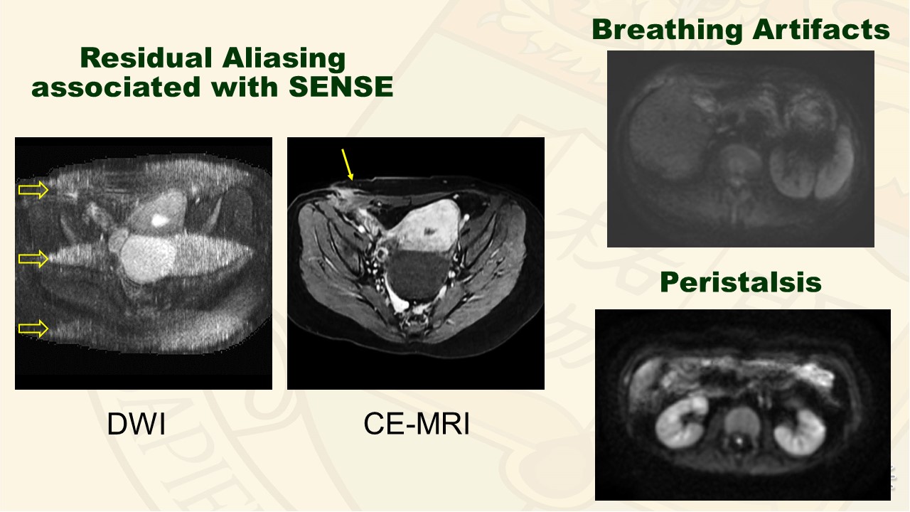 Slide #25
 Slide #26
 Slide #27
 Slide #28
 Slide #29
 Slide #30