Gynecological Cancer: How to Incorporate DWI in My Protocol
Kirsi Hannele Härmä1
1Dep. of Diagnostic, Interventional and Pediatric Radiology, Department of Diagnostic, Interventional and Pediatric Radiology, Bern University Hospital, University of Bern, Bern, Switzerland
1Dep. of Diagnostic, Interventional and Pediatric Radiology, Department of Diagnostic, Interventional and Pediatric Radiology, Bern University Hospital, University of Bern, Bern, Switzerland
Synopsis
Diffusion weighted MRI (DW-MRI) was emerging strongly during the past decade in female pelvic imaging, including gynecologic cancer. In not only distinguishing and detecting tumors and metastatic sites, but also serving as biologic marker with usable parameters predicting therapy response and risk of recurrence. From this point of view, DW-MRI is able to guide the choice of patient management directly influencing patient outcome. Further, pregnant women benefit from the DW-MRI as it offers a sensitive staging tool without use of contrast media and ionizing radiation. Radiologists should be active in communicating the evolving possibilities of DWI among the referring colleagues.
 Slide #1
 Slide #2
 Slide #3
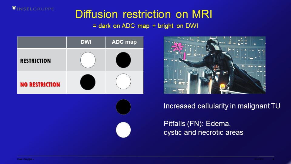 Slide #4
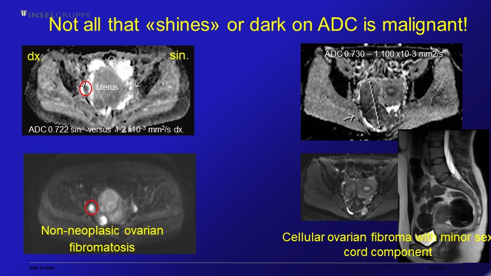 Slide #5
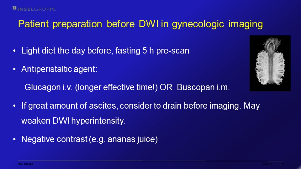 Slide #6
 Slide #7
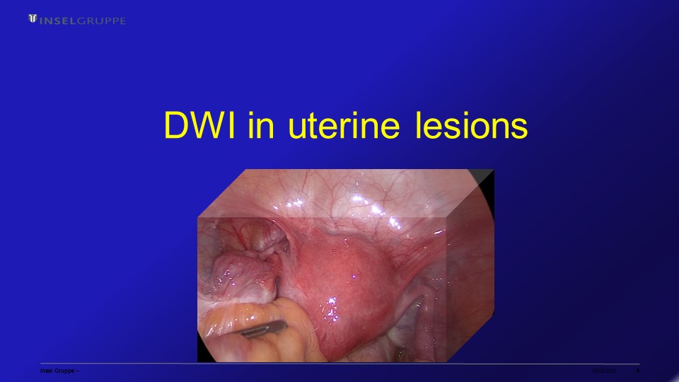 Slide #8
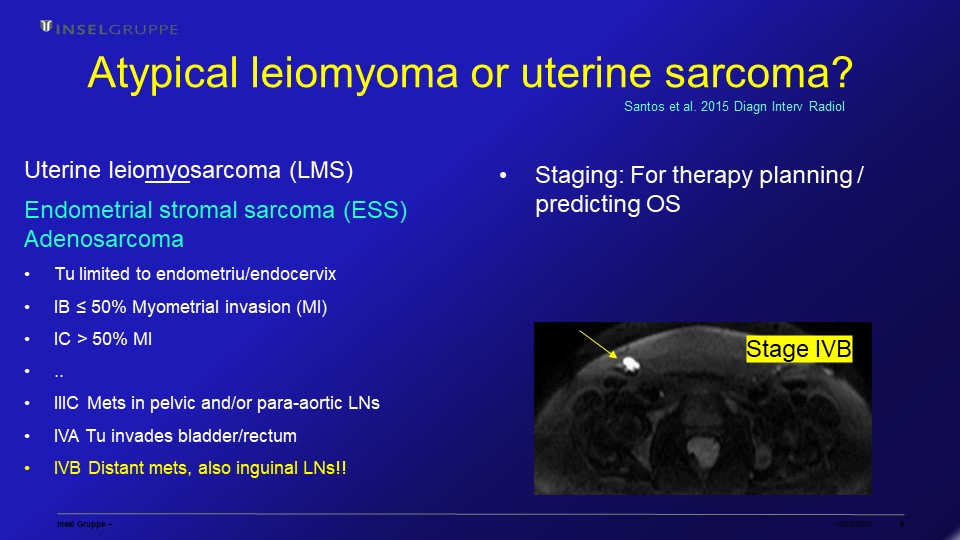 Slide #9
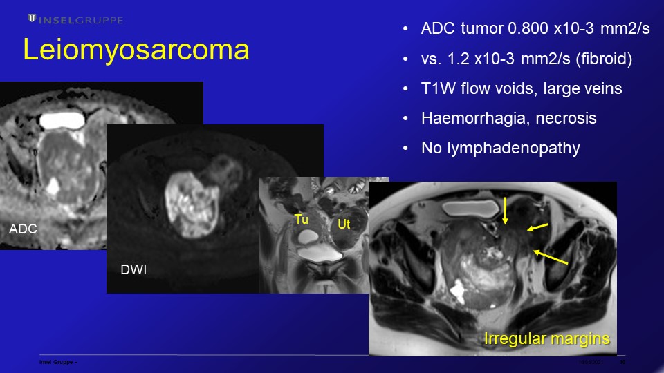 Slide #10
 Slide #11
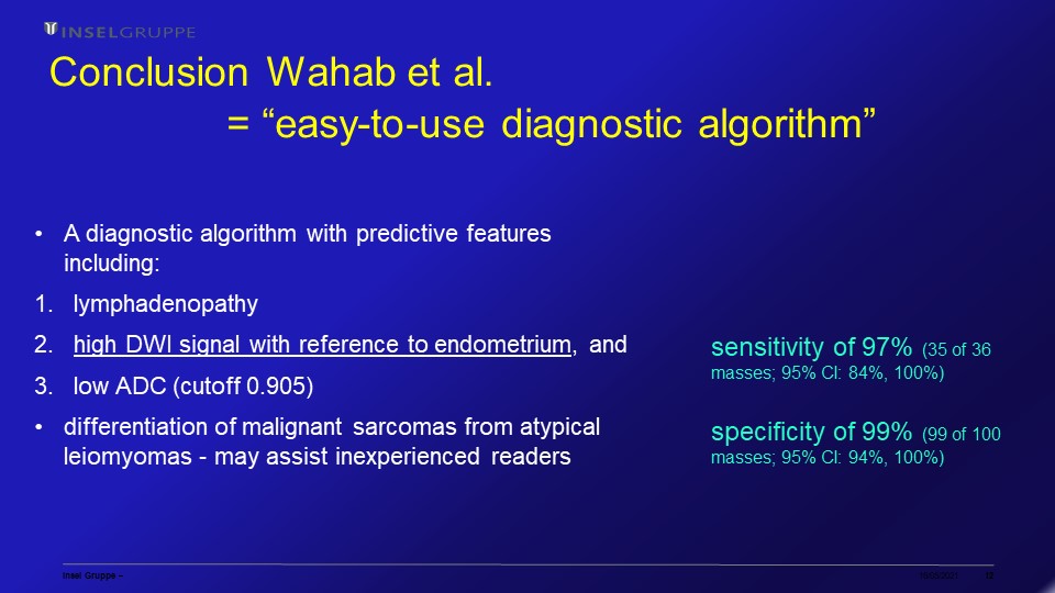 Slide #12
 Slide #13
 Slide #14
 Slide #15
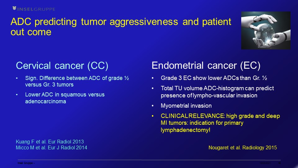 Slide #16
 Slide #17
 Slide #18
 Slide #19
 Slide #20
 Slide #21
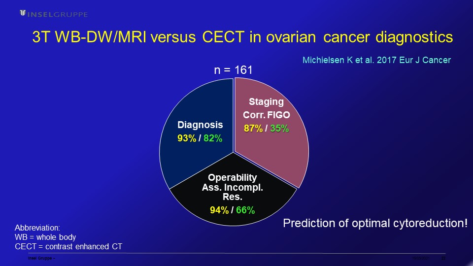 Slide #22
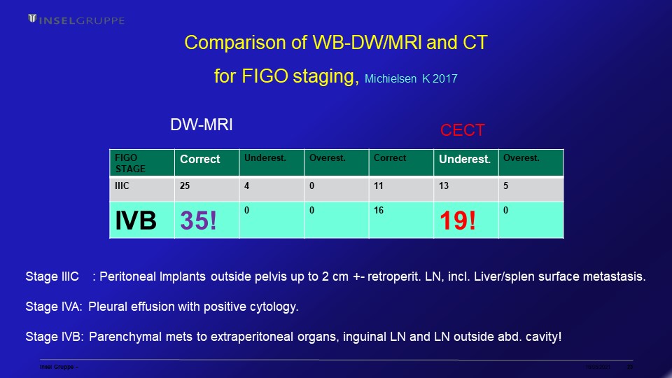 Slide #23
 Slide #24
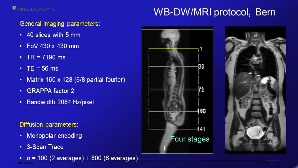 Slide #25
 Slide #26
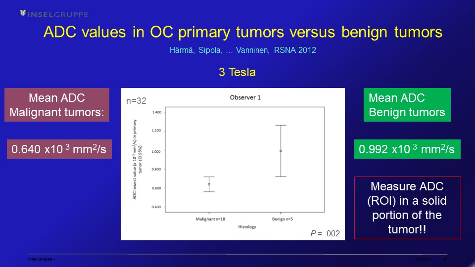 Slide #27
 Slide #28
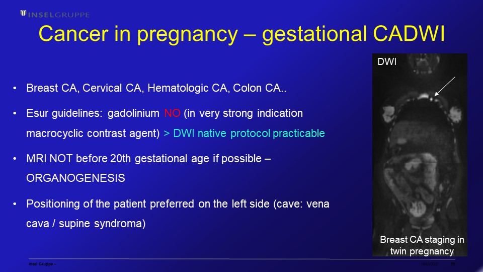 Slide #29
 Slide #30