Imaging in Regenerative Medicine
Erik M. Shapiro1
1Michigan State University, East Lansing, MI, United States
1Michigan State University, East Lansing, MI, United States
Synopsis
This educational lecture will provide a multimodal view of imaging in regenerative medicine. Indeed, the vastness of regenerative medicine requires a complete toolbelt to properly survey the completeness of research and development topics, and clinical methodologies for following treatments. This lecture will survey the uses of multimodal imaging in regenerative medicine, focusing on the benefits of individual imaging methodologies for probing specific research and development questions, and will provide my opinion on how these various imaging technologies might be used clinically.
Regenerative medicine is the branch
of medicine that develops methods to regrow, repair or replace
damaged or diseased cells, organs or tissues. Regenerative medicine seeks
to be long-term curative, differentiating itself from other clinical therapies
that focus primarily on treating symptoms of diseases or mitigating impact of
injurie. Regenerative medicine currently includes four main foci:
1)
In situ tissue engineering
2)
Cellular therapies
3)
Ex situ tissue engineering, essentially comprising artificial organs
4)
Hybrid biological devices
The
use of advanced imaging technologies has increased significantly in the past
two decades and has revolutionized patients’ care by enabling earlier and more
accurate diagnoses. Indeed, for MRI alone, in the USA there are ~ 13,500 MRI
sites performing over 35 million MRI scans annually, ~15,000 CT scanners
performing 92 million CT scans (2019 numbers, OECD.org), and ~ 2400 PET
installations performing ~ 2 million PET procedures (2015 numbers, IMVinfo.com)
illustrating the instrumental role that medical imaging plays in our lives.
Essentially, no critical medical decisions are taken without relying on some
sort of imaging. Furthermore, biomedical imaging plays a crucial role in
research and development of novel therapeutics, including in regenerative
medicine, at all stages from idea conception through preclinical testing
through translational and clinical trials.
Imaging
in regenerative medicine can be broken into many subcategories, but it may be
useful to distinguish at first between biomedical imaging during research and
development of new therapeutic approaches and clinical imaging. In general,
preclinical imaging is performed strictly using preclinical imaging systems in
cellular and small animal subjects, while clinical implementation of
regenerative medicine is or will be performed on humans, with imaging taking
place using clinical systems. Key milestones for using imaging in regenerative
medicine at the preclinical stage include the major facets of the development
phase, but also safety considerations. Key milestones of clinical imaging
include surgical planning, definition of implantation site, and long-term
monitoring.
Still
speaking generally, and independent of preclinical or clinical imaging, imaging
in regenerative medicine can also be divided into imaging native biomarkers or
exogenous imageable substances. Native imageable biomarkers would include basic
structural and anatomical features one expects to replace or repair, metabolic
signatures one expects to measure in function organs, and functional actions
one expects to see in normally functioning organs. Exogenous imageable
substrates can include impregnated imageable atoms or nanoparticles or can
include systemically delivered, internally accumulated chemicals, mediated by
normal or engineered metabolic machinery, or via indirectly or directly
detected engineered reporter genes.
Though
this is an MRI conference, this educational lecture will provide a multimodal
view of imaging in regenerative medicine. Indeed, the vastness of regenerative
medicine requires a complete toolbelt to properly survey the completeness of
research and development topics, and clinical methodologies for following
treatments. This lecture will survey the uses of multimodal imaging in
regenerative medicine, focusing on the benefits of individual imaging
methodologies for probing specific research and development questions, and will
provide my opinion on how these various imaging technologies might be used
clinically.
Acknowledgements
No acknowledgement found.References
No reference found.Figures
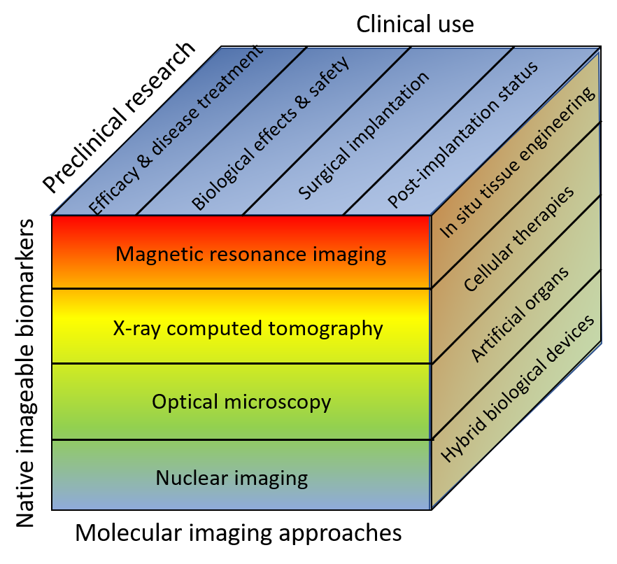
Figure 1