Complementary Modalities: DECT
1Mayo Clinic, Rochester, MN, United States
Synopsis
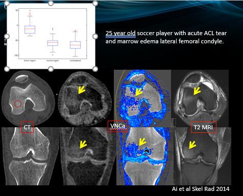
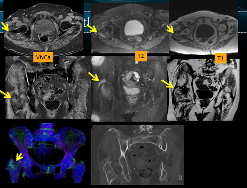
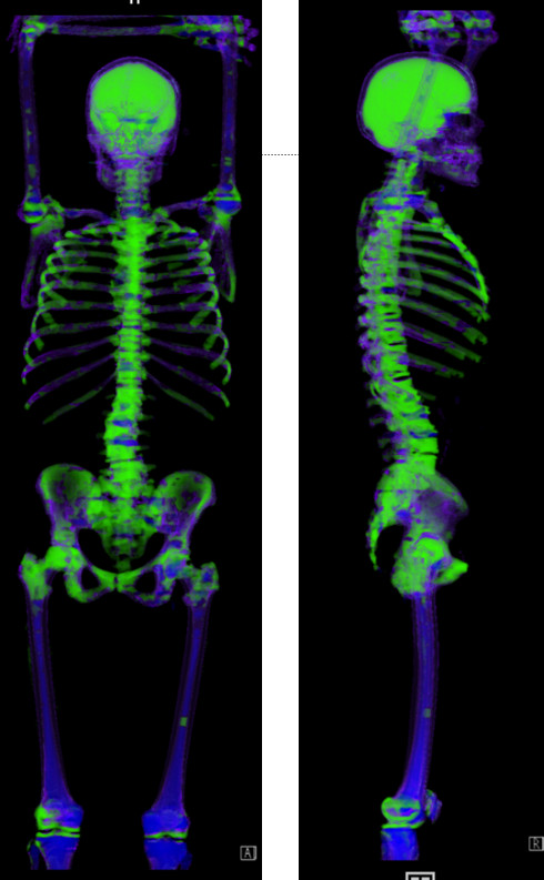
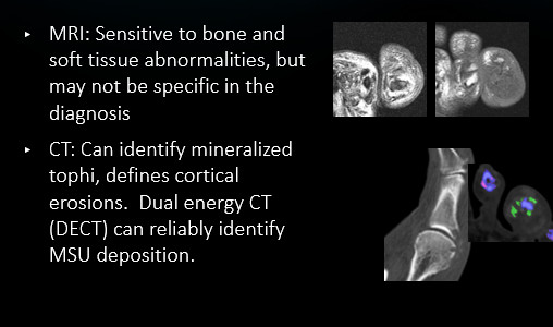
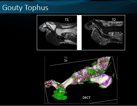
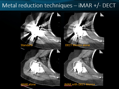
Dual-energy CT provides material composition information. Removal of the calcium signal (virtual non-calcium VNCa) from cancellous bone allows visualization of the marrow space. Studies have shown high sensitivity and specificity compared to MR in cases of trauma in the scaphoid, hip, knee, and ankle (fig 1,2). VNCa imaging can demonstrate marrow infiltration in cases of metastases and myeloma (fig 3). DECT allows identification of MSU crystals in cases of suspected gout with high sensitivity and specificity compared to aspiration (fig 4,5). Monoenergetic DECT with iterative metal artifact reduction can improve image quality in cases with significant metal artifact (fig 6).
Acknowledgements
I wish to thank Drs Baffour, Fletcher and McCollough for their valuable contributions to this work.References
1. Geijer M, Dunker D, Collin D, Gothlin JH. Bone bruise, lipohemarthrosis, and joint effusion in CT of non-displaced hip fracture. Acta Radiol. 2012;53(2):197-202.
2. Sanders TG, Medynski MA, Feller JF, Lawhorn KW. Bone contusion patterns of the knee at MR imaging: footprint of the mechanism of injury. Radiographics. 2000;20 Spec No:S135-151.
3. Ai S, Qu M, Glazebrook KN, et al. Use of dual-energy CT and virtual non-calcium techniques to evaluate post-traumatic bone bruises in knees in the subacute setting. Skeletal Radiol. 2014;43(9):1289-1295.
4. Pandey R, McNally E, Ali A, Bulstrode C. The role of MRI in the diagnosis of occult hip fractures. Injury. 1998;29(1):61-63. 7.
5. McCollough CH, Leng S, Yu L, Fletcher JG. Dual- and Multi-Energy CT: Principles, Technical Approaches, and Clinical Applications. Radiology. 2015;276(3):637-653.
6. Reagan AC, Mallinson PI, O'Connell T, et al. Dual-energy computed tomographic virtual noncalcium algorithm for detection of bone marrow edema in acute fractures: early experiences. J Comput Assist Tomogr. 2014;38(5):802-805.
7. Guggenberger R, Gnannt R, Hodler J, et al. Diagnostic performance of dual-energy CT for the detection of traumatic bone marrow lesions in the ankle: comparison with MR imaging. Radiology. 2012;264(1):164-173.
8. Kellock TT, Nicolaou S, Kim SSY, et al. Detection of Bone Marrow Edema in Nondisplaced Hip Fractures: Utility of a Virtual Noncalcium Dual-Energy CT Application. Radiology. 2017;284(3):922.
9. Suh CH, Yun SJ, Jin W, Lee SH, Park SY, Ryu CW. Diagnostic performance of dual-energy CT for the detection of bone marrow oedema: a systematic review and meta-analysis. Eur Radiol. 2018;28(10):4182-4194.
10. Mallinson PI, Coupal TM, McLaughlin PD, Nicolaou S, Munk PL, Ouellette HA. Dual-Energy CT for the Musculoskeletal System. Radiology. 2016;281(3):690-707.
11. Issa G, Davis D, Mulligan ME. The Ability of Dual-Energy Computed Tomography to Distinguish Normal Bone Marrow From Metastases Using Bone Marrow Color Maps. J Comput Assist Tomogr. 2018;42(4):552-558. 39
12. Pache G, Krauss B, Strohm P, et al. Dual-energy CT virtual non-calcium technique: detecting posttraumatic bone marrow lesions--feasibility study. Radiology. 2010;256(2):617-624.
13. Gosangi B, Mandell JC, Weaver MF, Uyeda JW, Smith SE, Sodickson AD, Khurana B. Bone marrow edema at dual-energy CT: a game changer in the emergency department. Radiographics 2020;40: 859-874
14. Koch V, Muller FC, Gosvig K, Albrecht MH, Yel I, Lenga L, Martin SS, Cavallaro M, Wichmann JL, Mader C, D’Angelo T, Mazziotti S, Cicero G, Vogl T, Booz C. Incremental diagnostic value of color-coded virtual non-calcium dual =energy CT for the assessment of traumatic bone marrow edema of the scaphoid. Eur Radiol 2021https://doi.org/10.1007/s00330-020-07541-x
15. Kosmala A, Weng AM, Heidemeier A, Krauss B, Knop S, Bley TA, Petritsch B. Multiple myeloma and dual-energy CT: diagnostic accuracy of virtual non-calcium technique for detection of bone marrow infiltration of the spine and pelvis. Radiol 2018;286:205-213.
16. Bongartz T, Glazebrook KN, Kavros SJ, Murthy NS, Merry SP, Franz WB, Michet CJ, Akkara Veetil BM, Davis JM, Mason TG, Warrington KJ, Ytterberg SR, Matteson EL, Crowson CS, Leng S, McCollough CH. Dual-energy CT for the diagnosis of gout: and accuracy and diagnostic yield study. Ann Rheum Dis 2015;74:1072-1077.
17. Mallinson PI, Coupal T, Reisinger C, Chou H et al. Artifacts in dual-energy CT gout protocol: a review of 50 suspected cases with an artifact identification guide. AJR 2014;203:103-109
18. Mallinson P, Reagan AC, Coupal T, Munk PL, Ouellette H, Nicolaou S. The distribution of urate deposition within the extremities in gout: a review of 148 dual-energy CT cases. Skeletal Radiol 2014:43:277-281.
19. Payam M, Baffour FJ, ADkins MC, Yu L, McCollough CH, Fletcher JG, Glazebrook KN. benefits of iterative metal artifact reduction and dual-energy CT towards mitigating artifact in the setting if total shoulder prostheses. Skeletal Radiol 2021;50:51-58.
20. Petritsch B, Kosmala A, Weng AM et al. Vertebral compression fractures: third generation dual-energy CT for detection of bone marrow edema at visual and quantitative analyses. Radiol 2017;284:161-168
Figures