Frontiers in Noncontrast Imaging: DWI & Spectroscopy
Laura Marie Fayad1
1Johns Hopkins University, Baltimore, MD, United States
1Johns Hopkins University, Baltimore, MD, United States
Synopsis
This talks focuses on the benefits of incorporating the non-contrast techniques of DWI and MR spectroscopy into a clinical MRI protocol for the evaluation of musculoskeletal tumors. DWI is a fast technique that provides information on the cellularity of lesions. DWI is useful for defining tumor extent by differentiating perilesional inflammation from perilesional tumoral infiltration. DWI is also helpful for characterizing lesions for malignancy, distinguishing viable tumor from post-treatment fibrosis and necrosis, and for distinguishing tumor recurrence from nodular fibrosis following surgery. Proton MR spectroscopy offers metabolic biomarkers of malignancy that can be used for lesion characterization and treatment response.
 Slide #1
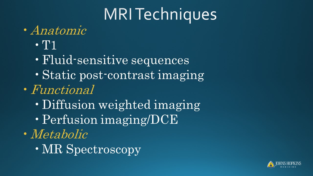 Slide #2
 Slide #3
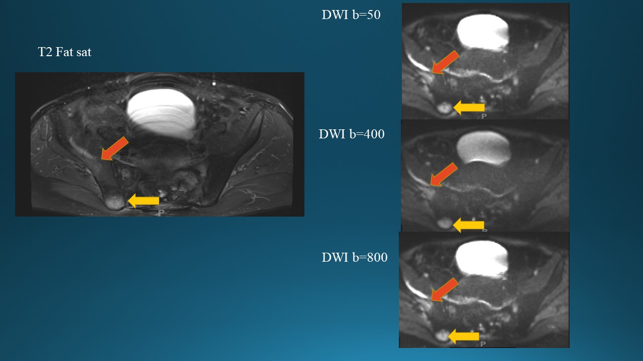 Slide #4
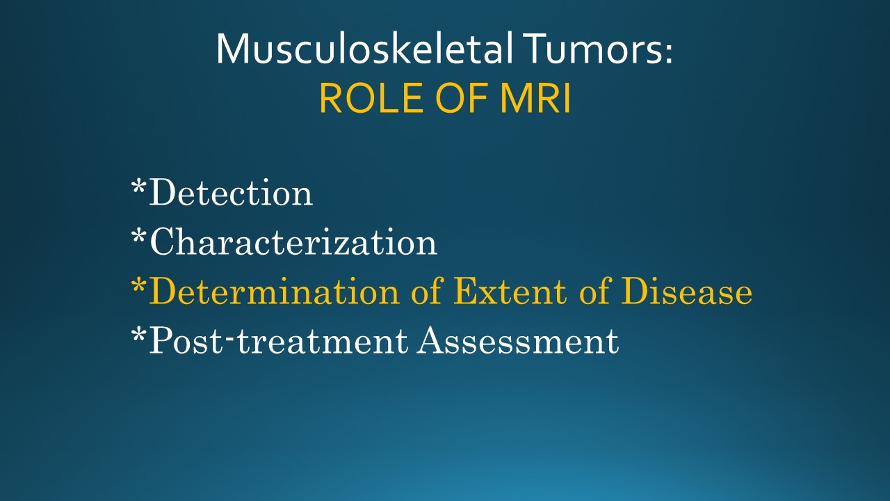 Slide #5
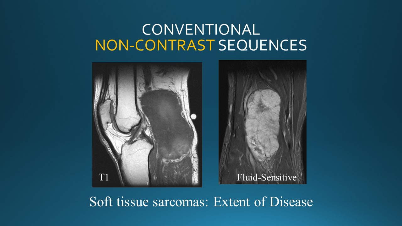 Slide #6
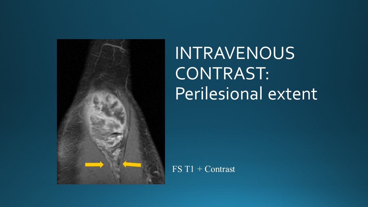 Slide #7
 Slide #8
 Slide #9
 Slide #10
 Slide #11
 Slide #12
 Slide #13
 Slide #14
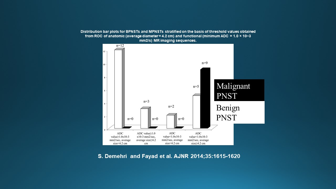 Slide #15
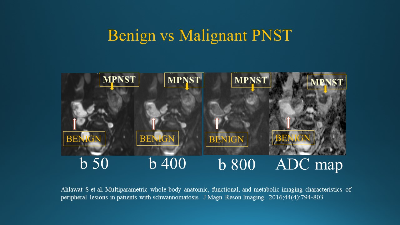 Slide #16
 Slide #17
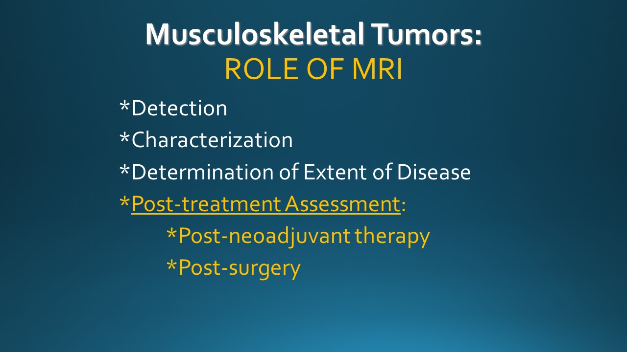 Slide #18
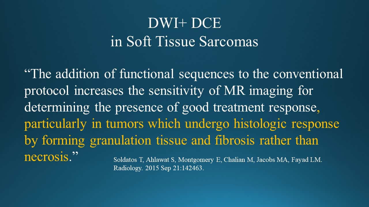 Slide #19
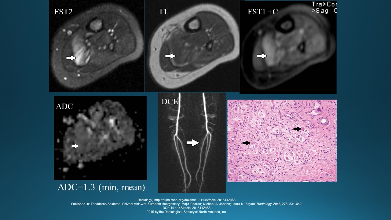 Slide #20
 Slide #21
 Slide #22
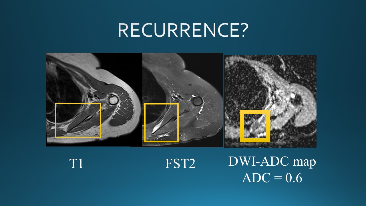 Slide #23
 Slide #29
 Slide #30