4339
fMRI based evaluation of yoga-induced changes in ischemic post-stroke patients1Department of NMR, All India Institute of Medical Sciences, New Delhi, India, 2Centre for Development of Advanced Computing, New Delhi, India, 3Manipal University, Jaipur, India, 4Department of Neurology, All India Institute of Medical Sciences, New Delhi, India
Synopsis
This fMRI study has evaluated for the first time, the functional changes effected by yoga in post-stroke (ischemic) recovery. Left hemisphere stroke patients (n=13) with motor deficits practiced Hatha yoga for one hour daily for six months under the supervision of certified yoga trainers. Pre-intervention, and 3 and 6 months post-yoga intervention assessments were carried out using fMRI at 3T and also clinically evaluated using NIHSS score. Results show positive response to yoga, reflected both in the significantly reduced NIHSS scores and the increased BOLD activity in the left pre-central gyrus region in the stroke patients.
Introduction
Stroke is a disease, which not only causes disability but also has social and economic consequences.1 Although there are a number of modalities like physiotherapy for post-stroke rehabilitation,2 there is growing interest in the therapeutic use of yoga.3,4 The objective of this study is fMRI based longitudinal evaluation of yoga-induced changes in post-stroke rehabilitation of ischemic stroke patientsMaterials and methods
Patient recruitment and assessment: Thirteen first-ever left hemisphere stroke patients (18-60 years) were enrolled for the study after written informed consent and with approval from the Institute Ethical Committee. All the patients had motor deficits with NIHSS (National Institute of Health Stroke Scale) score for left hemisphere < 15 and right hemisphere < 10. NIHSS scores were determined pre- and post-yoga (3 and 6 months) intervention.Yoga intervention and assessment: The patients practiced Hatha yoga for six months (one hour everyday) under the supervision of certified yoga trainers. The therapeutic response was assessed by the clinical scores.
MR studies: fMRI acquisition was carried out pre- and post-yoga (3 and 6 months) intervention at 3T (Ingenia, Philips). The fMRI studies involved unilateral and bilateral motor tasks (closing and opening the fist). Single-shot EPI (Echo Planner Imaging) sequence was used for the Blood Oxygen Level Dependent (BOLD) study with the following parameters: slices 35, slice thickness 4 mm, repetition time 2000 ms, echo time 30 ms, acquisition matrix size 76x74, number of dynamics - 180. fMRI data were analysed using the CONN toolbox based on Statistical Parametric Mapping [SPM 12 (Wellcome trust, London, UK)]. Time series data from pre-central gyrus region were extracted using brain atlas (Talairach Atlas Labels) and Automated Anatomical Labeling Atlas. Longitudinal comparative analysis of all the time series was carried out and the results presented as frequency distribution for each patient.
Results
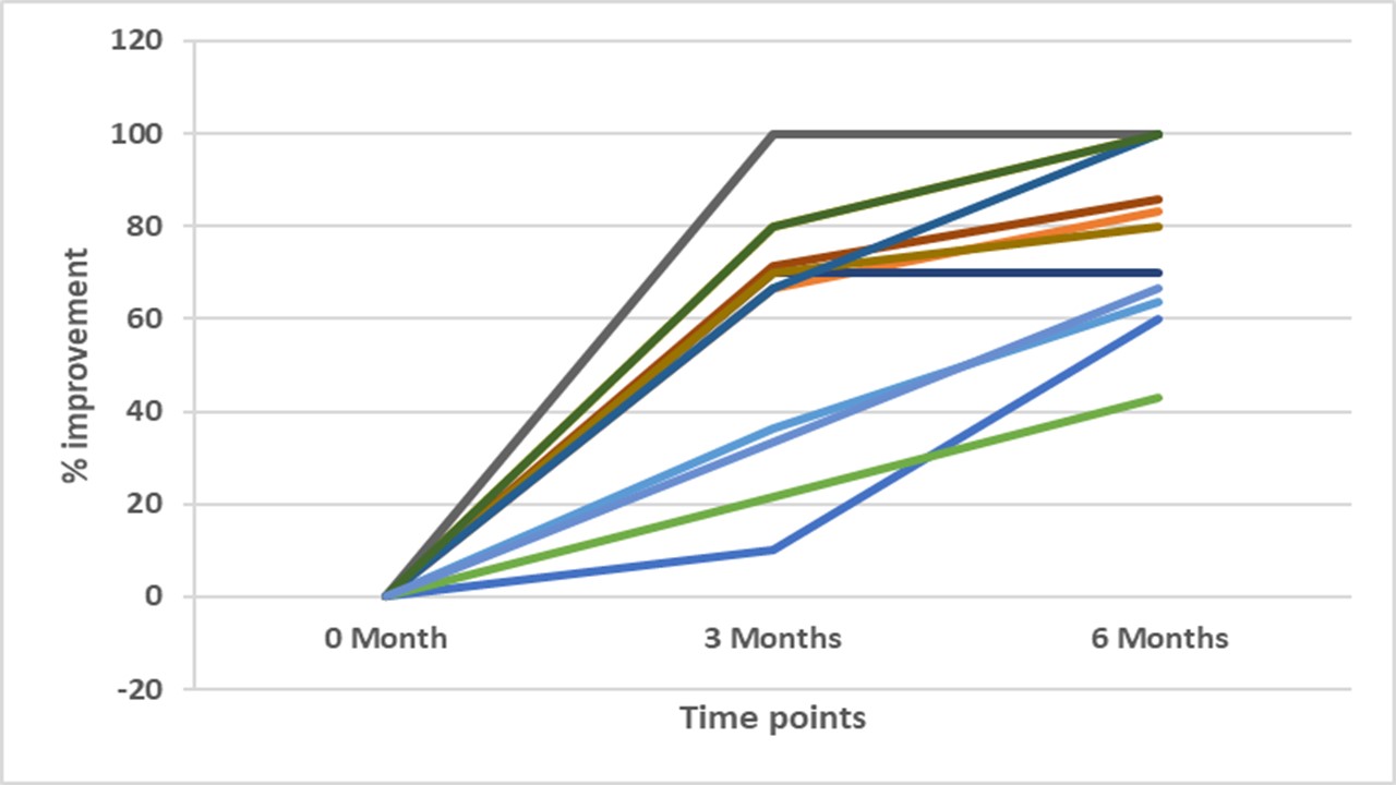
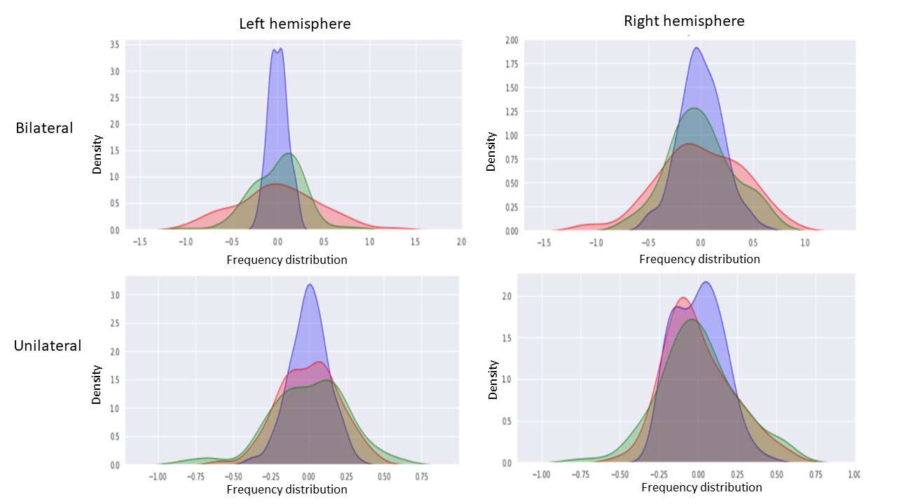
Longitudinal changes in clinical NIHSS score are shown for all patients as percentage improvement (Fig. 1). It shows that all the patients showed clinical improvement post-yoga intervention. This was also reflected in the fMRI activation changes observed in precentral gyrus. Frequency distribution of the BOLD activity from a patient who showed significant clinical improvement is shown as a representative data in Figure 2. The peak (purple) corresponding to the zero time point (pre-intervention) signifies that the BOLD activity was absent since most of the activity data was very close to the mean, namely zero, with very little standard deviation. In the 2nd (3 months post-intervention) and 3rd (6 months post-intervention) time points, the curves (red and green, respectively) are spread and not concentrated around zero indicating increase in BOLD activity, which in turn reflects clinical improvement. Although the group analysis showed significant improvement, there were individual variations to yoga intervention. Further in-depth analysis is under way.Conclusion
Stroke significantly affects the health of individuals and also has major societal impact including that of nation’s economy. In post-stroke rehabilitation, there is increasing interest in use of yoga in addition to the conventional physiotherapy. There are a number of reports on therapeutic benefits of yoga and its cost-effectiveness.3,4 With this in focus, the rigors of modern science have been combined with the ancient technique of yoga to understand the changes effected by it in ischemic stroke patients. The current fMRI study has evaluated for the first time, yoga-induced changes in functional activity in brain in post-stroke recovery. The preliminary results have shown that the improvement assessed by clinical scores reflected as changes in the pre-central gyrus activation. Although, all the patients have shown positive clinical response to yoga intervention, there were also individual variations in the response. These are interesting and warrants further in-depth studies and analyses.Acknowledgements
The work was supported by SATYAM scheme, Department of Science and Technology, Government of India.References
1. World Health Organization, Global Strategy for the Prevention and Control of Noncommunicable Diseases. Report by the Director General. A 53/14 Fift-third World Health Assembly, May 2000.
2.http://www.strokeassociation.org/STROKEORG/LifeAfterStroke/RegainingIndependence/PhysicalChallenges/Post-Stroke-Rehabilitation_UCM_310447_Article.jsp#.W-KLoKeB1Bw, American Stroke Association
3. Jeter PE, Slutsky J, Singh N, Khalsa SBS. Yoga as a therapeutic intervention: a bibliometric analysis of published research studied from 1967 to 2013. J Comp Alt Med, 2015: 1-7, 2015.
4. Sovova E, Cajka V, Pastucha D, et al. Positive effect of yoga on cardiorespiratory fitness: A pilot study. Intl J Yoga. 2015, 8: 134-138.
Figures