4328
Predicting the Neurodegeneration after Stroke using Disconnectivity Map1Institut de Bio-imagerie IBIO, Université de Bordeaux, Bordeaux, France, 2Neuroimagerie diagnostique et thérapeutique, CHU de Bordeaux, Bordeaux, France, 3Unité de soins intensifs neurovasculaires, CHU de Bordeaux, Bordeaux, France
Synopsis
We investigated remote effects of brain infarction. We previously demonstrated higher iron content (higher R2* values) in deep nuclei as a consequence of their disconnection. Here, we aimed at predicting such long term remote degeneration from the acute stage. Through a disconnectivity approach, we mapped the fibers that were likely to be disconnected by projecting the acute stroke masks on tractograms from 180 healthy volunteers. We showed that disconnected areas based on this approach were likely to show significantly higher iron content at follow-up. This validates the prediction of deep nuclei disconnection from acute stage through a disconnectivity approach.
Introduction
After brain infarction, damages are not only localized but also affect remote areas. How the deep gray matter is affected is still not well understood. Previously, we focused on iron deposition as a consequence of secondary degeneration within the deep gray matter, and we showed a significant increase of R2* in the thalamus and the substantia nigra ipsilateral to stroke as a possible consequence of their disconnection [1,2]. However, we could only show a correlation between stroke location and remote increase in iron content, but we could not directly and formally conclude that these two phenomena were linked through network disturbance. Traditionally, diffusion tractography has been used to identify fibers associated with regions of interest and lesions. Nevertheless, in most clinical settings, especially at the acute stage of stroke, it is impossible to obtain detailed tractography.Brain Connectivity and Behaviour toolkit (BCBtoolkit) is a software capable of estimating the probability of anatomical disconnection induced by a focal lesion [3]. Through BCBtoolkit we could directly test the possible connection between stroke and remote consequences within the deep gray matter as measured by higher iron content.
The aim of this study is to investigate the nonlocal effect of stroke on the substantia nigra and the thalamus.
Methods
Study Population and Image AcquisitionPatients admitted for an ischemic stroke between 2012 and 2015 were enrolled in a prospective study that included an initial (24 – 72h) and 1-year follow-up MRI. Patients were excluded based on the following criteria: dropout at 1-year follow-up, stroke only in the posterior fossa, direct involvement of thalamus or substantia nigra or microbleeds within these structures, severe artifacts on MRI. Overall, the study consisted of 179 patients for the analysis of the substantia nigra, and 158 patients for the thalamus.
All MRIs were performed on a 3T scanner (Discovery MR 750w, GE medical System) with the same protocol at baseline and at follow-up that included DWI, 3D T1 inversion-recovery-prepared fast spoiled gradient echo, 3D FLAIR, and 2D multi-echo fast gradient-echo (TR 775 ms; 8 TE, 4.3, 8.7, 13.0, 17.4, 21.7, 26.1, 30.5, 34.8 ms; flip angle, 20°; thickness, 4mm; matrix 320×320, FOV 240×240 mm).
All data were standardized in the MNI152 space. Strokes were delineated slice by slice based on DWI at baseline. We used an atlas to outline the substantia nigra and the thalamus [4,5].
Disconnectivity map
To define the distant consequences of stroke, we computed probability maps of disconnection for each patient using BCBtoolkit (Fig. 1). It identifies the tracts passing through the infarction (and therefore tracts that are likely to be disconnected) by using tractograms from 180 healthy subjects scanned at 7T as part of the Human connectome project. Then, we measured the overlap between the disconnected tracts and the masks of substantia nigra and of thalamic nuclei (medial, lateral, and posterior groups). According to the amount of voxels in the overlap, we classified the patients as having their substantia nigra or their thalamus “not disconnected,” “mildly disconnected,” or “severely disconnected.”
Quantification of Iron
R2* values were obtained by a voxel-by-voxel nonlinear least-squares fitting as a mono-exponential signal decay model from the multi-echo T2*. We defined the asymmetry index (AI) of R2* of the substantia nigra or thalamus as follows:
$$Asymmetry\ Index=\frac{\ {R2}_{disconneced\ side}^\ast-\ {R2}_{conneced\ side}^\ast}{{R2}_{disconneced\ side}^\ast+\ {R2}_{conneced\ side}^\ast}\times100$$
Statistical Analyses
We compared the AI between the baseline and the 1-year follow-up scan, using Wilcoxon singed-rank test. We also conducted voxel-based morphometry (VBM) analysis of R2* value in the substantia nigra and the thalamus, comparing the connected and disconnected sides. Finally, the impact of disconnectivity was tested with linear regression analysis.
Results
In the substantia nigra, and the medial & lateral thalamus, severely disconnected patients showed significantly higher AI of R2* at 1-year follow up, compared to baseline (Fig 2). The mildly disconnected group did not show any significant difference.In VBM analysis, the lateral area of the substantia nigra, lateral area of the posterior thalamus, lateral area of the medial thalamus, and lower area of the lateral thalamus showed significantly higher R2* values in the disconnected side compared with the connected side (Fig 3). We also found a few voxels with significant lower R2* values in the upper medial area in each thalamic group.
In multivariable linear regression, the variable “severely disconnected” was an independent predictor of high AI of R2* at 1-year follow-up in the substantia nigra, the medial thalamus, and the lateral thalamus (Fig 4) together with the infarction volume and AI at baseline.
Discussion/Conclusion
The disconnectivity map is based on tractograms from healthy subjects. Therefore whether this strategy could really reflect the status of the tracts in patients could be questioned. However, in this work, we have been able to estimate the effect of the network damage alternatively thanks to the quantification of iron increase on the long term follow up in disconnected areas. The finding that disconnected areas are those in which iron will accumulate on follow up is an important validation of the disconnectivity approach. It brings the opportunity to speculate on the status of remote areas very early after the occurrence of the infarction. The clinical consequences of such disconnection are currently investigated.Acknowledgements
The study was supported by public grants from the French Agence Nationale de la Recherche within the context of the Investments for the Future Program, referenced ANR-10-LABX-57 and named “TRAIL” (Translational Research and Advanced Imaging Laboratory). The cohort was funded by a public grant from the French government (PHRC programme hospitalier de recherche clinique inter-régional).References
- Linck PA, Kuchcinski G, Munsch F, Griffier R, Lopes R, Okubo G, et al. Neurodegeneration of the Substantia Nigra after Ipsilateral Infarct: MRI R2* Mapping and Relationship to Clinical Outcome. Radiology. 2019 May;291(2):438–48.
- Kuchcinski G, Munsch F, Lopes R, Bigourdan A, Su J, Sagnier S, et al. Thalamic alterations remote to infarct appear as focal iron accumulation and impact clinical outcome. Brain. 2017 Jul 1;140(7):1932–46.
- Foulon C, Cerliani L, Kinkingnéhun S, Levy R, Rosso C, Urbanski M, et al. Advanced lesion symptom mapping analyses and implementation as BCBtoolkit. Gigascience. 2018 Mar 1;7(3):1–17.
- Su JH, Thomas FT, Kasoff WS, Tourdias T, Choi EY, Rutt BK, et al. Thalamus Optimized Multi Atlas Segmentation (THOMAS): fast, fully automated segmentation of thalamic nuclei from structural MRI. Neuroimage. 2019 Jul 1;194:272–82.
- Keuken MC, Forstmann BU. A probabilistic atlas of the basal ganglia using 7 T MRI. Data Brief. 2015 Sep;4:577–82.
Figures
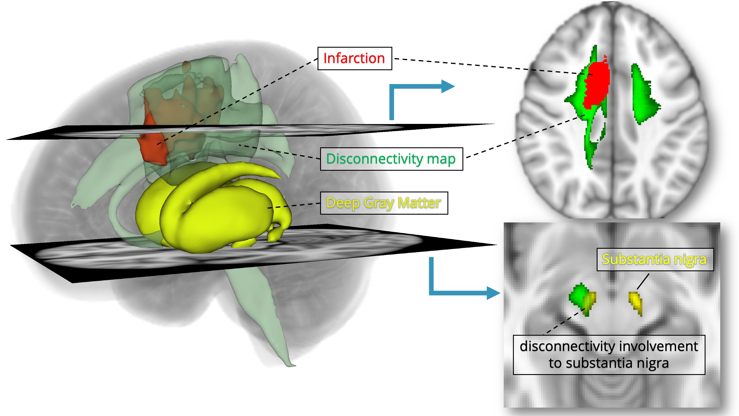
Figure 1. Overview of a representative disconnectivity map. Disconnectivity map (green) is calculated using the lesion mask of the infarction (red), based on the tractograms of healthy subjects. The degree of overlap of the disconnectivity map and masks of deep nuclei was used to estimate the remote effect of the infarction.
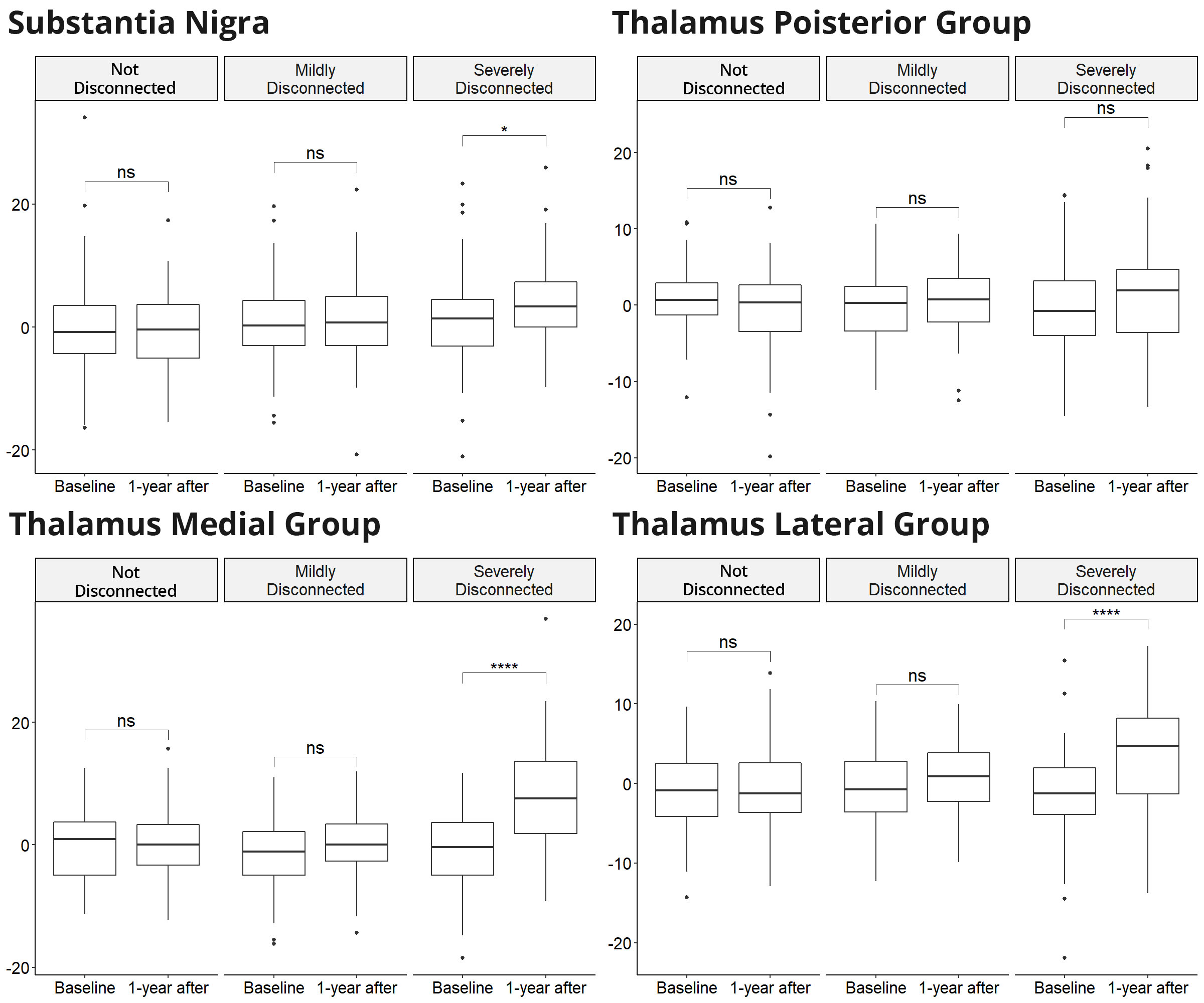
Figure 2. Comparison of the asymmetry index of R2* among “Not Disconnected,” “Mildly Disconnected,” and “Severely Disconnected” nuclei. Severely disconnected nuclei showed a significant increase of R2* at 1-year follow-up in the substantia nigra and the medial & lateral nuclei of the thalamus.
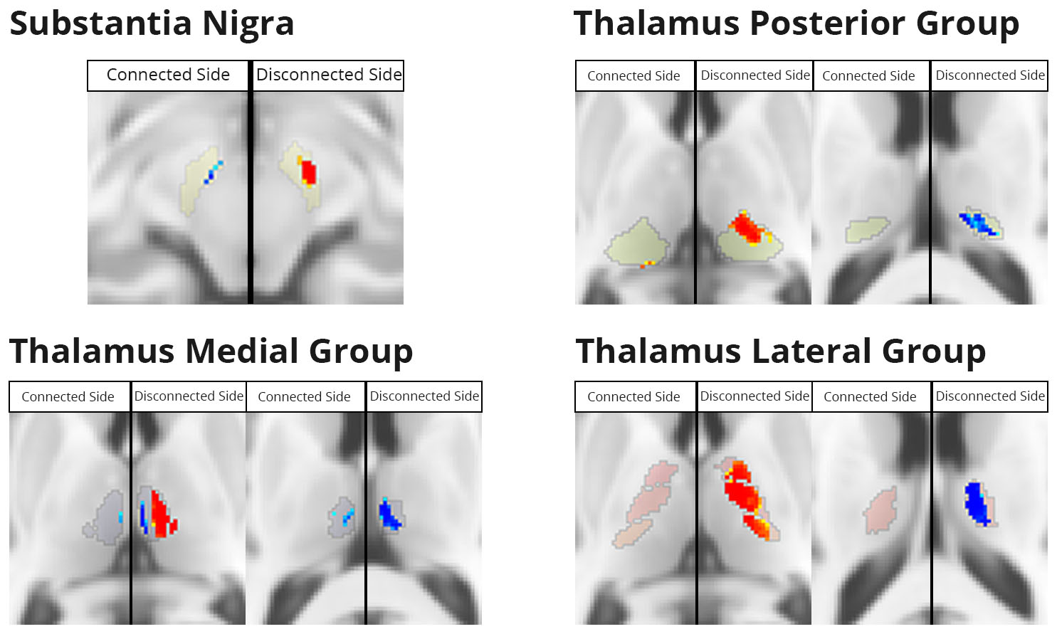
Figure 3. Voxel-based morphometry analysis for the R2* value in the substantia nigra and the thalamus. The red area means a significant increase of the R2* value at 1-year follow-up compared with that at baseline. The blue area means a significant decrease at 1-year follow-up.
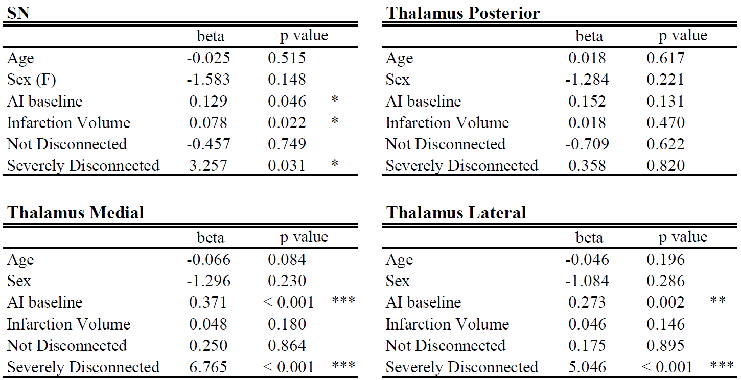
Figure 4. Multivariate linear regression analysis for the prediction of the asymmetry index at 1-year follow-up. The state of “Severely Disconnected” and the asymmetry index at baseline are the significant predictor. The infarction volume is significant only in the substantia nigra.