4319
TractLearn: comparison with the General Linear Model for optic pathways exploration1Neuroradiology, Grenoble University Hospital, Grenoble, France, 2School of Biomedical Engineering, The University of Sydney, Sydney, Australia, 3Ophthalmology, Grenoble University Hospital, Grenoble, France, 4Neurology, Grenoble University Hospital, Grenoble, France, 5IRMaGe, Inserm US 17, CNRS UMS 3552, Grenoble, France, 6Pixyl Medical, Grenoble, France, 7Sydney Imaging, Sydney, Australia
Synopsis
TractLearn was recently proposed for tract-based MRI quantitative analyses, based on Riemannian distances between anatomical structures. It allows to detect joint quantitative variations in a group of voxels, and in theory to decrease the number of false negatives compared with the General Linear Model (GLM). TractLearn also takes advantage of a manifold approach to capture controls variability as standard reference. Here we aim at comparing the performance of TractLearn with the GLM in detecting optic nerve voxel alteration, using the side of visual impairment as clinical reference.
INTRODUCTION
Modern approaches for microstructural characterization based on Diffusion-Weighted Imaging often extract a collection of metrics per structure of interest. Statistics are then usually based on the General Linear Model (GLM), which relies on a unique reference standard (ie. the Euclidean mean from a control group) for quantitative abnormalities detection. TractLearn has recently been proposed as a Riemannian alternative[1] in which distances in a manifold subspace after nonlinear dimension reduction allowed to detect joint statistical variations in a group of voxels and potentially be more sensitive than GLM in detecting abnormalities. Using Euclidean Mean as reference could also erroneously classify as abnormal a voxel linked to anatomic variation or modified by the acquisition condition (e.g. artifacts) rather than altered by the disease process itself. The absence of gold standard made difficult to conclusively compare TractLearn with GLM in our previous work. In this context, a unique opportunity is provided by anterior optic pathways exploration, because focal lesions on optic nerves track-weighted imaging (TWI) reconstructions due to unilateral optic neuropathy were found linked to ipsilateral visual impairment [2]. Here, we illustrate the benefits of TractLearn on a population referred for optic neuropathy, and assess the number of altered voxels in each optic nerve, as detected using TractLearn, in comparison with GLM. The secondary objective was to evaluate the necessity for statistical correction for multiple comparison, given that TractLearn studies anatomical structure as global objects rather than as independent voxels.METHODS
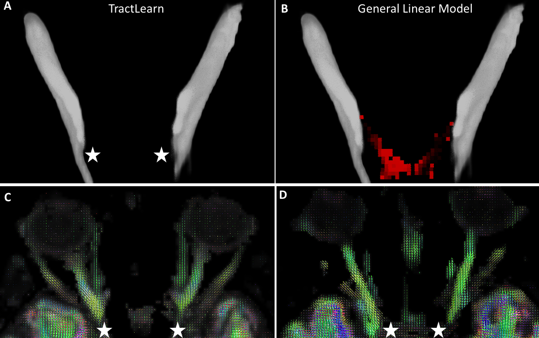
30 healthy volunteers and 30 patients with optic neuropathy were scanned on a 3T Philips Achieva- dStream MRI scanner. Diffusion MRI data (b=1000 s/mm2, 60 directions)[2] were analyzed using Single-Shell 3-tissues CSD[3] in MRtrix3[4] to compute fibre orientation distributions (FODs). We manually drew one ROI per optic nerve, and performed volumetric tractography (1000 streamlines initiated from seed-points randomly distributed throughout orbital area, using optic nerves as inclusion ROI). Finally, track-density maps [5] were used for spatial correspondence with a group-specific population template, and TWI of the FOD amplitude (TW-FOD) used as quantitative metric[6](Figure 1)TractLearn: we use the strategy developed in[1] to localize an anomaly, summarized as follow: · Dimensionality reduction using Umap[7] to convert collection of quantitative values from the optic nerves into a 3-dimensional point in the manifold subspace based on a Riemaniann approximation.
Atlas construction from the healthy control dataset:Y=f(x)+ε. Y being MRI data in real space, x the corresponding point in the reduced space and ε the residuals (Figure 2).
GLM: the manifold approach was replaced by an Euclidean mean of all track-weighted maps coming from controls, which was further back projected in the subject space:Y=Euclidean Mean + εGLM.
We consider that the residuals ε and εGLM are representative of any abnormalities present in a new patient when it is greater than the model variability learned during a leave-one-out procedure on controls.
We consider as false positive any abnormality detected in the contralateral optic nerve in patients. We have also compared the number of altered voxels detected in the clinically relevant optic nerve, using either GLM or TractLearn. Finally, we compare Bonferroni correction and False Discovery Rate (FDR) for multiple comparison with the absence of correction for multiple comparison (p<0.05).
RESULTS
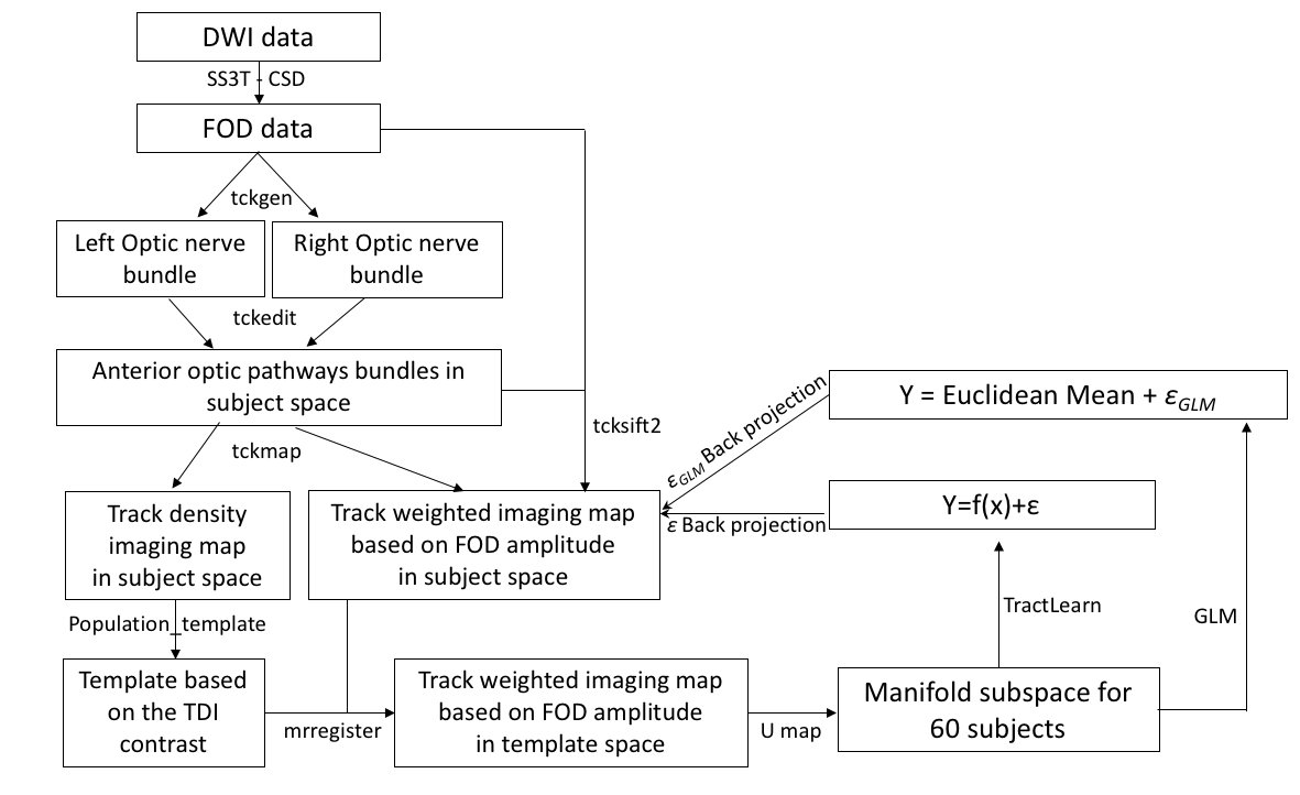
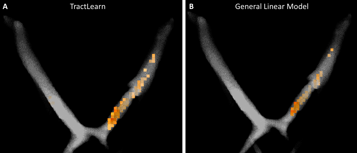
GLM identified altered voxels in 10, 11 and 21 patients out of 30 on the clinically injured-side using respectively the Bonferroni, FDR and no correction for multiple comparison, respectively. TractLearn identified altered voxels in 12, 13 and 23 patients out of 30 on the clinically injured-side using respectively the Bonferroni, FDR and no correction (Figure 3).Using TractLearn, we did not identify any abnormal voxels in contralateral optic nerves of the 30 patients while GLM detects abnormal voxels in the optic canal in 3 cases (Figure 4), regardless of correction strategy (Bonferroni, FDR or none). All 3 cases were linked to susceptibility artifacts making impossible to reconstruct fibres between the posterior part of the optic nerves and the optic chiasm.
Using TractLearn, we significantly identified more altered voxels in injured optic nerves in comparison with GLM (p<0.01) (Figure 5).
DISCUSSION
TractLearn demonstrates high specificity in detecting altered voxels in patients with unilateral optic neuropathy and a significantly higher number of altered voxels in clinically relevant optic nerves in comparison with GLM. TractLearn and GLM have close diagnostic performance in terms of number of patients with optic nerve lesion on the clinically-injured side (13 vs 11), although GLM was the only one to erroneously detect contralateral optic nerves abnormalities.We hypothesize that is linked to a severe local disease process allowing GLM to detect the most severely injured voxels despite the previously demonstrated important Standard Deviation in comparison with TractLearn. We believe that in diseases with more global and subtle quantitative variation, TractLearn could increase the gap with GLM. While we cannot confirm in vivo which voxels are biologically altered, we hypothesize that TractLearn gives a more accurate representation of the lesion size and open new horizons for diagnosis/prognosis purpose.
Finally, using FDR or Bonferroni correction for thresholding the Z scores maps led to false negative in clinically injured nerves. It is an interesting new advantage of our Riemaniann framework that needs to be confirmed in further studies.
Acknowledgements
The computation server used benefitted from the ERATRANIRMA project, by Agence Nationale de la Recherche [grant ANR-12-EMMA-0056]. This work was also supported by funding from the National Health and Medical Research Council of Australia and the Australian Research Council.References
- Attyé A, Renard F, Baciu M, et al (2020) TractLearn: a geodesic learning framework for quantitative analysis of brain bundles. medRxiv 2020.05.27.20113027. https://doi.org/10.1101/2020.05.27.201130272.
- Attyé A, Jean C, Remond P, et al (2018) Track-weighted imaging for neuroretina: Evaluations in healthy volunteers and ischemic optic neuropathy. J Magn Reson Imaging JMRI. https://doi.org/10.1002/jmri.259413.
- Dhollander T, Connelly A (2016) A novel iterative approach to reap the benefits of multi-tissue CSD from just single-shell (+b=0) diffusion MRI data4.
- Tournier J-D, Smith R, Raffelt D, et al (2019) MRtrix3: A fast, flexible and open software framework for medical image processing and visualisation. NeuroImage 202:116137. https://doi.org/10.1016/j.neuroimage.2019.1161375.
- Calamante F, Tournier J-D, Heidemann RM, et al (2011) Track density imaging (TDI): validation of super resolution property. NeuroImage 56:1259–1266. https://doi.org/10.1016/j.neuroimage.2011.02.0596.
- Calamante F (2017) Track-weighted imaging methods: extracting information from a streamlines tractogram. Magma N Y N. https://doi.org/10.1007/s10334-017-0608-17.
- McInnes L, Healy J, Melville J (2018) UMAP: Uniform Manifold Approximation and Projection for Dimension Reduction. ArXiv180203426 Cs Stat
Figures
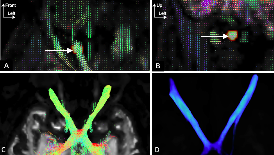
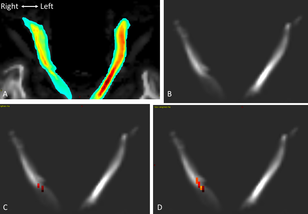
Tractography pipeline, made in two steps because a global tractographic approach including both optic nerves ROIs in one time would favor fibre generation in the healthy optic nerve over fibres in the diseased optic nerve. A-B: Axial and coronal FOD images show spherical ROIs (white arrow) to locate the optic nerve. C: Tractogramm as obtained using tckedit. D: Track file conversion based on TDI. The template was computed based on individual track-density maps to allow a perfect matching between optic nerves structures, which was not achieved via a FOD-guided non-linear registration.