4312
High angular resolution diffusion MRI tractography-based thalamic parcellation of postmortem fetal brain in the second trimester
Sheng-Min Huang1, Kuan-Hung Cho1, Koping Chang2, Pei-Hsin Huang2,3, and Li-Wei Kuo1,4
1Institute of Biomedical Engineering and Nanomedicine, National Health Research Institutes, Miaoli, Taiwan, 2Department of Pathology, National Taiwan University Hospital, Taipei, Taiwan, 3Graduate Institute of Pathology, National Taiwan University College of Medicine, Taipei, Taiwan, 4Institute of Medical Device and Imaging, National Taiwan University College of Medicine, Taipei, Taiwan
1Institute of Biomedical Engineering and Nanomedicine, National Health Research Institutes, Miaoli, Taiwan, 2Department of Pathology, National Taiwan University Hospital, Taipei, Taiwan, 3Graduate Institute of Pathology, National Taiwan University College of Medicine, Taipei, Taiwan, 4Institute of Medical Device and Imaging, National Taiwan University College of Medicine, Taipei, Taiwan
Synopsis
Proper fiber connections between cerebral areas are particularly essential to build brain functions. As a central relay, the thalamus plays an important role in regulating diverse brain functions. Here, we aimed to investigate the thalamo-cortical connections and thalamic parcellation of ex vivo fetal brains in the second trimester using high-resolution postmortem diffusion MRI tractography on 3T. Diffusion images with 333 µm isotropic voxel size were acquired and tractography-based thalamic parcellation was performed. The segregated thalamic patterns were found as similar as adult’s ones reported previously. Our results showed the thalamic development and demonstrated the capability of postmortem diffusion MRI tractography.
Introduction
Benefit from multiple image contrasts, MRI has become a favored tool in discovering the prenatal development of human brain 1-4. Being able to probe water molecular diffusion within complex tissues, diffusion MRI (dMRI) has been widely employed to characterize the white matter (WM) pathways. WM fiber bundles such as callosal fibers and thalamo-cortical tracts have been reconstructed to depict the development of human WM pathways 3,5. Although several WM bundles have been reconstructed in fetal brains, it remains unclear if these tracts are correctly connected to the corresponding cortex projection sites or not. As axon proning is an essential step for neuronal development 6, proper fiber connections or projections between cerebral areas are particularly essential to build brain functions. As a central relay, the thalamus plays an important role in regulating diverse brain functions. Here, we aimed to investigate the thalamo-cortical connections and thalamic parcellation of ex vivo fetal brains in the second trimester using high-resolution postmortem diffusion MRI tractography on 3T. Diffusion images with 333 µm isotropic voxel size were acquired and tractography-based thalamic parcellation was performed.Materials & Methods
One postmortem human fetal brain (23-week gestational age) was obtained with full parental consent and all the experiments followed the IRB approval. The ex vivo brain was formalin-fixed and immersed in formalin to avoid air interference during scan. All MR experiments were performed on a custom-built 3T MRI system equipped with a high-strength gradient coil (Resonance Research Inc., 675 mT/m and 11.5-cm bore size) 7. A dedicated solenoid RF coil was designed and integrated with a 3D-printed sample container for optimizing the signal-to-noise ratio. A high angular resolution dMRI (HARDI) sampling scheme with 60 encoding directions and maximum b-value of 2000 s/mm2 was performed by using a 3D multi-shot segmented EPI acquisition. The sequence parameters were FOV of 64x64x64 mm3, MTX of 192x192x192, TR/TE of 500/66 ms, Δ/δ of 32/18 ms, and views per segment of 32, yielding the isotropic voxel size of 333 µm. The frontal, parietal, occipital, temporal lobes and bilateral thalamic regions were manually drawn and confirmed by two physicians. Q-ball imaging reconstruction was carried out by using DSI studio (http://dsi-studio.labsolver.org/). A deterministic fiber tracking algorithm (angular threshold = 45 degrees, step size = 0.16 mm, normalized quantitative anisotropy threshold = 0.05, track length = 5 – 100 mm, and 1 million seeds) 8 was utilized to find the thalamo-cortical tracts connecting the thalamic regions and four pre-labelled cerebral lobes. Each thalamic voxel may contain several tracts corresponding to different lobes and the lobe which has the maximum number of tract connections would be assigned as the primary lobe corresponding to this voxel. All analyses were performed using Matlab (MathWorks, MA, USA).Results
Figure 1 shows the averaged diffusion-weighted images (DWIs) along three orthogonal planes. Several anatomical structures can be visually identified, including cortical plate (CP), thalamus, corpus callosum (CC), and several sulci. Figure 2 shows the thalamo-cortical tracts connecting the thalamus and those four pre-labelled lobes. Compared with the frontal, parietal, and temporal lobes, the occipital lobe has less tracts connecting to the thalamus. The tractography-based thalamic parcellation is shown in figure 3. At first glance, a topological pattern of thalamo-cortical connections is clearly shown. Although some thalamic voxels are lack of tract connections, the parcellation still shows well-organized symmetric patterns in bilateral thalamic regions.Discussion
To our best knowledge, this is the first study to employ postmortem diffusion MRI and tractography-based approach to parcellate the thalamus of ex vivo fetal brains. The topological pattern of segregated thalamic regions was found as similar as adult’s one reported previously 9. Our results clearly showed the thalamic development in the second trimester and demonstrated the capability of postmortem diffusion MRI tractography. It is also interesting to observe that less thalamic-cortical tracts connecting the thalamus and occipital lobe were reconstructed. Since the visual system is not activated until late third trimester, the tracts connecting the thalamus and occipital lobe may be immature in this gestational age. A further investigation on other fetal brains in later gestational ages is needed to address this issue. Methodologically, technical improvements including HARDI sampling scheme, sequence parameter optimization, reconstruction approach, and fiber tracking algorithms are needed to refine the robustness of parcellation and solidify the results by examining the reproducibility on other fetal brains.Acknowledgements
This work was supported by the grant from Ministry of Science and Technology (MOST-109-2221-E-400-001-MY2 for LWK and MOST-109-2811-E-400-500-MY2 for SMH) and National Health Research Institutes (NHRI-BN-109-PP-06 and NHRI-BN-109-SP-11 for LWK, and NHRI-EX109-10903NI for PHH).References
- Huang, H., et al., Anatomical characterization of human fetal brain development with diffusion tensor magnetic resonance imaging. J Neurosci, 2009. 29(13): p. 4263-73.
- Vasung, L., et al., Quantitative and Qualitative Analysis of Transient Fetal Compartments during Prenatal Human Brain Development. Front Neuroanat, 2016. 10: p. 11.
- Wilkinson, M., et al., Migration Pathways of Thalamic Neurons and Development of Thalamocortical Connections in Humans Revealed by Diffusion MR Tractography. Cereb Cortex, 2017. 27(12): p. 5683-5695.
- Vasung, L., et al., Ex vivo fetal brain MRI: Recent advances, challenges, and future directions.Neuroimage, 2019. 195: p. 23-37.
- Vasung, L., et al., Spatiotemporal Relationship of Brain Pathways during Human Fetal Development Using High-Angular Resolution Diffusion MR Imaging and Histology. Front Neurosci, 2017. 11: p. 348.
- Low, L.K. and H.J. Cheng, Axon pruning: an essential step underlying the developmental plasticity of neuronal connections. Philos Trans R Soc Lond B Biol Sci, 2006. 361(1473): p. 1531-44.
- Cho, K.H., et al., Development, integration and use of an ultra-high-strength gradient system on a human-size 3 T magnet for small animal MRI. PLoS One, 2019. 14(6): p. e0217916.
- Yeh, F.C., et al., Deterministic diffusion fiber tracking improved by quantitative anisotropy. PLoS One, 2013. 8(11): p. e80713.
- Najdenovska, E., et al., In-vivo probabilistic atlas of human thalamic nuclei based on diffusion- weighted magnetic resonance imaging. Sci Data, 2018. 5: p. 180270.
Figures
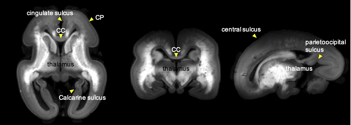
The averaged DWIs along three orthogonal planes. Several anatomical structures can be identified, including cortical plate (CP), thalamus, corpus callosum (CC), and several sulci.
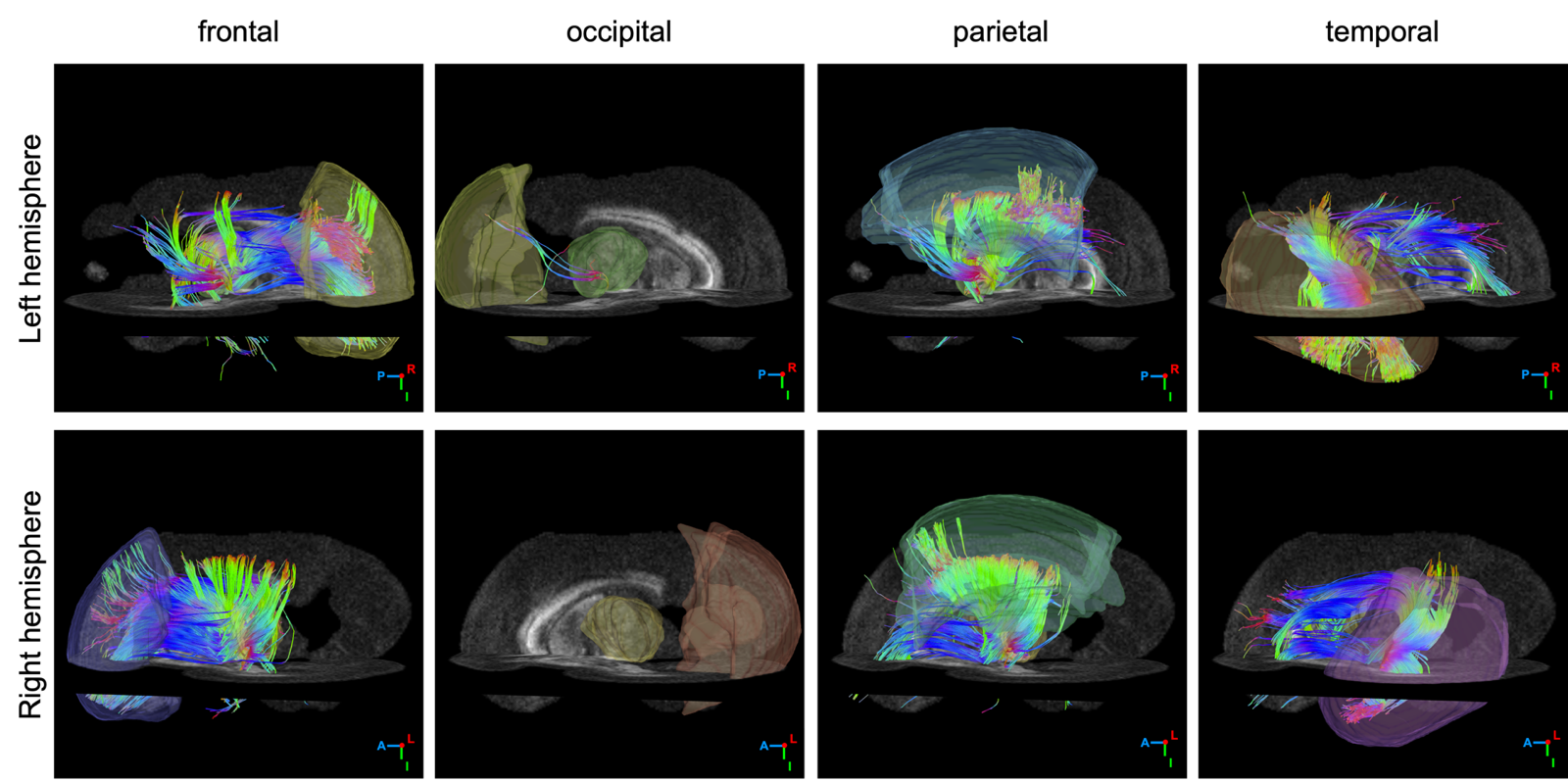
The thalamo-cortical tracts connecting the thalamus and four pre-labelled lobes, including the frontal, occipital, parietal, and temporal lobes. The occipital lobe has less tracts connecting to the thalamus, compared with the other three lobes.
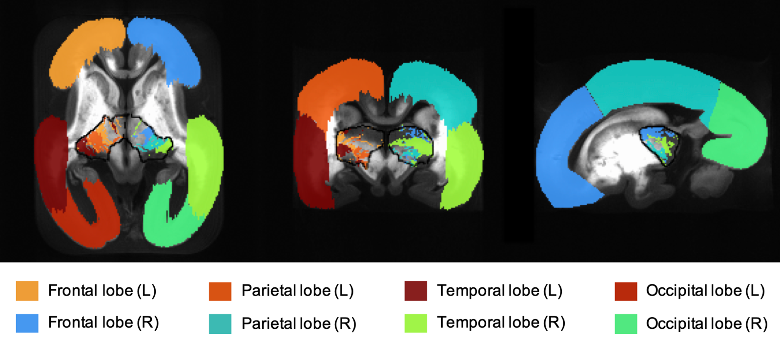
The tractography-based thalamic parcellation. The boundaries of bilateral thalamic regions are outlined in black. Each lobe and its associated voxels within the thalamus are color-coded as the same color. Note that no thalamo-cortical tracts were found in those thalamic voxels without colored overlay.