4308
Classification of bipolar patients using a macroscopic dispersion measure of the white matter tractogram1Institute of Biomedical Engineering, Boğaziçi University, Istanbul, Turkey, 2Istanbul Medipol University, Istanbul, Turkey, 3Coretech Labs Inc., San Diego, CA, United States
Synopsis
Imaging based bio-markers are important in classification of psychiatric disorders for accurate diagnosis. We evaluated the performance of using a macroscopic dispersion measure of the white matter tractogram in the random forest classifier for identifying bipolar subjects. The macroscopic dispersion of the white matter tractogram enables increased performance of the bipolar/normal classification. Multidimensional scaling plots support that the macroscopic changes along the brain white matter improves the discrimination of bipolar and normal groups.
INTRODUCTION
Imaging-based biomarkers are important in the classification of psychiatric disorders for an accurate diagnosis. Diffusion-based biomarkers are proposed to understand the microstructural brain abnormalities, however, there is a limited number of diffusion MRI methods employed to study mental disorders [1]. In this study, we evaluated the performance of using a novel macroscopic dispersion measure of the white matter tractogram in the random forest classifier (RFC) for the separation of bipolar and normal subjects.METHODS
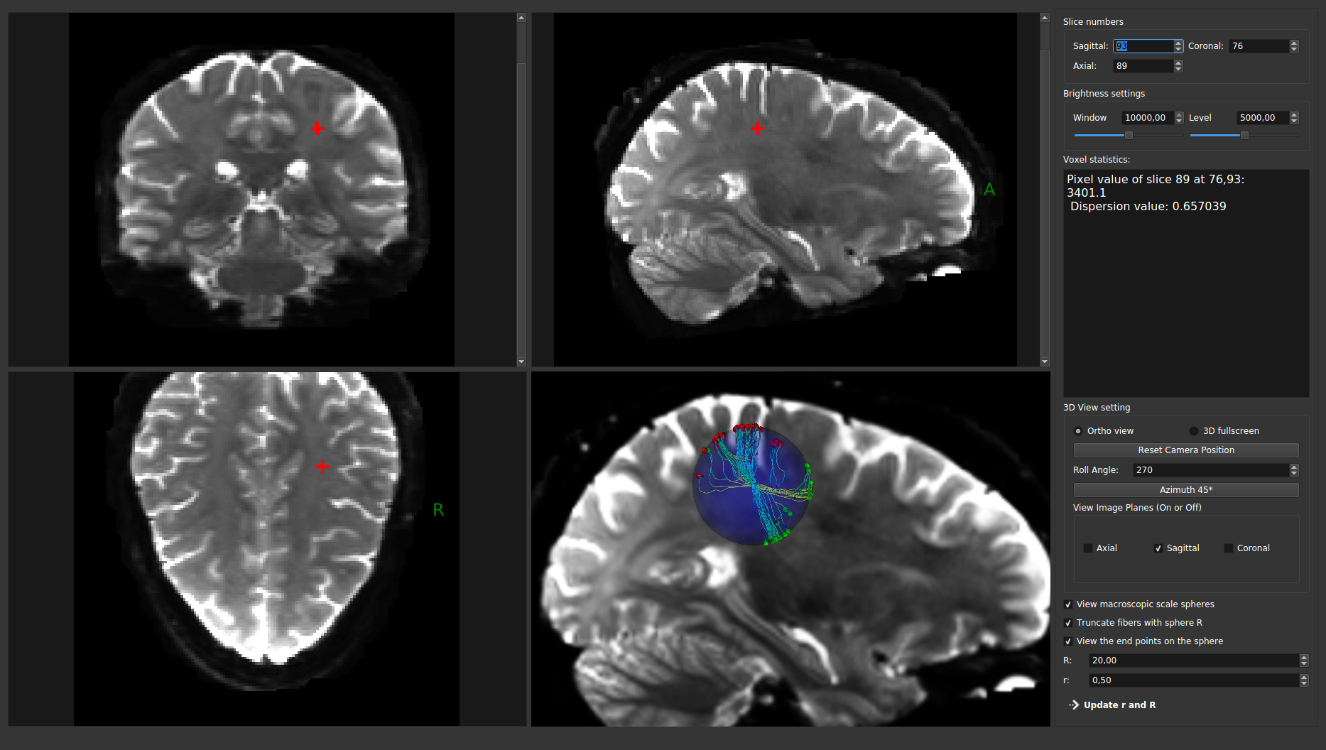
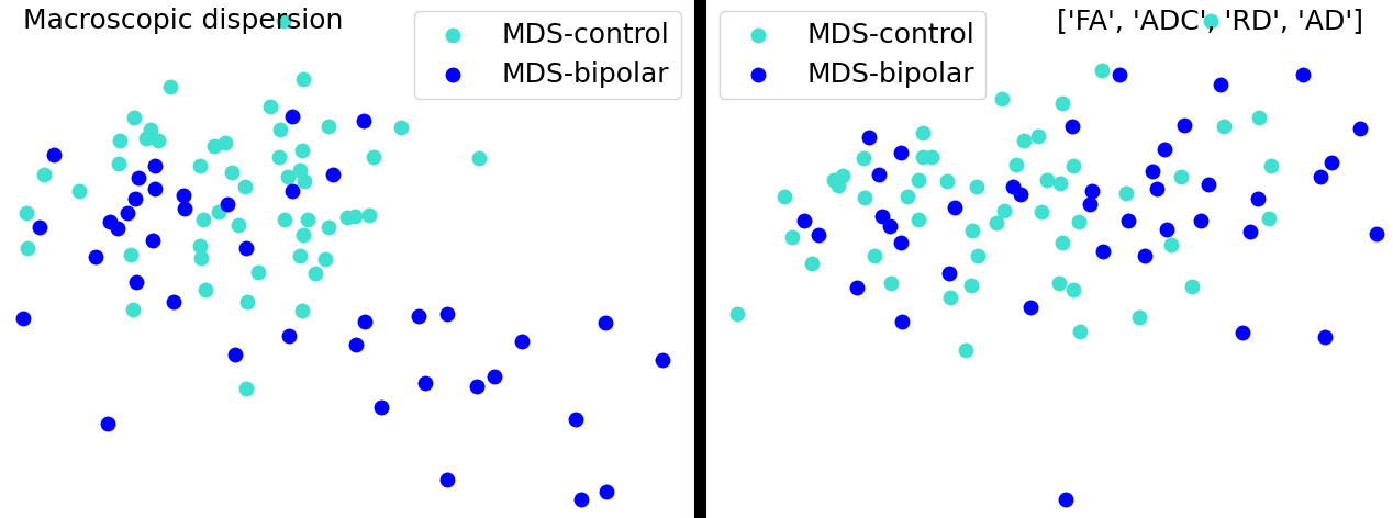
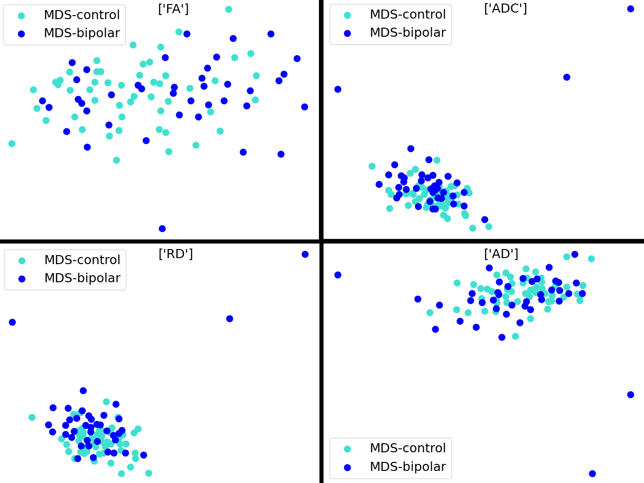
Previously published real diffusion MRI dataset [2], which has 40 bipolar patients and 52 healthy controls was used for the validations.In our previous work [3], we demonstrated macroscopic dispersion characteristics of the white matter tractogram in a group of bipolar patients. Macroscopic dispersion metric computed from a tractogram for each voxel. This method utilizes a truncation sphere which is located at the voxel of interest (VOI) and truncates the fibers passing through the VOI. The macroscopic dispersion of the VOI is formulated as the normalized variances of the start and end points of the truncated fibers: $$$\delta_p = \frac{1}{R^2} (\sigma^{2(s)}+\sigma^{2(e)})$$$. We released a publicly available graphical user interface tool for the proposed macroscopic dispersion. It can be downloaded from https://bitbucket.org/\_ali\_demir/ (Fig.1). Given a tractogram and volume spacings (resolution), $$$\delta$$$ is sampled by iterating the value of $$$p$$$ for the real coordinate centers of each voxel to form a unique dispersion map of the given tractogram.We evaluated the classification performance of the proposed macroscopic dispersion metric using the random forest classifier implemented in scikit-learn project [4]. Computed brain maps were registered to the MNI152 space to perform statistical computations. We formed the feature vector using the metric statistics of frontal associative white matter regions, such as the callosal body, cingulum, inferior occipitofrontal fascicles, and superior longitudinal fasciculus. Julich brain atlas [5] was used to label anatomical regions. 10-fold cross-validation was performed to report the classification performance. Qualitative validation was demonstrated using multidimensional scaling (MDS) [6] of the feature vector showing estimated Euclidean distances in 2-dimensional space. Each data from the normal or bipolar group represented at a hypothetical 2D point on the MDS plot, which shows the degree of separation regarding the given metric.
RESULTS
The results compared the classification performance of the given diffusion metrics (FA, ADC, RD, AD) and the macroscopic dispersion. Accuracy statistics were reported with the mean and the standard deviation of 10-fold cross-validation. The classification accuracy of RFC using the macroscopic dispersion feature was $$$(74\pm12)\%$$$, whereas the accuracy of RFA using features as the combination of diffusion metrics was $$$(61\pm15)\%$$$. RFA was also tested for each diffusion metric separately and the accuracy statistics were $$$(48\pm16)\%$$$ for FA, $$$(64\pm14)\%$$$ for ADC, $$$(62\pm12)\%$$$ for RD, and $$$(56\pm16)\%$$$ for AD. MDS plots of these features were shown in Fig.2 and Fig.3 comparing the pairwise Euclidean distances of the features. The MDS plot of the macroscopic dispersion shows a separated distribution of the bipolar group.CONCLUSION
The macroscopic dispersion of the white matter tractogram enables increased performance of the bipolar/normal classification. MDS plots support that the macroscopic changes along the brain white matter improves the discrimination of bipolar and normal groups.Acknowledgements
This work was supported in part by EU 7th framework - TUBITAK Co-Funded Brain Circulation Scheme 2236 grant 291762/112C003.References
1. O. Pasternak, S. Kelly, V. J. Sydnor, M. E. Shenton, Advances in microstructural diffusion neuroimaging for psychiatric disorders, NeuroImage 182(2018) 259 – 282.
2. A. M. Uluğ, M. Özkan, , P. B. Kingsley et. al., Multi-contrast z-score comparison discriminates patients with psychiatric disorders from controls, Proc. Intl.Soc. Mag. Reson. Med. 23 (2015) 3561.
3. A. Demir, M. Özkan, A. M. Uluğ, A macro-structural dispersion characteristic of brain white matter and its application to bipolar disorder, IEEE Transactions on Biomedical Engineering (2020) 1–1.
4. F. Pedregosa, G. Varoquaux, A. Gramfort, V. Michel et. al., Scikit-learn: Machine learning in Python, Journal of Machine Learning Research 12 (2011)2825–2830.
5. M. Axer, K. Amunts, D. Grassel, C. Palm, J. Dammers, H. Axer, U. Pietrzyk, K. Zilles, A novel approach to the human connectome: Ultra-high resolutionmapping of fiber tracts in the brain, NeuroImage 54 (2) (2011) 1091 – 1101.
6. I. Borg, P. J. F. Groenen, Modern Multidimensional Scaling - Theory and Applications, Springer Series in Statistics, 1997.
Figures