4259
FMRI of Post High-Frequency Focused Ultrasound Ablation of ViM Shows Reduced Ipsilateral Thalamic and Cortical Motor Activation1Imaging Institute, Cleveland Clinic Foundation, Cleveland, OH, United States, 2Neurological Institute, Cleveland Clinic Foundation, Cleveland, OH, United States
Synopsis
High Intensity Focused Ultrasound (HIFU) in now entering clinical practice, for example to treat essential tremor (ET) by creating small lesions in the thalamus. Due to small size of treatment lesions, treatment success depends critically on targeting, which is classically done using measurements and landmarks. We explore an alternative method using functional imaging to guide targeting and assess efficacy, specifically using 7T task-related fMRI. We present preliminary data of the patterns of BOLD activation in a set of patients before and after HIFU.
Introduction
High Intensity Focused Ultrasound (HIFU) is a new treatment for essential tremor (ET) using an MR guided array of 1024 transducers transmitting extracranial ultrasound at 650 kHz to a focal spot to thermally ablate the ventral intermediate nucleus of the thalamus (ViM). The size of the thermal cavity is a sphere about 6 mm in diameter, while the size of ViM is slightly larger, around 5x7 mm. If the HIFU lesion is misaligned to extend into adjacent thalamic nuclei, unwanted side effects can occur such as ataxia or parasthesias. In addition, treatment effectiveness of reduced tremor is impaired when the HIFU is off target. Currently, positioning is guided by a long experience using brain measurements, which is fairly constant among all adults. Alternative methods use direct targeting using structural imaging to thalamic landmarks. Nevertheless, given the precision possible with the modern HIFU technology, there is great utility in providing a target evidenced by functional imaging, rather than numbers or structure. In addition to targeting is functional imaging for efficacy. We propose to use 7T task-related fMRI to address two clinical problems using HIFU: (1) initial targeting of the HIFU lesion; (2) objectively evaluating the effect of a HIFU procedure by computing a functional imaging metric. We proposed using a finger tapping paradigm to visualize corresponding thalamic activation in patients before the HIFU procedure; and conversely to visualize the change in activation after HIFU procedure. We hypothesize that the focus of thalamic activation corresponds to the desired target of ViM, and that a HIFU lesion will reduce this activation.Methods
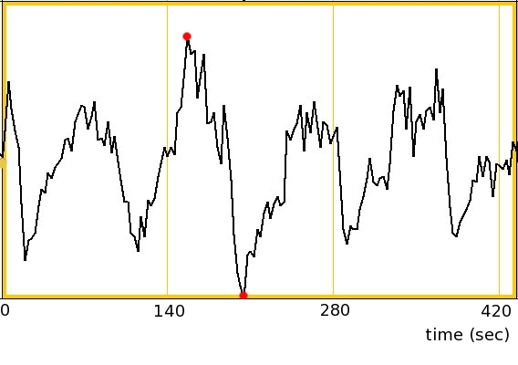
Three patients with essential tremor were scanned twice at 7T (Siemens MAGNETOM), before and after unilateral HIFU ablation of ViM. Using a 32-channel receive, single transmit head coil (Nova, Inc.), fMRI was acquired using a simultaneous multi-slice EPI protocol (TR=2.8 sec, 1.2x1.2x1.5mm3). An anatomic volumetric T1-weighted image was acquired using a MP2RAGE [1] sequence. Head-motion and physiologic noise were corrected using SLOMOCO [2] and PESTICA [3]. A 4mm spatial blur was also applied. Finger tapping was completed in a block design with four sets of 44.8sec tapping with equal rest periods in between. Subjects were instructed to do bi-lateral finger tapping using all fingers. A sample time series of the finger tapping data from the right motor cortex is shown in Figure 1.The finger tapping data was analyzed using 3dDeconvolve. The T1s images were aligned to the EPI using a linear transformation. The EPI aligned T1w image was then non-linearly transformed using ANTS [4] to a T1-weights template in Talairach space. This transformation was applied to the finger tapping data. Two of the three subjects had left-side lesions, the other had a right-side lesion. The subject with the right-side lesion was flipped right/left such that all the lesions were on the left side. Resulting difference maps of before and after treatment were created.
Results
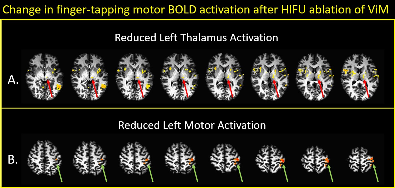
The mean difference maps show reduced activation after HIFU treatment in regions: treated region of thalamus, and ipsilateral motor regions of the precentral gyrus. Figure 2 shows statistical difference maps of these two regions (A: thalamus; B: precentral gyrus), specifically pre-HIFU minus post-HIFU. Map p-value thresholds are 0.07 and 0.02, corrected, respectively. The reduced activation post-HIFU is notable asymmetric with no contralateral activation at these thresholds. Other motor related regions also showing reduced BOLD activity are the left globus pallidus, portions of left putamen, and a focus likely representing the sub-thalamic nucleus.Discussion
The clinical efficacy of HIFU ablation of ViM for essential tremor is tremendous and immediate, with patients leaving the procedure tremor-free; and for the first time, often after many years, able to hold a cup a coffee or write their signature. This is the first study at 7T to examine the BOLD fMRI response to this treatment, in a cohort of 3 patients who were scanned both before and after a HIFU procedure. As might be expected, there is reduced BOLD activity at the treated thalamus, not only at the site of the ablation, but in adjacent tissue of the thalamus. In addition, there is reduced activation in the ipsilateral motor regions of the precentral gyrus.Conclusion
The functional MRI effects of HIFU ablation of ViM are readily apparent using a finger-tapping paradigm, averaged over 3 essential patients. Specifically, there is asymmetrically reduced BOLD activation co-localizing with the treated ViM nucleus and also the ipsilateral motor cortex.Acknowledgements
The authors gratefully acknowledge the support of Tobias Kober and Siemens Healthineers, Inc. for technical support.References
[1] Marques JP, Kober T, Krueger G, et al. MP2RAGE, a self bias-field corrected sequence for improved segmentation and T1-mapping at high field. Neuroimage. 2010;49(2):1271-1281.
[2] Beall EB, Lowe MJ. Isolating physiologic noise with independently determined spatial measures. Neuroimage. 2007;37(4):1286-300.
[3] Beall EB, Lowe MJ. SimPACE: generating simulated motion corrupted BOLD data with synthetic-navigated acquisition for the development and evaluation of SLOMOCO: a new, highly effective slicewise motion correction. Neuroimage. 2014;101:21-34.
[4] Li M, Lin J, Koenig K, Lowe M. Automatic nonlinear transformation of 7T MRI brain image to Talairach stereotaxic space. Presented at: Magnetic Resonance in Medicine. Honolulu, HI, USA; April22-27, 2017.
Figures