4237
Kinematic MRI Tracking of Wrist Carpal Bones1Department of Radiology, Medical College of Wisconsin, Milwaukee, WI, United States, 2Department of Neurosurgery, Medical College of Wisconsin, Milwaukee, WI, United States
Synopsis
In this study, a methodology is developed for kinematic tracking and profiling of free-moving wrist carpal bones. The derived kinematic profiles consist of joint metrics computed and tracked during free wrist motion. Such metrics are hypothesized to be potentially valuable for diagnosis and management of joint dysfunction. Here, an advanced 4D MRI protocol is deployed to capture frames of unconstrained moving joints which are registered to a high-resolution fixed volume using a novel slab-to-volume boundary registration method. Using this registration approach on a sample subject-dataset, concept demonstrations of a selection of metrics describing scaphoid-lunate kinematic motion are presented.
Introduction
A particularly challenging image registration problem is the mapping of a single or few 2D frames to a given volumetric (3D) reference image1. Here, this challenging problem is tackled in the context of kinematic sub-joint bone imaging, where only few moving frames (forming a slab structure) are available at each dynamic position2. In this study, a novel 4D MRI acquisition of the wrist is described and deployed to perform kinematic tracking of individual carpal bones during unconstrained wrist movement. In this paradigm, the acquired moving slab frames are registered to a high-resolution complete static volume that is used to define reference coordinates for kinematic tracking.Methods
MRI: Imaging data were collected on a GE Premier 3T MRI scanner using a 16 channel large flex coil were used to capture dynamic and static images of the wrist. A healthy test subject was recruited into a locally approved IRB protocol and provided written consent to participate. The subject’s arm was fixed with padding to give necessary range of motion for required tasks (i.e. ulanr-radial deviation in this feasibility study). No motion constraints were utilized. Instead, visual cues were used to pace the motion and the subject were trained prior to the exam using same visual cues. Static fixed images were acquired using a 3D SPGR sequence with 0.6mm isotropic voxel with acquisition matrix size of 256x256x76. Other acquisition parameters were TE=1.7ms, TR=5.3ms, FA=10$$$^{\circ}$$$, NEX=1, BW/Pixel=417Hz. An additional zero-echo time acquisition of identical resolution was acquired for future potential use in tracking cortical bone structures. Finally, 32 dynamic volumes with temporal resolution of 2.8s were acquired using 3D SPGR series with 0.78x0.78x2.5mm voxel size with 128x128x6 acquisition matrix size. The other acquisition parameters were TE=1.2ms, TR=4ms, FA=10$$$^{\circ}$$$, NEX=1, BW/Pixel=977Hz. The visual guidance utilized to direct subject motion indicated 3 cycles of motion in the 90 second acquisition. This rate of motion was found to cause minimal motion artifacts in the described dynamic 3D acquisition.Registration: A novel approach to register sparse moving frames of carpal bones with the high-resolution MRI fixed volumes was developed. This model works on the boundary point cloud of segmented carpal bones, applies a rigid transformation, and minimizes a cost function matching the points at the boundaries. 6 degrees of freedom (DOF) rigid-body registration were applied. The fixed, $$$F$$$ and moving sub-volumes (slabs), $$$M$$$, were transformed into discrete points of their boundaries and used to minimize $$$O=F[\theta,{\bf e},T_z](x_i,y_i,z_i)-M(x_j'+X_c,y_j'+Y_c,z_j')$$$, where $$$(x_i,y_i,z_i)$$$ are boundary points at a transferred/rotated position. $$$(x_j',y_j',z_j')$$$ denote boundary points of the moving slab. Shifting $$$x$$$- and $$$y$$$-coordinates by the difference between the in-plane center of the fixed and the moving images $$$(X_c,Y_c)$$$, bypasses the search on the transfer degrees $$$T_{x,y}$$$ and results in a rapid registration approach.
Results and Discussions
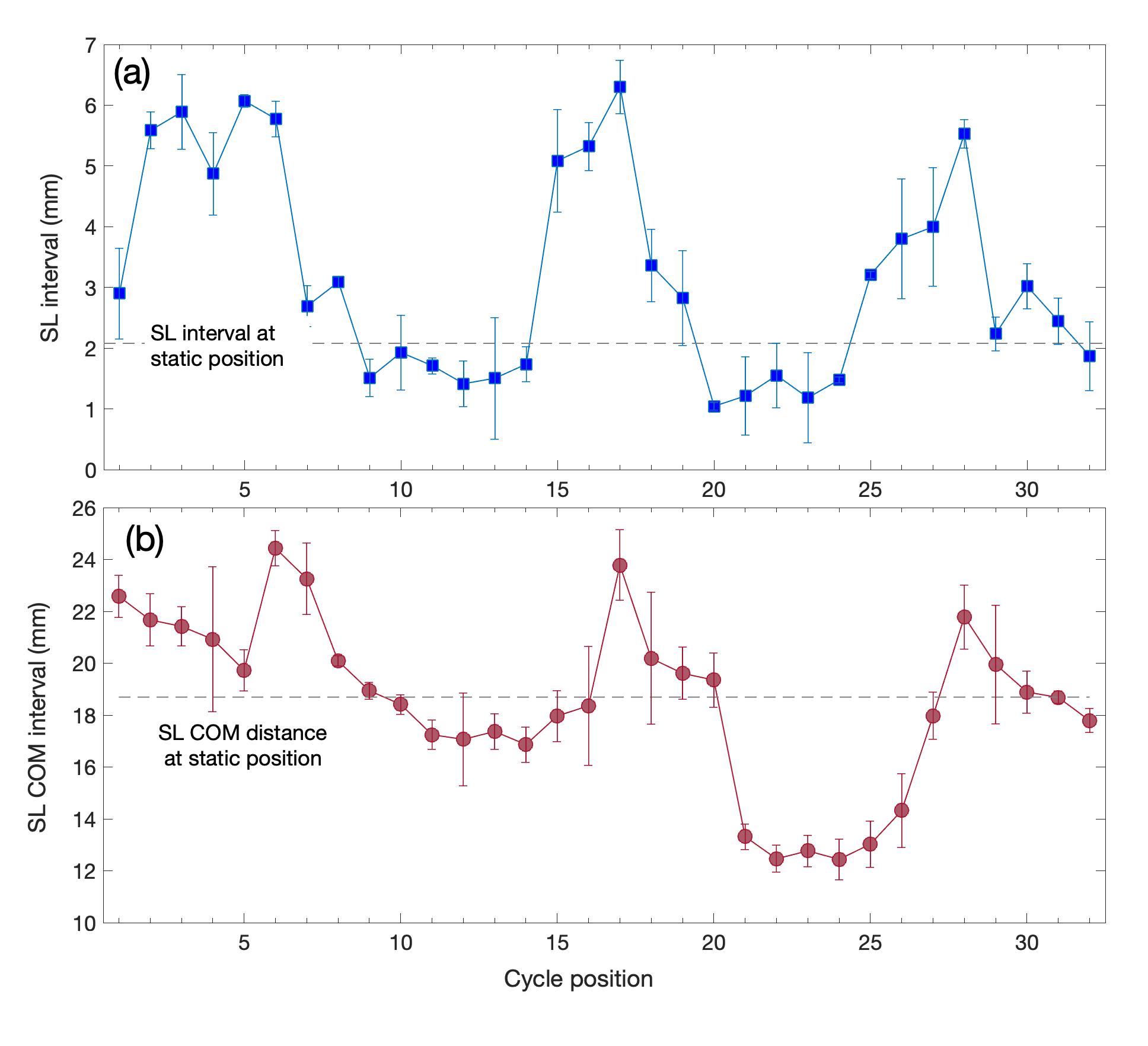
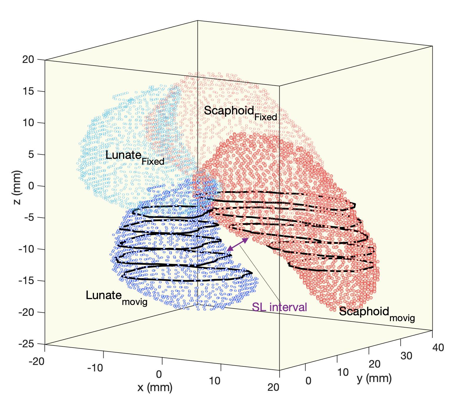
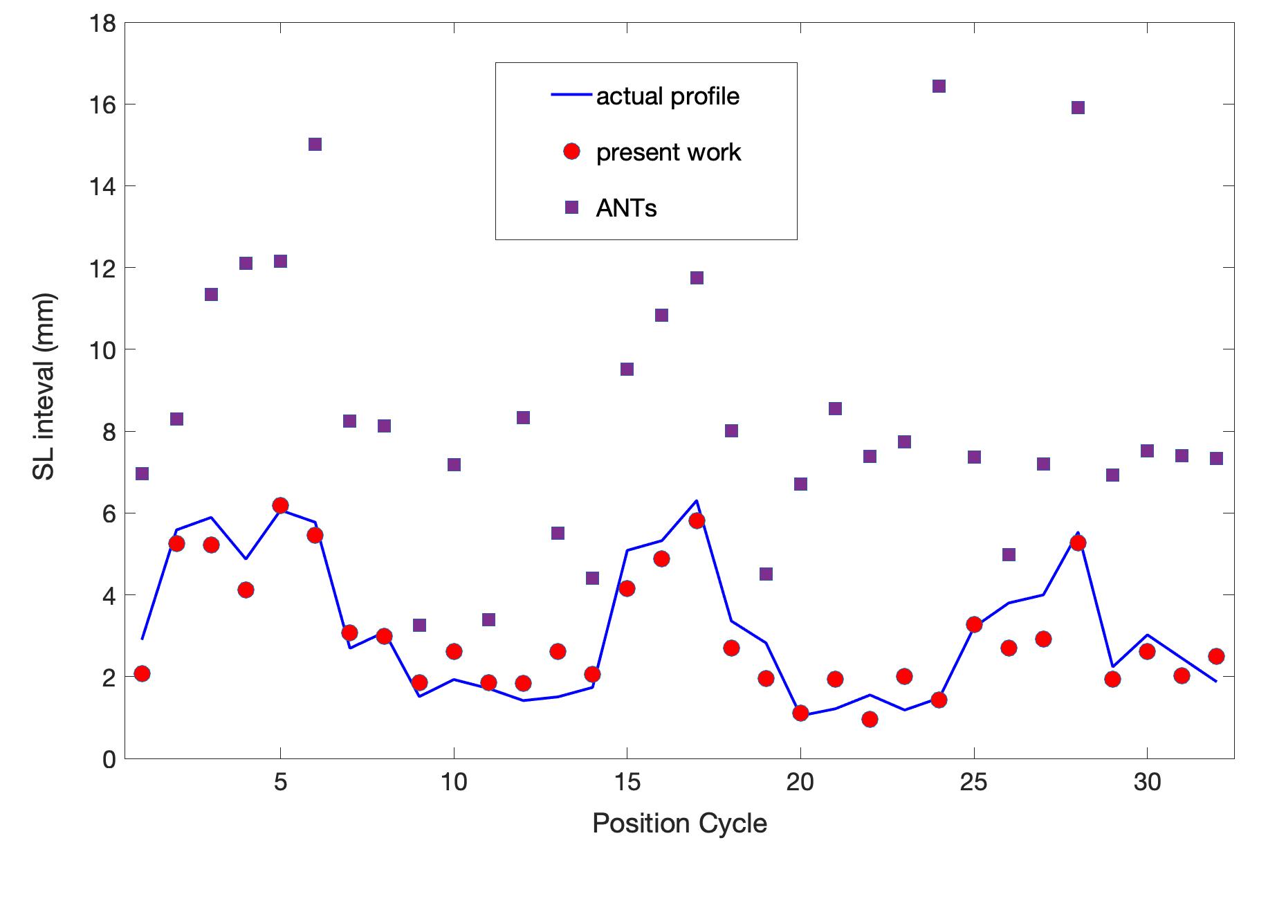
Using the dynamic registration of the static 3D volumes, a wide variety of metrics can be extracted and utilized for kinematic profiling. In this proof-of-concept demonstration, the kinematics of the Scaphoid-Lunate (SL) interval during an ulnar/radial deviation was investigated. During the 32 MRI cycle positions, volunteers were asked to move their wrist three times from a complete ulnar/radial deviation process. Figure 1 shows the resulting profiles of (a) SL interval and (b) SL center of mass distance. The results in (a) illustrate two points with a gap of ~2 mm (dashed line) at the static position (using high-resolution images and accounting for the cortical bone gap in the SPGR image contrast). This statically-defined gap is then navigated using the registered 3D volumes. In this analysis, an error estimate is also performed by registering artificial moving frames (with moving parameters estimated from real data) to the fixed volumes.The performance of the developed registration approach is visualized in Fig. 2 for one motion cycle. The black solid lines depict the boundaries of the moving frames, which are well-mapped to the 3D high-resolution volume. Figure 3 compares our results for the SL-interval kinematics with those obtained using the well-known and utilized registration software package ANTs3. While ANTs is accepted by the MRI community for efficient volume-to-volume registration problems, our results show that for our particular case of slab-to-volume registration, its accuracy is not enough to properly obtain the desired kinematic metric tracking of wrist carpal bones.
Conclusion
Through the use of an advanced 4D MRI acquisition technique and a novel boundary-based slab-to-volume registration approach, these results have demonstrated the feasibility of kinematic metric tracking of unconstrained wrist motion. In this demonstration study, sample kinematic profiling the scaphoid-lunate gap has been presented. Many more metrics across several more wrist motions can be tracked in similar fashion. Future work will continue to deploy these new technologies to explore more complete kinematic tracking and profiling of unconstrained wrist motion.Acknowledgements
This study was supported by NIH R21AR075327. The content is solely the responsibility of the authors and does not necessarily represent the official views of the NIH.References
1. Ferrantea E, Paragios N. Slice-to-volume medical image registration: a survey. Medical Image Analysis 2017; 39:101-123.
2. Foster B H, Shaw C B, et al. A principal component analysis-based framework for statistical modeling of bone displacement during wrist maneuvers. Journal of Biomechanics 2019; pages 1–9.
3. Avants B B, Tustison N J, et al. A reproducible evaluation of ANTs similarity metric performance in brain image registration. Neuroimage 2011; 54(3):2033–2044.
Figures