4223
Quantitative Assessment of cartilage composition using MR T1ρ and T2 imaging 10 years Post ACL-Reconstruction1Dept. of Biomedical Engineering, Lerner Research Institute, Cleveland Clinic, Cleveland, OH, United States, 2Dept. of Biomedical Engineering, Case Western Reserve University, Cleveland, OH, United States, 3Program of Advanced Musculoskeletal Imaging (PAMI), Cleveland Clinic, Cleveland, OH, United States, 4Dept. of Diagnostic Radiology, Imaging Inst., Cleveland Clinic, Cleveland, OH, United States, 5Dept. of Orthopaedic Surgery, Orthopaedics and Rheumatology Inst., Cleveland, OH, United States, 6Dept. of Radiology, Vanderbilt University, Nashville, TN, United States, 7Dept. of Orthopaedics and Rehabilitation, Vanderbilt University, Nashville, TN, United States, 8Dept. of Radiology, Wright Ctr. Of Innovation in BioMed. Imaging, The Ohio State University, Columbus, OH, United States
Synopsis
This is a preliminary report for the MOON multivendor multisite 10 year follow up using quantitative MR imaging techniques for patients following ACL reconstruction. Imaging protocol was harmonized between sites and T1ρ and T2 imaging sequences that were cross-validated between sites and vendors were applied. Phantom data showed excellent intra-site repeatability and small inter-site variations. Significantly elevated cartilage T1ρ and T2 were observed in operated knees compared to contralateral knees. The preliminary results demonstrated feasibility of combining multi-site large cohort study with advanced quantitative MRI, with harmonized protocols and rigorous quality control for both data acquisition and processing.
Background and Purpose
Patients with Anterior Cruciate Ligament (ACL) injuries have a high risk of developing post-traumatic osteoarthritis (PTOA) despite ACL reconstruction (ACLR). Previous studies lacked a long term follow up (over 10 years post-treatment) and large sample sizes. MRI mapping techniques such as T1ρ and T2 imaging have been used for detecting early degeneration of soft tissues (such as articular cartilage) in OA. The goal of this study is to evaluate the joint degeneration at 10 years after ACLR using quantitative MRI in a large cohort, the Multicenter Orthopedic Outcomes Network (MOON) onsite cohort.Methods
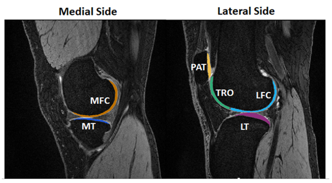
A total of 209 patients will be studied in this multivendor multisite prospective cohort study. The patient cohort is the MOON onsite cohort at 10-year after ACLR. Inclusion/Exclusion criteria for patients include 22-45 years old; ACL tear during a sport; no previous knee injury at the time of initial enrollment; no graft reconstruction during follow-up; no history of surgery on the contralateral knee at the time of initial enrollment. In this preliminary report, 15 patients (age 35.8±5.4 years; 6 female) were studied at 10 years after ACLR, along with 4 healthy controls (36.8±11 years; 2 female). Data were collected from two sites using 3T MR systems (Siemens SkyraFit with a 1Tx/15Rx knee coil and Philips Ingenia with a 1Tx/16Rx knee coil respectively). The 3D MAPSS T1ρ and T2 imaging sequences have been previously cross-validated between sites and different MR systems5. The imaging protocol of the study was harmonized between sites. The imaging protocol of human subjects included Sag, Cor, Ax turbo-spin-echo (TSE) morphological imaging, high resolution 3D Dual Echo Steady State (DESS) imaging (TR/TE=17.55/6.02ms, resolution=0.36x0.46x0.6mm3), 3D MAPSS T1ρ and T2 imaging (TR/TE=6.51/3.24ms, resolution = 0.44x0.88x4mm3, spin-lock frequency = 500Hz and time of spin-lock = [0, 10, 40, 70] ms for T1ρ, Prep TE = [0, 20.064, 40.128, 60.192] ms for T2 imaging). Agarose gel phantoms (2%, 3%, 4%, weight/volume) were scanned for quality control. Data were transferred to one site for image quality control and for centralized quantitative analysis. DESS, T1ρ-weighted, and T2-weighted images were registered to the first echo of the T1ρ-weighted images. Cartilage compartments including medial/lateral femoral condyle (MFC/LFC), medial/lateral tibial plateau (MT/LT), trochlear (TRO), and patellar (PAT) cartilage, were automatically segmented on the registered DESS image by an in-house deep learning algorithm3 as shown in Figure 1. T1ρ and T2 maps were obtained by a voxel-wise two parameter mono-exponential fitting. To evaluate the difference in T1ρ and T2 between the operated and contralateral knees, two-tailed paired t-tests were performed under 95% confidence intervals. Spearman correlation coefficients of T1ρ and T2 values were calculated to investigate the relationship between the two measures.Results
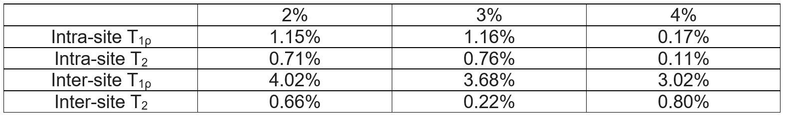
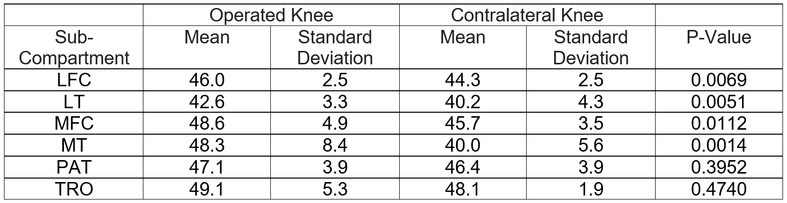
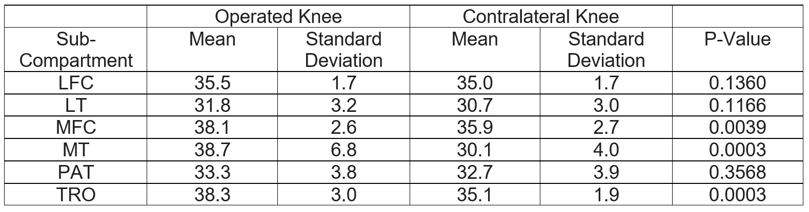
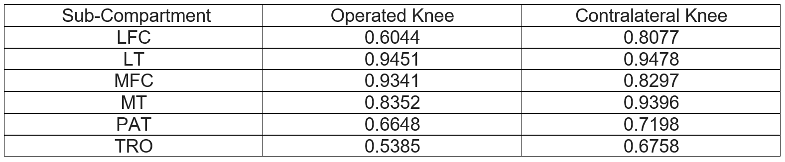
In phantoms, the intra-site coefficient of variation (CV) of repeated scans ranged from 0.17-1.16% for T1ρ and 0.11-0.76% for T2 (Table 1), suggesting excellent intra-site repeatability. The inter-site CV ranged from 3.02-4.02% for T1ρ and 0.22-0.80% for T2 (Table 1), which is within the range of our previous cross-validation study5. In patients, T1ρ values were significantly elevated in LFC, LT, MFC and MT, while T2 values were significantly elevated in MFC, MT and TRO of the operated knees compared to the contralateral knees (Table 3). The Spearman correlation coefficients between T1ρ and T2 values ranged from 0.68-0.95 in contralateral and 0.54-0.95 in operated knees (Table 4). The operated knees tended to have lower correlation between T1ρ and T2 compared to the contralateral knees.Discussion
Studies on PTOA after ACLR with long term follow ups (> 10 years) in the literature have been primarily limited to radiographs and patient reported outcomes. The only MRI study with following ACLR long term had 7 patient follow ups after 8 years and 2 patient follow ups after 10 years6. The long-term soft tissue degeneration are largely unknown in patients after ACLR. The preliminary results of our study showed the feasibility of combining a multi-site large cohort study with advanced quantitative MRI, harmonized protocols and rigorous quality control for both data acquisition and data processing. Our preliminary reports showed excellent intra-site repeatability and small inter-site variation in phantoms. Traveling volunteers will be recruited for the study to evaluate the inter-site variations in human subjects. In patients, significantly elevated T1ρ and T2 in the operated compared to the contralateral knees was observed, indicating degeneration in the operated knees. The results are consistent with reports of shorter-term studies in patients after ACLR7. Interestingly, although both T1ρ and T2 showed significant differences between the operated and contralateral knees in the medial side (MFC and MT), only T1ρ, but not T2, were significantly elevated in the lateral side (LFC and LT); while in TRO, T2, but not T1ρ, were significantly elevated. Furthermore, we observed low correlation between T1ρ and T2 in some compartments of the operated knees, such as TRO. These results suggested that T1ρ and T2 may provide complementary information related to PTOA development and progression. The findings need to be confirmed with data collected from more patients. More healthy controls will be enrolled and compared with both knees from patients. In future analysis, we will also correlate T1ρ and T2 measures with morphological lesion grading, and with patient outcomes and functions.Acknowledgements
The study is supported by NIH/NIAMS R01 AR075422.References
1. Ambellan F, Tack A, Ehlke M, Zachow S. Automated segmentation of knee bone and cartilage combining statistical shape knowledge and convolutional neural networks: Data from the Osteoarthritis Initiative. Med Image Anal. 2019 Feb;52:109-118. doi: 10.1016/j.media.2018.11.009. Epub 2018 Nov 17. PMID: 30529224.
2. Bae JH, Hosseini A, Wang Y, Torriani M, Gill TJ, Grodzinsky AJ, Li G. Articular cartilage of the knee 3 years after ACL reconstruction. A quantitative T2 relaxometry analysis of 10 knees. Acta Orthop. 2015;86(5):605-10. doi: 10.3109/17453674.2015.1039426. PMID: 25854533; PMCID: PMC4564784.
3. Gaj, S, Yang, M, Nakamura, K, Li, X. Automated cartilage and meniscus segmentation of knee MRI with conditional generative adversarial networks. Magn Reson Med. 2020; 84: 437– 449. https://doi.org/10.1002/mrm.28111
4. Pedoia V, Su F, Amano K, Li Q, McCulloch CE, Souza RB, Link TM, Ma BC, Li X. Analysis of the articular cartilage T1ρ and T2 relaxation times changes after ACL reconstruction in operated and contralateral knees and relationships with bone shape. J Orthop Res. 2017 Mar;35(3):707-717. doi: 10.1002/jor.23398. Epub 2016 Sep 19. PMID: 27557479; PMCID: PMC6863081.
5. Kim J, Mamoto K, Lartey R, Xu K, Tanaka M, Bahroos E, Winalski CS, Link TM, Hardy PA, Peng Q, Botto-van Bemden A, Liu K, Peters RD, Wu C, Li X. Multi-vendor multi-site T1ρ and T2 quantification of knee cartilage. Osteoarthritis Cartilage. 2020:Online ahead of print.
6. Potter HG, Jain SK, Ma Y, Black BR, Fung S, Lyman S. Cartilage injury after acute, isolated anterior cruciate ligament tear: immediate and longitudinal effect with clinical/MRI follow-up. Am J Sports Med. 2012;40(2):276-85. Epub 2011/09/29. doi: 10.1177/0363546511423380. PubMed PMID: 21952715.
7. Theologis AA, Haughom B, Liang F, Zhang Y, Majumdar S, Link TM, Ma CB, Li X. Comparison of T1rho relaxation times between ACL-reconstructed knees and contralateral unoperated knees. Knee Surg Sports Traumatol Arthrosc. 2014 Feb;22(2):298-307. doi: 10.1007/s00167-013-2397-z. Epub 2013 Jan 31. PMID: 23370983; PMCID: PMC3692610.
Figures