4220
Quantitative T2 mapping MRI near metal: comparison of metal artifact reduction techniques1Radiology, Eastern Chiba Medical Center, Tonage, Japan, 2Division of Health Sciences, Graduate School of Medical Sciences, Kanazawa University, Kanazawa, Japan, 3Philips Japan, Tokyo, Japan, 4General Medical Services, Chiba University Graduate School of Medicine, Chiba, Japan, 5Orthopaedic Surgery, Eastern Chiba Medical Center, Chiba, Japan, 6Faculty of Health Sciences, Institute of Medical, Pharmaceutical and Health Sciences, Kanazawa University, Kanazawa, Japan
Synopsis
Cartilage degeneration can be evaluated non-invasively by qualitative T2 mapping MRI. Although cartilage evaluation using T2 mapping is desirable in cases which open reduction and internal fixation for fractures around the knee joint and osteotomy for knee osteoarthritis, evaluation is often difficult due to the existence of metal artifacts. Theoretically, metal artifacts are suppressed by metal artifact reduction techniques such as VAT and SEMAC. Therefore, we evaluated the usefulness of T2 mapping using VAT or SEMAC for cartilage around the knee joint that was metal-fixed after surgery.
PURPOSE
The progression of knee osteoarthritis has a close relation to cartilage degeneration. In recent years, cartilage degeneration can be evaluated non-invasively by qualitative T2 mapping MRI1. On the other hand, there are cases in which open reduction and internal fixation for fractures around the knee joint and osteotomy for knee osteoarthritis have poor postoperative performance due to the progression of degeneration of residual cartilage. Although cartilage evaluation using T2 mapping is desirable in such cases, evaluation is often difficult due to the existence of metal artifacts. Theoretically, metal artifacts are suppressed by metal artifact reduction techniques such as VAT and SEMAC2. However, there is still no study that combines existing metal artifact reduction techniques and quantitative MRI sequences. The purpose of this study was to evaluate the usefulness of T2 mapping using VAT or SEMAC for cartilage around the knee joint that was metal-fixed after surgery.METHODS
All subjects were examined with 3.0T whole-body clinical system (Ingenia CX, Philips Healthcare) and 16ch knee coil. The study was approved by the local IRB, and written informed consent was obtained from all subjects. We compared T2 values and standard deviation among three techniques of T2 mapping (Multi-echo TSE, Multi-echo TSE using VAT and Dual-echo TSE using SEMAC) in sucrose phantom of various T2 values, and knee cartilage near metal of two patients. The operations of two patients were high tibial osteotomy and tibial plateau fracture, both of which had titanium alloy plate placed around the knee joint [Fig.1]. Imaging parameters for T2 mapping were; Sagittal, voxel size=0.5×0.5×3.5mm3, FOV=150×150mm2 and 23 slices. Multi-echo TSE and Multi-echo TSE VAT; band width=291.4 and 641.0Hz/px, TR=2000ms, TE=10, 20, 30, 40, 50, 60, 70, 80ms, and total acquisition time=5m54s. Dual-echo TSE SEMAC; band width=641.0Hz/px, TR=2000ms, TE=16, 80ms, and total acquisition time=11m24s.RESULTS AND DISCUSSION
Figure 2 shows the results of T2 values measurement in the sucrose phantom. T2 values of Multi-echo TSE and Multi-echo TSE VAT were almost the same. Dual-echo TSE SEMAC tended to be overestimated below 55ms and underestimated above 55ms. Figures 3 and 4 shows the T2 mapping of the knee cartilage in two patients. In Multi-echo TSE, the metal implant was greatly deformed due to susceptibility artifact, and image distortion were observed in the lower leg. Multi-echo TSE VAT and Dual-echo TSE SEMAC improved image uniformity and distortion compared to Multi-echo TSE. T2 values of two patients were the general values in Multi-echo TSE and Multi-echo TSE VAT3, however it tended to be overestimated in Dual-echo TSE SEMAC [Fig.5]. Comparing the T2 values of the cartilage in the patella and tibia, standard deviation increased on the tibia near the metal, but it was decreased by Multi-echo TSE VAT.CONCLUSION
T2 mapping by Multi-echo TSE combining VAT technique on the cartilage near metal after surgery was effective in reducing standard deviation of T2 values and metal artifacts.Acknowledgements
No acknowledgement found.References
1. Nieminen MT, et al. T2 relaxation reveals spatial collagen architecture in articular cartilage: a comparative quantitative MRI and polarized light microscopic study. Magn Reson Med. 2001 Sep; 46(3): 487-93.
2. Khodarahmi I, et al. Metal About the Hip and Artifact Reduction Techniques: From Basic Concepts to Advanced Imaging. Semin Musculoskelet Radiol. 2019 Jun; 23(3): 68-81.
3. Mosher TJ, et al. Human articular cartilage: influence of aging and early symptomatic degeneration on the spatial variation of T2--preliminary findings at 3T. Radiology. 2000 Jan; 214(1): 259-66.
Figures
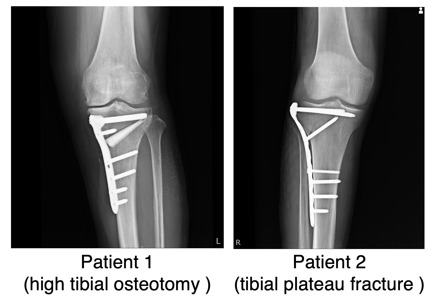
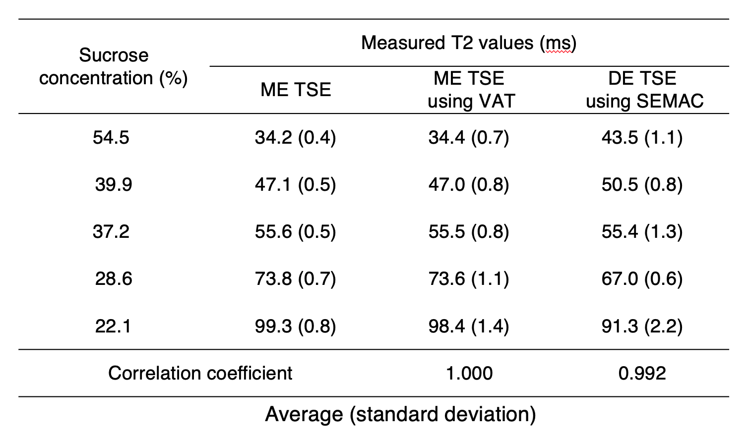
Fig.2 Measurement of T2 values in sucrose phantom.
ME, Multi-echo; DE, Dual-echo
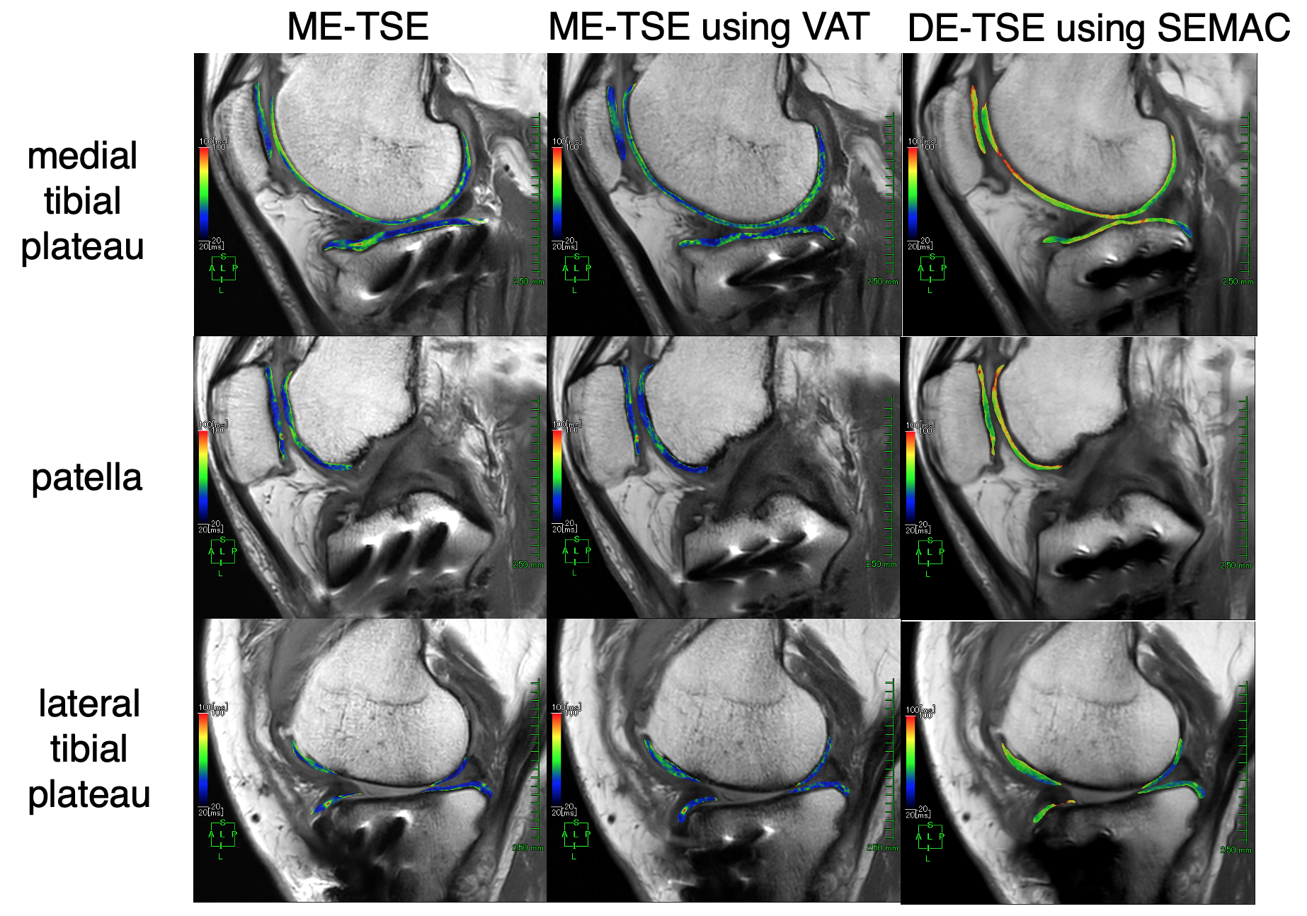
Fig.3 T2 mapping of knee cartilage near metal plate in the tibial plateau (patient 1).
ME, Multi-echo; DE, Dual-echo
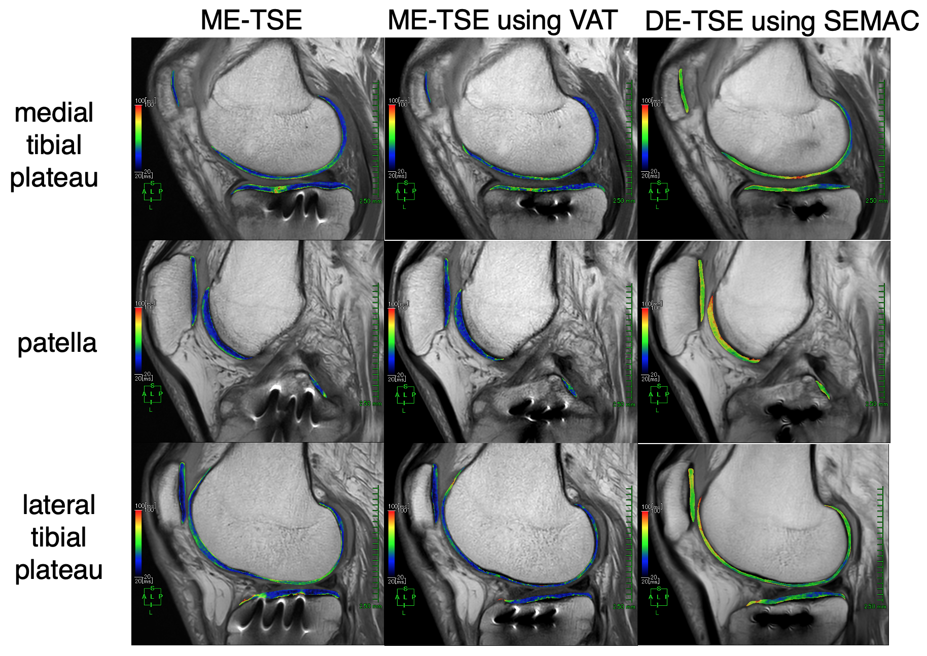
Fig.4 T2 mapping of knee cartilage near metal plate in the tibial plateau (patient 2).
ME, Multi-echo; DE, Dual-echo
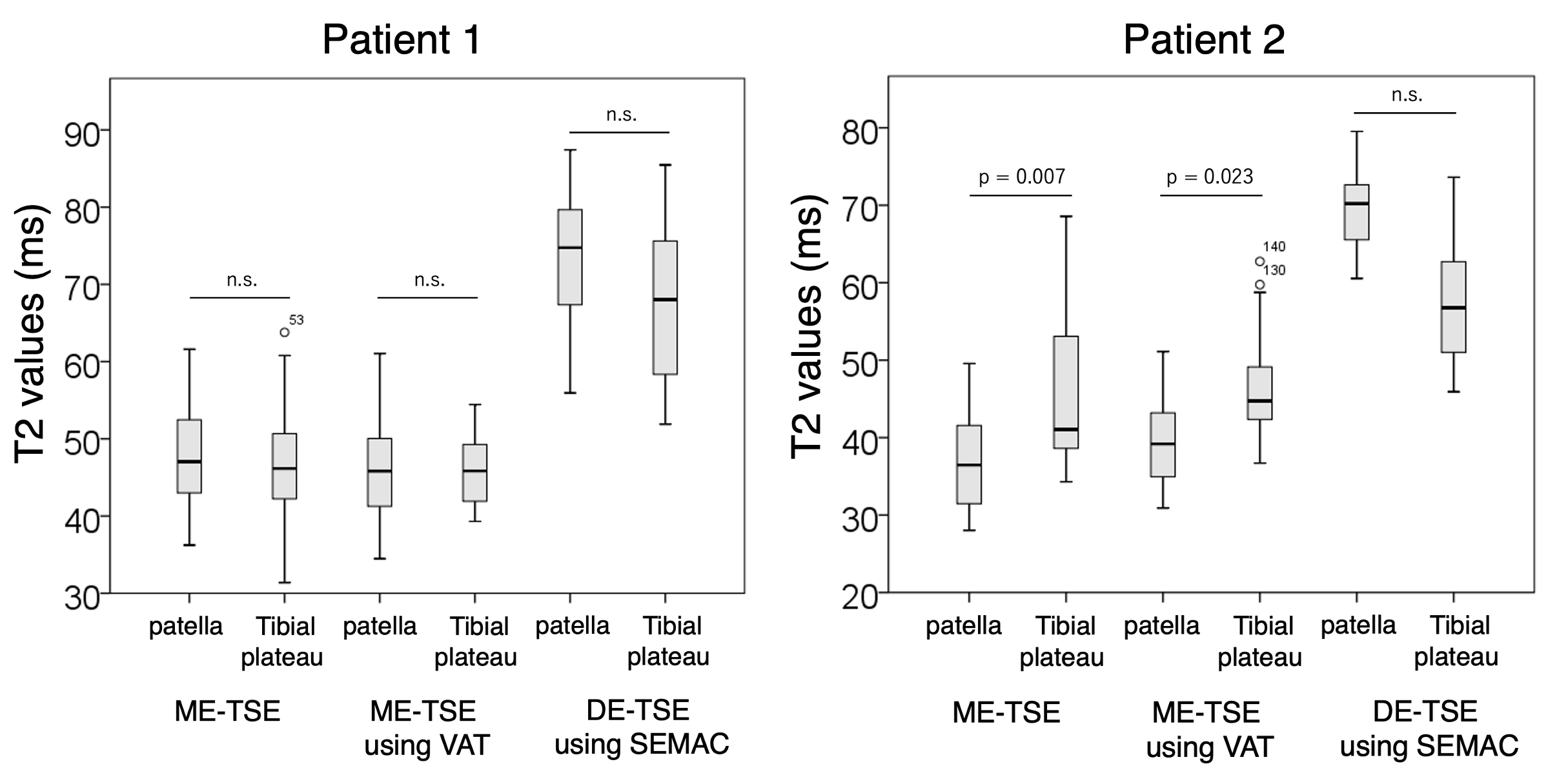
Fig.5 Measurement of T2 values of knee cartilage in two patients.
ME, Multi-echo; DE, Dual-echo