4209
Monitoring changes of knee with Amateur Marathon athletes using Synthetic Magnetic Resonance Imaging :A preliminary study1The Fifth Affiliated Hospital of Sun Yat-sen University, Zhuhai, China, 2MR Research, GE Healthcare, Beijing, China
Synopsis
To explore the value of Synthetic magnetic resonance imaging (Sy MRI) in the quantitative monitoring of knee joint structural changes and cartilage of amateur marathon runners before and after the whole marathon. Twenty-six knee joints of healthy volunteers who took part in the marathon were scanned by Sy MRI technology one week before the race and within 48 hours after the race. The values of T1, T2 and PD of knee cartilage in whole and different regions before competition were higher than those of 48 hours after the marathon, and the difference was statistically significant
Introduction
Marathon is very common, and it brings various sports injuries, especially knee joint injuries1-3.Previous literature has shown that Synthetic magnetic resonance imaging (Sy MRI) can obtain quantitative images of morphology and function in one scan, and has good consistency with conventional MRI in knee joint4,5.This reasearch was to explore the value of Sy MRI in the quantitative monitoring of knee joint structural changes and cartilage of amateur marathon runners before and after the whole marathon.Methods
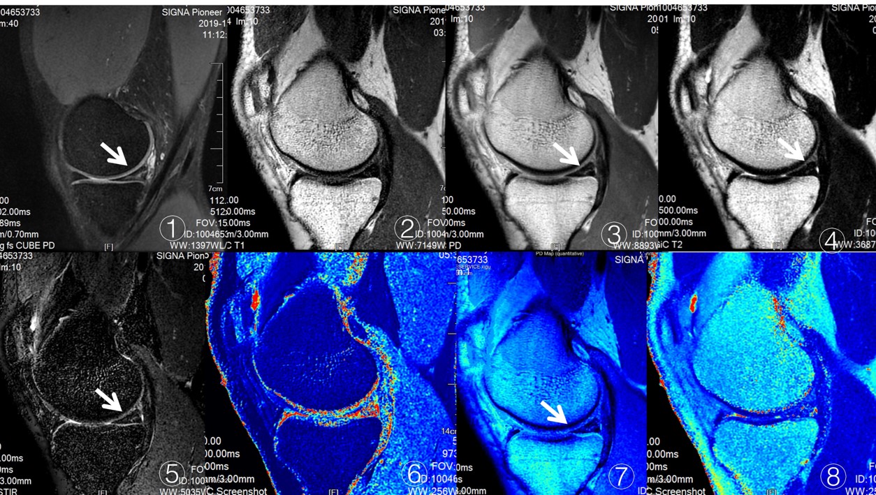
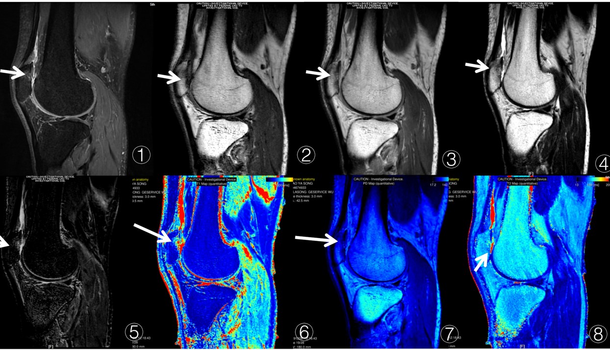
Twenty-six knee joints of healthy volunteers who took part in the marathon were scanned by Sy MRI technology one week before the race and within 48 hours after the race. The conventional contrast weighted images Sy MRI-T1WI, Sy MRI-T2WI, Sy MRI-IW, Sy MRI-STIR and T1, T2, PD maps were obtained. 3D-CUBE-PD sequence was used as a reference to evaluate the detection of knee joint lesions6. The dynamic monitoring of knee articular cartilage before and after the whole marathon was carried out by Sy MRI quantitative maps, and the value of monitoring the dynamic changes of extracellular matrix of cartilage was evaluated. The knee joint articular surface was divided into CMFC, PMFC, CLFC, PLFC, MTP, LTP, Patella and Trochlear. The consistency between groups was tested by intra-group correlation coefficient (ICC). Paired t-test was used to compare the values of T1, T2 and PD of cartilage before and after competition.Results
The two radiologists had good consistency in the measurement of T1, T2 and PD values of Sy MRI (ICC > 0.91). Except for the increase of knee joint effusion, there was no significant change in knee joint structure before and after exercise. The detection rate of Sy MRI sequence for cartilage injury and joint effusion was 100%, and the detection rate of meniscus lesions was 77.8%. The values of T1, T2 and PD of knee cartilage in whole and different regions before competition were higher than those of 48 hours after the marathon, and the difference was statistically significant (P < 0.001).Discussion
In this study, it was found that the conventional contrast weighted images and quantitative pseudo-color maps obtained by SyMRI had a higher detection rate of knee joint lesions.This study found that SyMRI can reflect the dynamic changes of global and local knee cartilage before and after the whole marathon, and the T1, T2 and PD values of knee cartilage after exercise are lower than those before exercise, and there is statistical significance.
Conclusion
Synthetic MRI technique has a good display of knee joint structural lesions, and is of high value in monitoring the dynamic changes of knee cartilage extracellular matrix in amateur marathon athletes.Acknowledgements
NOReferences
[1] Fredericson M, Misra AK. Epidemiology and aetiology of marathon running injuries[J]. Sports Med, 2007, 37(4⁃5):437⁃439.
[2] Alentorn⁃Geli E, Samuelsson K, Musahl V, et al. The association of recreational and competitive running with hip and knee osteoarthritis: a systematic review and meta⁃analysis [J]. J Orthop Sports Phys Ther, 2017, 47(6): 373⁃390.
[3] Fredericson M, Misra A K. Epidemiology and Aetiology of Marathon Running Injuries[J]. Sports Medicine, 2007, 37(4-5):437-439.
[4] Kumar N M, Benjamin F, Stern S E, et al. Synthetic MRI of the Knee: Phantom Validation and Comparison with Conventional MRI[J]. Radiology, 2018:173007-.
[5] Marion Roux, Tom Hilbert, Mahmoud Hussami, etal. MRI T2 Mapping of the Knee Providing Synthetic Morphologic Images: Comparison to Conventional Turbo Spin-Echo MRI[J]. Radiology. 2019;293(3):620-630.
[6] Richard Kijowski, Kirkland W. Davis, Michael A. Woods, M. Knee joint: comprehensive assessment with 3D isotropic resolution fast spin-echo MR imaging--diagnostic performance compared with that of conventional MR imaging at 3.0 T[J]. Radiology, 2009, 252(2):486.
Figures