4208
A Combined Solid-State 1H and 31P Magnetic Resonance Imaging to Assess Bone Mineral and Matrix Densities in Rat bones1Carl J. Shapiro Department of Orthopaedic Surgery, Beth Israel Deaconess Medical Center, Boston, MA, United States, 2Harvard Medical School, Boston, MA, United States, 3Massachusetts General Hospital, Charlestown, MA, United States, 4Department of Orthopaedic Surgery, Boston Children's Hospital, Boston, MA, United States, 5Department of Radiology, Massachusetts General Hospital, Charlestown, MA, United States
Synopsis
Bone matrix and mineral densities (BMD) are important parameters to identify bone diseases such as osteoporosis and osteomalacia. Micro-CT, CT, and DXA scans provide bone mineral densities but not bone matrix. In this study, a non-invasive, radiation-free, and clinically proven combined 1H/31P MRI method was developed to measure bone matrix and mineral densities from rat bones from the same volume-of-interest sequentially in a single session. A custom-designed home-made double-tuned single volume coil was designed for 7T, and 1H/31P ZTE rat bone images were obtained, auto-registered, bone matrix and mineral densities were computed quantitatively, and osteoporosis and osteomalacia were successfully identified.
Methods: One set of three cylindrical pellets of polyethylene glycol (PEG) mixed with silicon dioxide at 25%, 50%, and 100% PEG weight fraction for 1H and a second set of three cylindrical pellets of hydroxyapatite (HA) mixed with PEG at 25%, 50% and 100% HA weight fraction for 31P and a dual calibration phantom pellet of PEG and HA mixed in 1:1 weight fraction were prepared for density calibrations. A double-tuned single solenoid coil was built for the sequential acquisition of 1H and 31P rat femur imaging. VAPOR saturation segment was included in the 1H-ZTE for water+fat suppressions; the pulse lengths and bandwidths in VAPOR were optimized for efficient suppression. Initially, 1H ZTE imaging was carried with water +fat suppression acquiring short T2 bone matrix signals with SW=250000Hz, TR=20ms, No. of averages=8, radial projections=13,030, FOV=30×30×30, and matrix size=64×64×64, resulting in 1H images in 50 minutes. Immediately after completing 1H imaging, 31P ZTE imaging was started on the same rat femur with SW=200000Hz, TR=1000ms, No. of averages=2, and the rest of the parameters were the same as 1H acquisition, resulting in 31P images in 7 hours. The dual phantom was imaged in all 1H and 31P rat femur experiments to serve as a calibration phantom. This cycle of 1H and 31P imaging was repeated on control (CTR), ovariectomized (OVX), and partially nephrectomized (NFR) groups of rat femurs with optimized TR values from Ernst angle calculations because of the extremely long 1H (9s) and 31P (67s) T1s of rat femurs at 7T. 1H and 31P derived bone matrix and mineral image densities were converted to PEG and HA equivalent mass densities (g.cm-3) according to the linear regression relationships. The B1 field intensity variations due to coil profiles were corrected with 1H/31P B1 maps obtained with homogeneous (Water/HA) phantoms. Additionally, micro-CT and gold standard gravimetric analyses were conducted and compared with the MRI derived bone matrix and mineral densities.
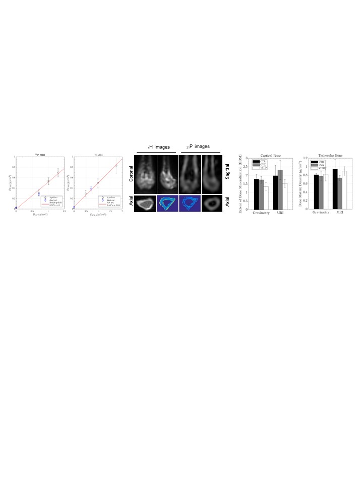
Results: Rat bone matrix and mineral densities were successfully measured with ZTE imaging with 0.468mm resolution and SNR of 25-30 and 12-17 respectively. A significant linear-relationship was confirmed between MRI densities and physical densities of the three-density PEG/HA phantoms with 3-point fit in Fig.1(a). The dual-phantom was used to convert the signal-intensity in the bone image to density with conversion factors. Fig.1(b) shows 1H (top-left row), 31P (top-right row) images and the bottom-row in color shows the same slice 1H and 31P images. The EBM computed by ZTE imaging is in good agreement with gravimetric analysis shown in Fig.1(c).
Discussion: The 1H/31P signals acquired from the three-density 1H/31P pellets confirmed the linearity with their physical densities which is a linear test exploited to compute bone matrix and mineral densities along with dual phantom. The Dual phantom facilitated quantitative matrix and mineral densities in rat bones. Optimization of matrix size, acquisition time, and bandwidth provided 1H/31P rat bone images with optimum SNR and spatial resolution to compute matrix and mineral densities. The double-tuned single solenoid coil with a better filling factor and multiple advantages(8-10) and dual phantom are suitable for rat femur imaging to measure matrix and mineral densities quantitatively.
Conclusion: 1H and 31P rat femur images were successfully acquired sequentially in the same session for auto co-registration of the volume-of-interest to compute bone matrix and mineral densities for estimating EBM accurately. Solid-state 1H and 31P imaging were feasible on ex-vivo rat bone specimens at 7T with the custom-designed RF coil and optimization of imaging parameters. We expect that this method can be extended to humans to measure bone matrix and mineral densities non-invasively.
Acknowledgements
This work has been supported by the National Institutes of Health (AN: K99/R00 AR057093), and internal grants from the Carl J. Shapiro Department of Orthopaedic Surgery at BIDMC (EKR) and Boston Children’s Hospital Orthopaedic Surgery Foundation (BDS). MR Imaging was supported by the Small Animal Imaging Facilities at Boston Children's Hospital and the Athinoula A. Martinos Center for Biomedical Imaging at Massachusetts General Hospital, Boston, MA.References
References:
1. Haihui Cao, et al., “Quantitative 31P NMRS and 1H MRI measurements of bone mineral and matrix density in rat models” Bone, 2010, 46, 1582-1590.
2. Yaotang Wu, et al., “Water- and Fat-Suppressed Proton Projection MRI (WASPI) of Rat Femur Bone” Magn. Reson. Med., 2007, 57:554-567.
3. Yaotang Wu, et al., “Bone Matrix Imaged In Vivo by WASPI MRI of Animal and Human Subjects” J. Magn. Reson. Imaging, 2010, 31:954-963.
4. Robson MD, et al., “Magnetic resonance: an introduction to ultrashort TE (UTE) imaging. J. Comput. Assist. Tomogr. 2003; 27(6): 825–846.
5. Wu Y et al., “Bone Mineral Imaged In Vivo by 31P Solid State MRI of Human Wrists” J. Magn. Reason. Imaging, 2011, 34, 623-633.
6. Weiger M, et al., “MRI with zero echo time: hard versus sweep pulse excitation”. Magn. Reson. Med 2011;66:379–389
7. Idiyatullin D et al., “Fast and quiet MRI using a swept radiofrequency”. J Magn. Reson. 2006; 181:342–349
8. Victor Kassey, et al. “Design and Development of a Versatile Double Tuned R.F Probe Head Coil for Quantification of Bone Mineral and Matrix, ORS 2018, New Orleans.
9. Victor Kassey, et al. “Bone Matrix Density of Rat Bone with Water and Fat suppressed Solid State 1H ZTE MRI at 7T, ORS 2019, Austin, TX.
10.Victor Kassey, et al. “Bone Mineral Density of Rat Bone with 31P ZTE MRI using home-built MRI coil at 7T, ORS 2019, Austin, TX.
Figures