4196
Half Fourier Acquisition Single Shot Turbo Spin Echo Diffusion Encoding with Transition between Pseudo-Steady States for 3T1Donders Institute, Radboud University, Nijmegen, Netherlands
Synopsis
Half-Fourier acquisition single-shot turbo spin-echo diffusion-weighted (HASTE DW) is a clinically established magnetic resonance imaging tool for the detection of small lesions, especially, primary and relapsing cholesteatoma at 1.5T field. HASTE DW imaging at high field strengths (3T and above) is challenging due to the intrinsic high radio frequency power deposition of the method, resulting from the extended echo train. We present a HASTE DW with smooth transition between pseudo-steady states, especially developed for 3T imaging, which features a low specific absorption rate and high quality images, without any loss of sensitivity.
Introduction:
The value of half-Fourier acquisition single-shot turbo spin-echo diffusion-weighted magnetic resonance imaging (HASTE DW MRI) has been proven in the evaluation of cholesteatomatous tissue in the middle ear at 1.5T in the recent decade because of its great promise for a rapid, distortion free, and highly reliable results1. However, HASTE sequences have an inherently high radio frequency (RF) power deposition arising from the large number of 180° refocusing RF pulses used in the extended echo train. This is especially problematic at: (1) high spatial resolutions where longer echo trains are acquired, and (2) at high field strengths where the power deposition per pulse is higher. For this reason, the clinical application of HASTE DW is limited to 1.5T field.Availability of HASTE DW pulse sequence at 3T scanners is appealing for the more accurate preoperative detection of small lesions because of the potentially two-fold higher signal-to-noise ratio (SNR) that could be attained. In this work, we report on a HASTE DW pulse sequence with smooth transition between pseudo-steady states (TRAPS), which can be safely used at 3T scanners without associated imaging consequences. This method provides: (1) a significantly lower SAR level, (2) the same level of sensitivity, and (3) similar quality images as compared to the default pulse sequence at the same field strength.
Theory:
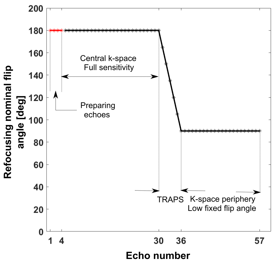
Echo time (TE) is inherently long in HASTE DW method due to a long block of time reserved for the inserted diffusion preparation segment. As a result, a centre-out phase encoding scheme is followed to achieve the shortest possible TE. The overall image gross structure and contrast are determined by the centre of k-space. Thus, it is crucial to preserve the full sensitivity for the central k-space lines. As shown in figure 1, in the presented approach, refocusing RF pulses with a nominal 180° flip angle are employed for the first 30 echoes (including preparing echoes). Afterwards, when odd and even echo parities equalize in magnitude, the nutation angle is slowly reduced through a ramp of 6 echoes to a 90° flip angle, which is then kept fixed until the end of the signal read out. Doing so, the greatest signal can be obtained if the magnetization is continuously in a pseudo-steady state2.Methods:
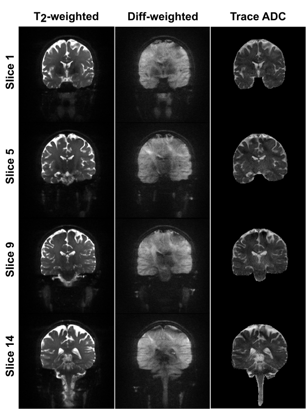
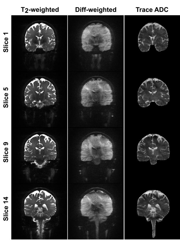
MRI in vivo scans were performed on a whole body 3T Siemens PrismaFit scanner (80 mT/m strength and 200 T/m/s slew rate) with a 32-channel receive-only head coil in a 25 years old healthy female subject. Data were collected with HASTE DW and TRAPS HASTE DW methods for b = 0 and b = 1000 s/mm2. The common parameters were as follow: TE/TR = 106/2000 ms, FOV = 220 * 220 mm2, matrix size of 192 * 192 with 70% phase resolution and GRAPPA = 3 accelerated acquisition with 12 integrated reference lines, 1.1 mm isotropic in-plane resolution, 14 coronal slices (2.5 mm), and rBW = 685 Hz/px. b = 0 datasets were collected with 4 averages and the total scan time of 112 s, and b = 1000 s/mm2 data were collected with 10 averages and the total scan time of 280 s using a 3D diagonal diffusion weighting scheme. Strong conventional fat saturation was used for all acquisitions.Results:
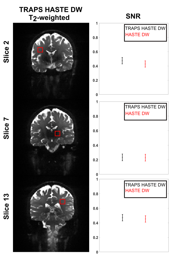
Example T2-weighted, DW, and trace apparent diffusion coefficient (ADC) maps are shown in figure 2 and figure 3 for HASTE DW and TRAPS HASTE DW, respectively, where a comparable quality can be observed over all slices for all contrasts. Albeit the short echo train acquired (only 57 echoes out of 134), the SAR level of HASTE DW was 53% versus 36% for TRAPS HASTE DW. In order to obtain comparable images with matched imaging parameters, for the purpose of this study, we utilized a high acceleration factor of 3 along with a 70% sampling on the phase encoding direction to keep the default method’s SAR level within the limits. Otherwise, the default method’s SAR level would have been 141%, against only 61% for TRAPS HASTE DW. Qualitative analyses did not reveal any new artifacts emerging on TRAPS HASTE DW images. The SNR analysis in sample regions of interest in the white matter did not show any significant difference between the two methods (i.e. no sensitivity loss for the developed method), with a systematic slightly higher SNR for TRAPS HASTE DW (figure 4).Discussion:
High resolution imaging at 3T with HASTE DW within clinically affordable scan times (i.e. TR ~ 2000 ms) is impossible without intensive k-space undersampling due to the SAR limitation. Hence, preserving the valuable higher SNR offered by a higher field strength would be very promising for the detection of small lesions in the size range of 2-3 mm. Preliminary results suggest TRAPS HASTE DW could be a powerful and reliable MRI tool for detecting and following small lesions like cholesteatoma at 3T where the enhanced SNR can improve the method’s predictive validity rate. Of course, a precise evaluation of the method’s diagnostic accuracy is necessary, which is undergoing.Acknowledgements
Netherlands Organisation for Scientific Research (NWO)
Siemens Healthineers Nederland
References
1. B. Henninger and C. Kremser, Diffusion weighted imaging for the detection and evaluation of cholesteatoma, World J. Radiol., 2017 May 28, 9(5): 217–222.
2. J. Hennig, M. Weigel, and K. Scheffler, Multiecho sequences with variable refocusing flip angles: optimization of signal behavior using smooth transitions between pseudo steady states (TRAPS), Magn. Reson. Med., 2003, 49 (3): 527–535.
Figures