4191
Interleaved single-shot EPI for geometric distortion improvement1School of Biomedical Engineering, Shanghai Jiao Tong University, Shanghai, China
Synopsis
EPI is a versatile MRI acquisition method, which can ultrafast yield an image and has been widely applied. However, EPI is well-known sensitive to B0 inhomogeneities and susceptibility effects, which leading to geometrical image distortions. In order to reduce the geometric distortion on EPI images, this work presents a new interleaved single-shot EPI method, in which ky-t line can be sampled with higher slope rate such that the geometric distortion can be alleviated. The experimental results of phantom and human brain at 3T demonstrate the capability of the new method.
Introduction
Echo planar imaging (EPI) is a powerful technique to acquire MRI image rapidly, which can produce a 2D image within a single shot. EPI has been widely used in the investigation of diffusion, perfusion and functional imaging. However, as EPI takes a few tens of milliseconds to complete k-space coverage, the nuisance phase caused by B0 inhomogeneities and susceptibility effects accumulates during this process, thus giving rise to geometric distortion on images. Several methods based on multi-shot acquisition, such Top-up, PSF-EPI and EPTI, have been proposed to address this issue.1-3 However, multi-shot acquisition is sensitive to the shot-to-shot phase variation, which may be induced by motion or pulsation. Here we present a new single-shot EPI method with interleaved phase encoding lines (interleaved single-shot EPI, isEPI), to reduce the phase error accumulation during spatial encoding, therefore improve the distortion on images.Methods
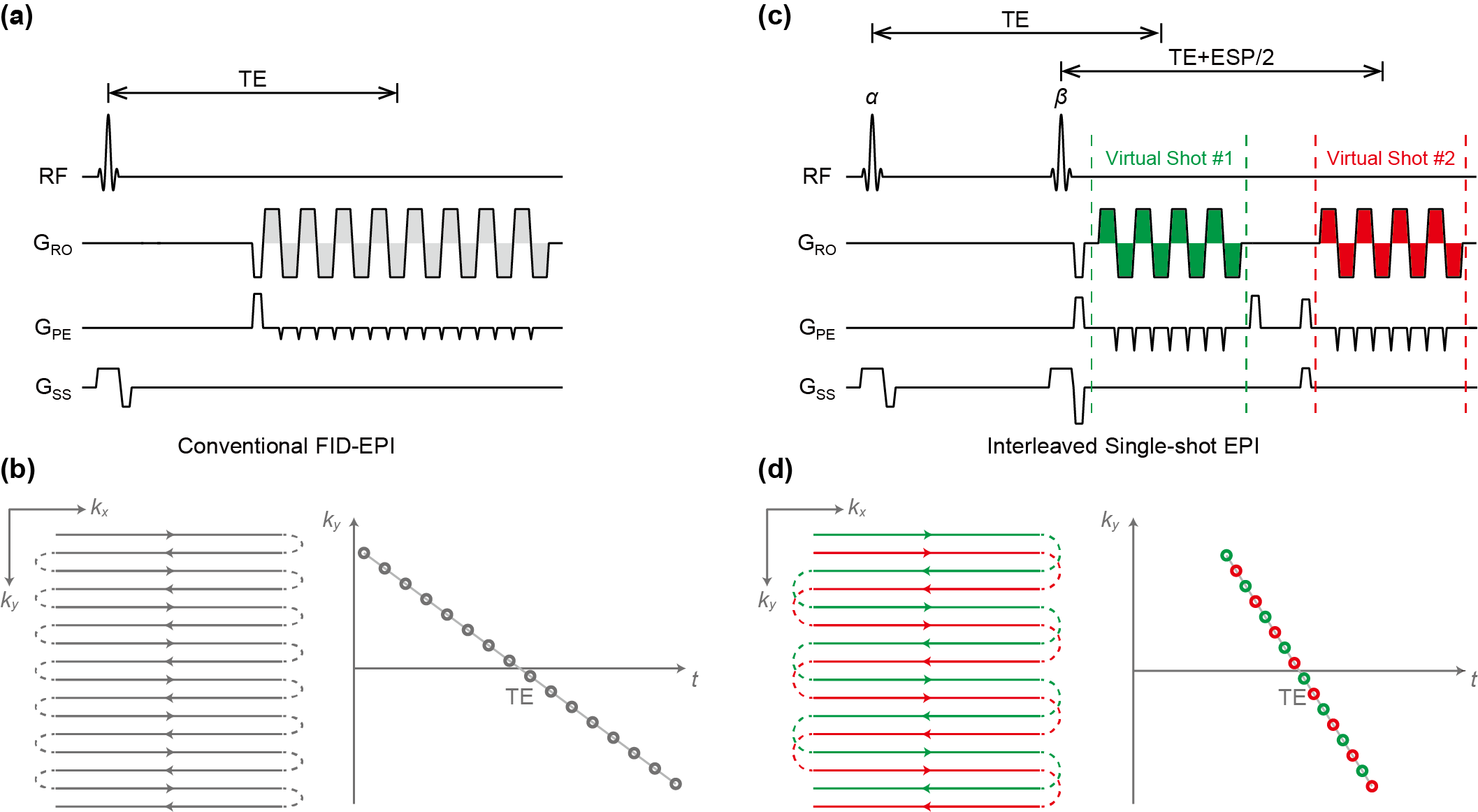
Conventional gradient-echo EPI (Fig. 1a) uses an RF for excitation and prolonged echo train to sample the k space (Fig. 1b, left), resulting in a consecutive ky-t space (Fig. 1b, right). Here, we use two RF pulses to stimulate two echo trains (Virtual Shot #1 and Virtual Shot #2) to sample k space (Fig. 1c). Different from single-shot multi-echo EPI methods for multiple-contrast acquisition,4 the two echo trains can be interleaved in the PE direction to cover the full k space (Fig. 1d), because the blip gradients are double-sized to produce a larger leap on ky space and the echo time make T2* decay consecutive along the PE direction. As shown in Fig. 1d, the interleaved ky-t line is 2-fold shortened in t direction compared to the conventional EPI. To make the amplitude of two visual shots equal, the flip angle of two RF pulses (α and β) should satisfy the condition $$$\cos^2\frac{\beta}{2}\sin\alpha=\sin\beta\cos\alpha$$$. All experiments were executed on a UIH uMR790 3T MRI scanner with a 24-channel head-neck coil (United Imaging Healthcare, Shanghai, China). The American College of Radiology (ACR) MRI phantom (J.M. Specialty Parts, San Diego, California) and a healthy human subject were scanned. The acquisition parameters for ACR phantom are as follows: FOV=220x220 mm2; slice thickness = 3 mm; TR/TE = 4000/74 ms; echo spacing = 0.61 ms; matrix size = 128 x 128 (64 PE lines for each echo train). The flip angle of α and β are set to 45° and 53°, respectively. To demonstrate the performance of isEPI, the currents in shimming coils were altered to yield an inhomogeneous magnetic field with 31 Hz line width on water resonance, while the line width is 10 Hz on shimmed water resonance. In human brain experiments, we use double-sized blip gradient for 2-fold acceleration and set TE = 44 ms. The slice thickness is 4 mm for human brain, and the other settings are same to ACR phantom. Conventional gradient-echo single shot EPI and 2D FSE data were also obtained on the same phantom and human brain for the comparison.Results and Discussion
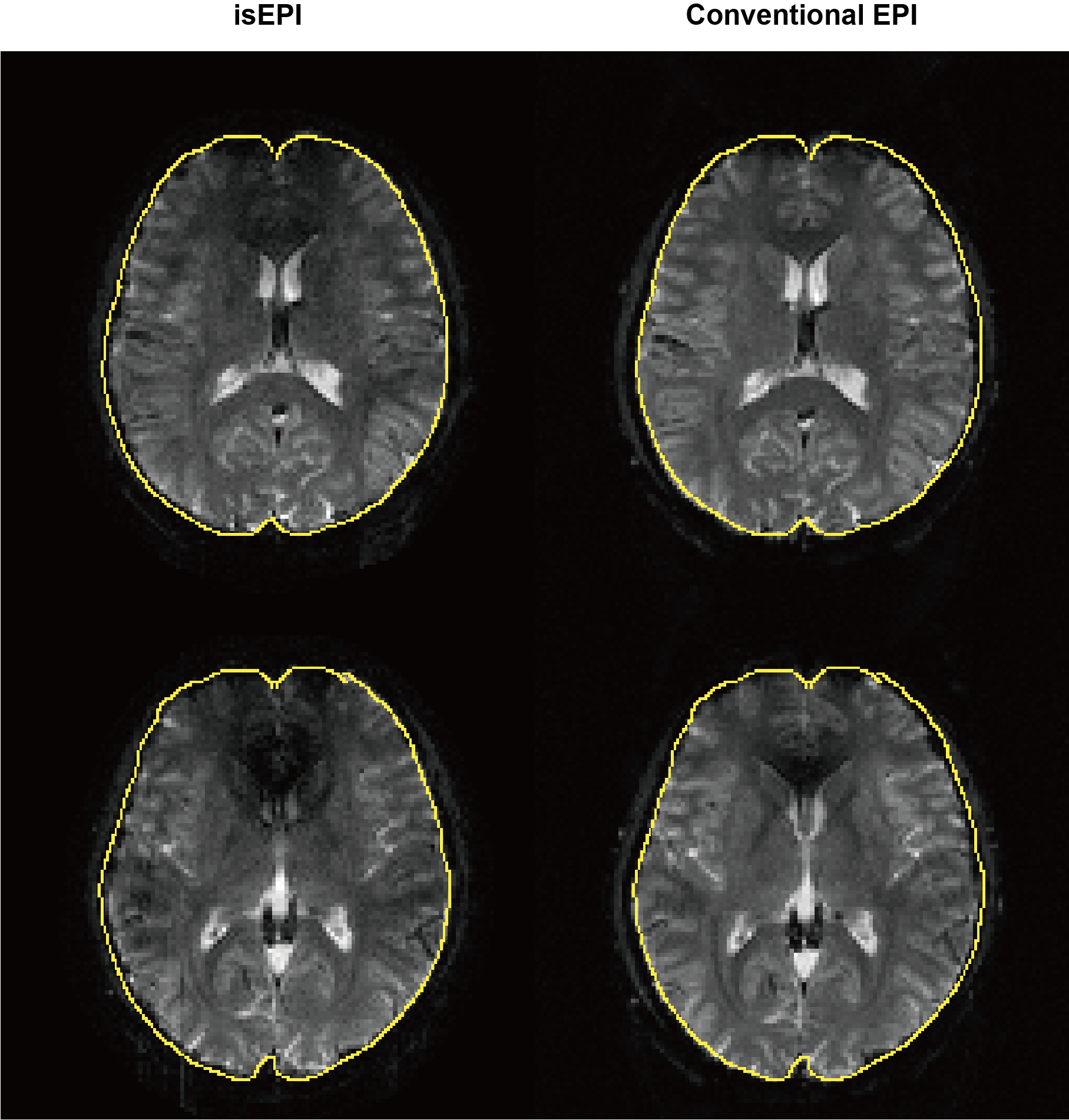
The experimental results of phantom in inhomogeneous field are listed in Fig. 2. The conventional EPI image is subjected to severe geometric distortion and localization shift along phase encoding direction. Compared to conventional EPI, the outlines of isEPI image are much closer to the boundaries of distortion-free FSE image, and much less geometric distortion. In the human brain experiments, the isEPI image (Fig. 3, left column) exhibits less geometric distortion than conventional EPI (Fig. 3, right column). The isEPI method shows the ability to reduce the geometric distortion due to T2* effects in inhomogeneous field, since the T2* phase accumulation is reduced. To obtain two echo trains with identical amplitude, we applied two RF pulses for stimulation in isEPI. The delay between two RF pulses is equal to the length of one echo train, leading to restriction on minimum TE. As the optimal flip angle of the two RF pulses is around 45° and 53° degree, the SNR in the isEPI is about half of that in conventional EPI which uses 90° RF pulse for excitation. Partial k-space technique or center-out k space trajectory may help to reduce the minimum TE and increase the signal intensity.5-6Conclusions
In this work, we present a new EPI method to improve the geometric distortion in EPI image. With two echo trains to cover the k space, the nuisance phase accumulation due to B0 inhomogeneities and susceptibility effects is reduced in ky-t space. The experimental results validate its ability to obtain images with reduced distortion by a single shot. It has the potential to be expanded to acquire diffusion weighting images or perfusion images.Acknowledgements
This work is supported by National Science Foundation of China (No. 62001290) and Sponsored by Shanghai Sailing Program (20YF1420900).References
1. Andersson JL, Skare S, Ashburner J. How to correct susceptibility distortions in spin-echo echo-planar images: application to diffusion tensor imaging. NeuroImage. 2003;20:870-888.
2. Zeng H, Constable RT. Image distortion correction in EPI: comparison of field mapping with point spread function mapping. Magn Reson Med. 2002;48:137-146.
3. Wang F, Dong Z, Reese T, et al. Echo planar time-resolved imaging (EPTI). Magn Reson Med. 2019;81:3599-3615.
4. Posse S, Wiese S, Gembris D, et al. Enhancement of BOLD contrast sensitivity by single-shot multi-echo functional MR imaging. Magn Reson Med. 1999;42:87-97.
5. Jesmanowicz A, Bandettini PA, Hyde JS. Single-shot half k-space high-resolution gradient-recalled EPI for fMRI at 3 Tesla. Magn Reson Med. 1998;40:754-762.
6. Chen X, Zhu A, Du Y. Centre-out (COEPI): A fast single-shot imaging technique with a short TE. Magn Reson Med. 2020;84:787-799.
Figures