4183
Volume-Selective 3D Ultrashort Echo-time Imaging1Biomedical Institute for Convergence at SKKU, Sungkyunkwan University, Suwon, Korea, Republic of, 2Department of Biomedical engineering, Sungkyunkwan University, Suwon, Korea, Republic of, 3Department of Intelligent Precision Healthcare Convergence, Sungkyunkwan University, Suwon, Korea, Republic of
Synopsis
Non-Cartesian radial sampling can preserve the spatial resolution without aliasing when under-sampling, but makes streak artifacts and internal structure identification difficult.Because C-UTE uses non-selective rectangular pulses to acquire projection data containing information about the entire object, setting the ROI smaller than the object further increases the under-sampling problem. the recently developed VS-UTE uses selective SINC pulses, and it's a method to minimize under-sampling problems. In this study, we demonstrate the ability of VS-UTE to maintain image quality when the number of projection views is under-sampled or when the imaging volume is chosen smaller than the imaging target.
Purpose
3D radial ultrashort echo-time (UTE) sequences are widely used because of the ability to detect short T2/T2* tissues and relative insensitivity to motion due to oversampling at low frequencies in the k-space1,2. When number of projection views are undersampled, such non-Cartesian radial sampling can preserve the spatial resolution without aliasing, but makes streak artifacts with internal structure identification degraded3,4. Setting the region-of-interest (ROI) smaller than the object to be imaged makes it worse. Unlike conventional 3D UTE(C-UTE), the recently developed volume-selective UTE (VS-UTE) allows spherical volume selection, which minimizes the undersampling problems by reducing the number of radial views needed to satisfy the Nyquist criterion. In previous studies, we demonstrated that VS-UTE can effectively reduce streak artifacts by blocking signals generated outside the FOV in phantom and lung imaging5. In this study, we show the ability of VS-UTE to maintain the image quality with high spatial resolution when number of projection views are undersampled or an imaging volume is selected smaller than the object to be imaged.Methods
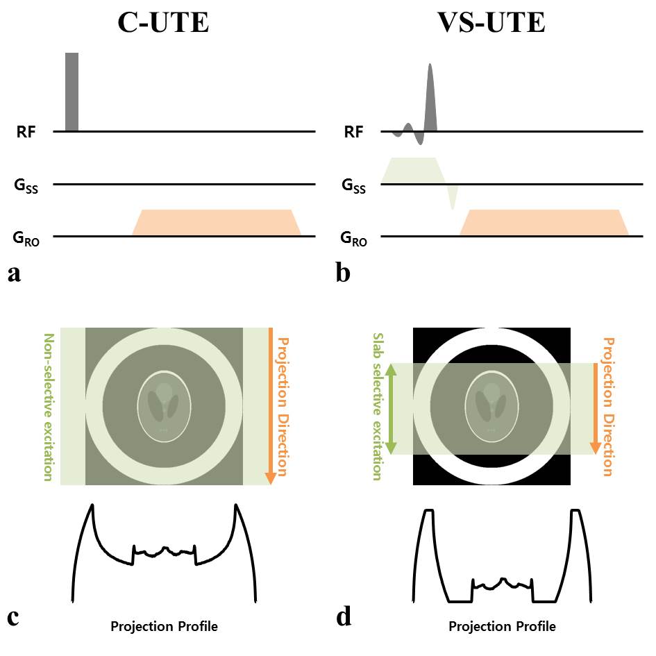
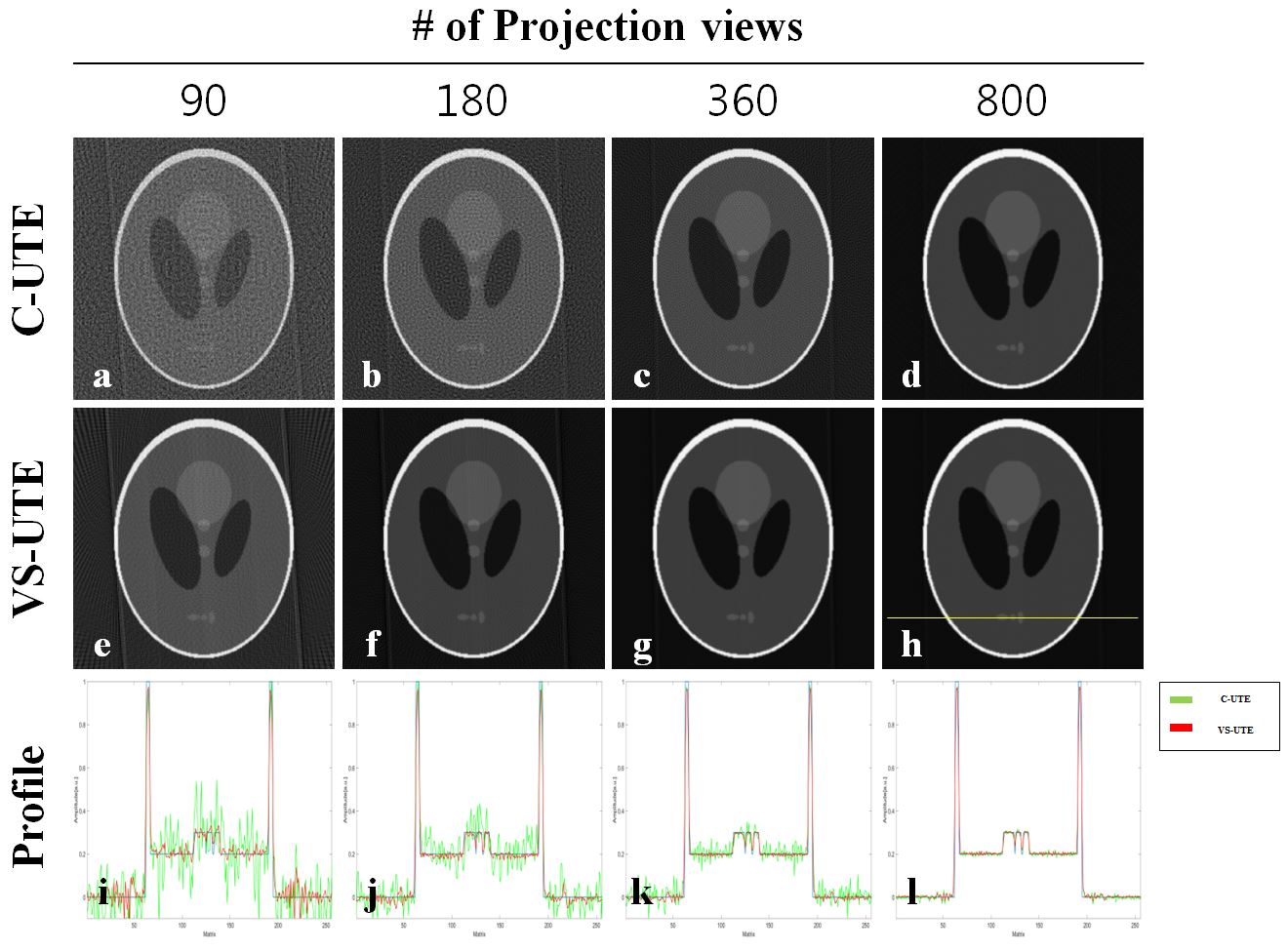
C-UTE acquires projection data which include the entire object’s information using a rectangular pulse (Fig.1a,c). In contrast, VS-UTE performs slab excitation by using a frequency-selective SINC pulse in the presence of a slab-selective gradient, and acquires the projection data in the same direction as the slab-selective direction, thereby including only information inside the selected volume (Fig.1b,d). Therefore, C-UTE requires more projection data to satisfy the Nyquist criterion than VS-UTE because C-UTE needs to cover the entire object, not the selected one.Simulation: Simulation of C-UTE and VS-UTE was performed for the shepp-logan phantom located inside the white concentric ring. Projection data was acquired with gridding after 2D inverse FFT. The slab-selective excitation was implemented by multiplying a rectangular function to the image. The simulation information is as follows: Matrix size = 512×512, number of projection views = 45, 90, 180, 360, 800.
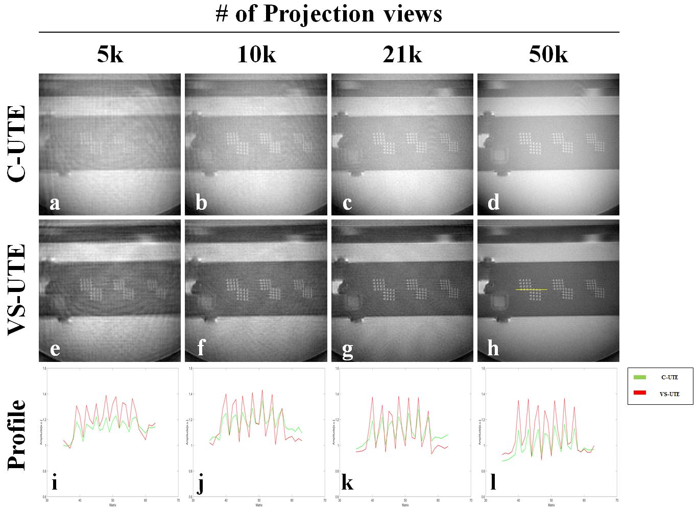
Experiment: VS-UTE and C-UTE were implemented and tested at 3T (PRISMA, Siemens). An 18-channel chest coil and 16-channel spine coil were used for signal reception in combination with a transmit body coil. To better evaluate image quality in terms of spatial resolution, the ACR phantom was scanned so that the resolution grids are included in the internal structure, and the ROI of C-UTE and VS-UTE was set smaller than the ACR phantom. Scan parameters were: TR = 3 ms, FOV = 100×100×100 mm3, matrix size = 128×128×128, isotropic resolution = 0.78 mm, flip angle = 5°, number of projection views = 3k, 5k, 9k, 10k, 20k, 30k, 50k, pulse duration (UTE/VS-UTE) = 20/150 us, TE (UTE/ VS-UTE) = 120 us.
Results and Discussion
Figure 2 shows the simulation images (a-h) and 1D profiles (i-l) obtained using C-UTE and VS-UTE acquisition schemes. The 1D profiles were obtained along the yellow lines. In case of C-UTE, as the number of projection views decreased to 90, severe streak artifacts appeared and significantly degraded image quality (clearly seen in both image and 1D profile) because signals from the outside of the shepp-logan phantom were involved in the projection data (Fig.2a-c). In contrast, VS-UTE was able to identify the internal structure much better than C-UTE with almost no streak artifacts (Fig.2e-g).Figure 3 shows the images including the resolutions grids and 1D profiles of the ACR phantom. The 1D profiles were obtained along the yellow lines. As the number of projections decreased below 21k, streak artifacts begin to appear in C-UTE images and it is difficult to identify the resolution grids if the number of projections was 5k (Fig 3. a-c). In addition, the overall contrast-to-noise ratio (CNR) of C-UTE was also much lower than VS-UTE. These are all because the projection data were contaminated by the signals from the outside of the ROI. In case of VS-UTE, there also existed some streak artifacts, but CNR was much better in VS-UTE than C-UTE including the case of 50k projection views. The resolution grids were also identifiable even when there were 5k projection views (Fig.3e-g).
Conclusion
We have demonstrated that volume-selective UTE (VS-UTE) can provide much better image quality than conventional 3D UTE (C-UTE), especially when number of projection views are undersampled with volume excitation smaller than the object to be imaged. The advantage of VS-UTE in maintaining the internal structure expression even with a small number of projection views can be more effective with sparse sampling or compressed sensing. A little expense for using VS-UTE is that the achievable minimum TE is slightly longer in VS-UTE than C-UTE because VS-UTE employs a SINC pulse, not a square pulse.Acknowledgements
This study was supported by National Research Foundation of Korea NRF-2020R1A2B5B02002676References
1. Bergin CJ, Pauly JM, Macovski A. Lung parenchyma: Projection reconstruction MR imaging. Radiology 1991;179:777-781.
2. Glover GH, Pauly JM. Projection Reconstruction Techniques for Reduction of Motion Effects in MRI. Magnetic Resonance in Medicine 1992;28:275–89.
3. Du J, Thornton FJ, Fain SB, Korosec FR, Browning F, Grist TM, Mistretta CA. Artifact reduction in undersampled projection reconstruction MRI of the peripheral vessels using selective excitation. Magn Reson Med 2004;51(5):1071-1076.
4. Macdonald J, Wieben O, Nagel SK, and Johnson KM. Artifact reduction in 3D radial imaging with out-of-volume saturation pulses. In Proceedings of the 24th Annual Meeting of ISMRM, Singapore, 2016. p. 1831.
5. Jinil Park, Jang-Yeon Park. Reducing Streak Artifacts in 3D Radial Imaging Using Volume-selective Signal Acquisition. In Proceedings of the 29th Annual Meeting of ISMRM, 2020. P. 2360.
Figures