4182
Phantom validation of a novel user-configurable ultrashort echo time (UTE) sequence1Division of Biomedical Engineering, University of Saskatchewan, Saskatoon, SK, Canada, 2Department of Mechanical Engineering, University of Saskatchewan, Saskatoon, SK, Canada, 3Research Collaboration Manager, Siemens Healthcare Limited, Oakville, ON, Canada, 4Department of Radiology, University of Manitoba, Winnipeg, MB, Canada
Synopsis
Ultrashort echo time (UTE) pulse sequences can capture the signal from short T2 tissues. Many UTE techniques have been developed for achieving an ultrashort TE (less than 0.1 ms) or enhancing the short T2 contrast. However, these approaches have not been extensively compared head-to-head due to the lack of a flexible UTE sequence that integrates the different approaches. Therefore, in this work, we developed a versatile UTE sequence that can directly compare UTE approaches and provide options for researchers to choose the appropriate UTE that fit the particular application.
Introduction
Ultrashort echo time (UTE) techniques allow for rapid acquisition of signal, delivering short-T2 contrast in MR images1-2. Hallmarks of UTE techniques include the use of a half-Sinc2 or rectangular Radiofrequency (RF) excitation pulses, non-Cartesian trajectories (radial3 and spiral4-5), and long-T2 contrast suppression. However, these approaches and the resulting images have not been systematically compared and evaluated, because there lacks a flexible UTE sequence that conveniently includes these options. Furthermore, this comparison and evaluation could provide valuable information and guidance for identifying the most appropriate UTE techniques for a particular application. Therefore, in this work, we developed a novel, versatile UTE pulse sequence and systematically evaluated the implemented UTE approaches in an in-house phantom incorporating a short T2 component.Methods
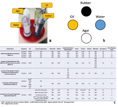
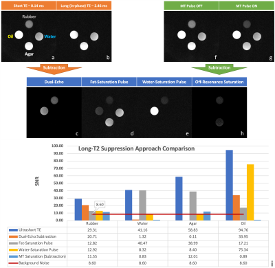
The UTE sequence was developed using IDEA (Siemens Healthineers, Erlangen, Germany) and evaluated on a three-tesla system (Prisma, VE11C, Siemens Healthineers, Erlangen, Germany). The sequence was designed to be user-configurable and includes options that can be changed on-the-fly. These include the choice of excitation pulse (half-Sinc or hard pulse), type of trajectory (radial or spiral), dimensionality (2D or 3D), and type of suppression technique (i.e.long -T2). To supplement the data analysis, we developed an offline UTE reconstruction pipeline based on the gridding algorithm in MATLAB (MathWorks, Natick, MA, USA) with an off-resonance artifact correction technique6 aiming to provide high-quality UTE images. To carry out the study, we constructed a phantom that contains rubber as the short-T2 component (whose T2 is ~0.3 ms) while water, oil, and agar as the long-T2 components (see Figure 1.a-b). To assess the performance of the sequence, we systematically evaluated and compared 1) the approaches for off-resonance artifact correction; 2) the combinations of common RF pulses and trajectories; 3) the comprehensive long-T2 suppression techniques (see details in Figure 1.c). We evaluated our results by visual inspection and Signal-to-Noise Ratio (SNR) calculation (using a normal image and a pure noise image).Results
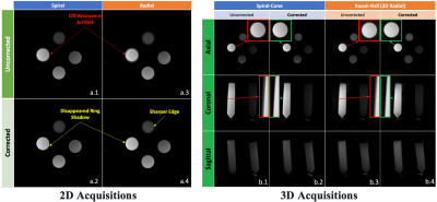
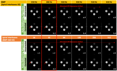
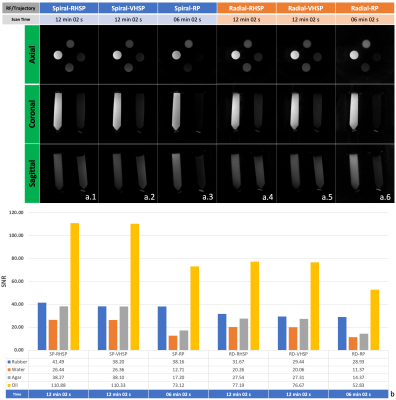
The auto-deblurring algorithm applied in 2D and 3D can successfully suppress the off-resonance artifact, characterized by the blurry edges, in the spiral and radial trajectories (Figure 2). The correction for the long-duration spiral trajectory relies on not only the deblurring algorithm but also some acquisition adjustments, for example, an increase of bandwidth-per-pixel (BWP) or the number of spiral interleaves (Figure 3). Among the combinations of the implemented RF pulses and trajectories (in 3D), the regular half-Sinc pulse (RHSP) with the spiral-cone trajectory provides overall higher SNRs for all material types, however, at cost of a longer acquisition time (Figure 4). Qualitative and quantitative comparisons between the implemented long T2 suppression techniques were performed (Figure 5).Discussion
In this work, we showed that our UTE sequence is versatile, efficient and can correct off-resonance artifacts in a phantom with a short-T2 component. For the artifact appearing in 2D radial, the automatic deblurring algorithm proposed by Noll et al.6 is sufficient for correction due to its normally short gradient readout (Figure 2.a., 3-4). Applying the same deblurring algorithm is less effective for the spiral trajectory with a longer gradient duration (lower BWP with fewer spiral-interleaves), as indicated by the red arrows in Figure 3. To overcome this deblurring issue for longer trajectories, our study shows that the increase of BWP or the number of spiral-interleaves, as a complementary measure (to the algorithm), can alleviate the off-resonance artifact on the acquisition side (see Figure 3.a.1-6 and c.1-6) and achieve a better correction outcome with the auto-deblurring algorithm (see Figure 3.b.1-6 and d.1-6). As for the 3D scans, the off-resonance artifact was observed to extend to other dimensions. This presented an opportunity for us to modify the deblurring algorithm, which was originally proposed in 2D, and extend it to three dimensions. The results suggest the extension of the algorithm to 3D was successful with the off-resonance artifact disappearing (Figure 2.b.1-4). The RF pulse comparison (Figure 4) shows the RHSP and Variable-rate selective excitation (VERSE)7 - modified Half-Sinc Pulse (VHSP) are similar in terms of image quality, SNR, and time efficiency. While these two pulses provided higher SNRs, the cost was an increase in scanning time when compared to the Rectangular Pulse (RP) due to their double-excitation scheme. In general, the spiral trajectory has an overall higher SNR for all materials than their counterparts for the radial trajectory (Figure 4). A possible explanation for this is that the spiral trajectory can provide better k-space coverage than the radial trajectory with matching scanning parameters. Lastly, the sequence has a comprehensive long-T2 suppression capability (Figure 5). The dual-echo subtraction method saturated the water and agar components thoroughly and suppressed the oil's signal partially (Figure 5.a-c). The fat-saturation pulse drastically saturated the signal from oil, although there was still some insignificant residual signal in the oil and rubber (Figure 5.d). The water-saturation pulse successfully saturated the water-based materials (i.e.water and agar) without any signal residual (Figure 5.e). The off-resonance saturation approach fully suppressed the water and oil signals while it only partially statured the agar component due to its magnetization transfer effect (Figure 5.f-h).Conclusion
This multi-featured UTE sequence can help to evaluate different UTE approaches and further provide valuable guidance for UTE imaging in various applications.Acknowledgements
The authors acknowledge our funding sources, Mitacs-Accelerate IT10545, IT19172 and Arthritis Society Young Investigator Operating Grant. The authors also thank Siemens Healthineers for their technical supports.
References
- Bydder GM. The Agfa Mayneord lecture: MRI of short and ultrashort T 2 and T 2* components of tissues, fluids and materials using clinical systems. The British journal of radiology. 2011 Dec;84(1008):1067-82.
- Gold GE, Pauly JM, Macovski A, Herfkens RJ. MR spectroscopic imaging of collagen: tendons and knee menisci. Magnetic resonance in medicine. 1995 Nov;34(5):647-54.
- Bergin CJ, Pauly JM, Macovski A. Lung parenchyma: projection reconstruction MR imaging. Radiology. 1991 Jun;179(3):777-81.
- Glover GH. Simple analytic spiral k‐space algorithm. Magnetic Resonance in Medicine: An Official Journal of the International Society for Magnetic Resonance in Medicine. 1999 Aug;42(2):412-5.
- Cline HE, Zong X, Gai N. Design of a logarithmic k‐space spiral trajectory. Magnetic Resonance in Medicine: An Official Journal of the International Society for Magnetic Resonance in Medicine. 2001 Dec;46(6):1130-5.
- Noll DC, Pauly JM, Meyer CH, Nishimura DG, Macovskj A. Deblurring for non‐2D Fourier transform magnetic resonance imaging. Magnetic Resonance in Medicine. 1992 Jun;25(2):319-33.
- Conolly S, Nishimura D, Macovski A, Glover G. Variable-rate selective excitation. Journal of Magnetic Resonance (1969). 1988 Jul 1;78(3):440-58.
Figures
