4180
A Short TR Adiabatic Inversion Recovery Zero Echo Time (STAIR-ZTE) Sequence with Interleaved Encoding and a Modulated RF Pulse for Myelin Imaging1University of California, San Diego, San Diego, CA, United States, 2GE Healthcare, San Diego, CA, United States, 3Veterans Affairs San Diego Healthcare System, San Diego, CA, United States
Synopsis
It is challenging to directly image myelin due to its extremely short T2* (<300µs at 3T). Adiabatic Inversion Recovery prepared Ultrashort Echo Time (IR-UTE) imaging has been proposed for direct myelin imaging in human brain. More recently, Short Repetition Time Adiabatic Inversion Recovery (STAIR) has been proposed as a novel contrast mechanism for myelin imaging with improved suppression of long T2 signal. In this study, we explored feasibility of STAIR based Zero Echo Time (STAIR-ZTE) combined with an amplitude- and phase-modulated RF pulse and interleaved Water- and Fat-Suppressed Proton Projection MRI (WASPI) for myelin imaging in human brain.
Introduction
It is challenging to directly image myelin due to its extremely short T2* (<300µs at 3T). Inversion Recovery prepared Ultrashort Echo Time (IR-UTE) imaging has been proposed for direct myelin imaging in human brain1–3. More recently, Short Repetition Time Adiabatic Inversion Recovery (STAIR) has been proposed as a novel contrast mechanism for myelin imaging with improved suppression of long T2 signal4. In this study, we explored feasibility of STAIR based Zero Echo Time (STAIR-ZTE) combined with an amplitude- and phase-modulated RF pulse and interleaved Water- and Fat-Suppressed Proton Projection MRI (WASPI) for myelin imaging in human brain.Methods
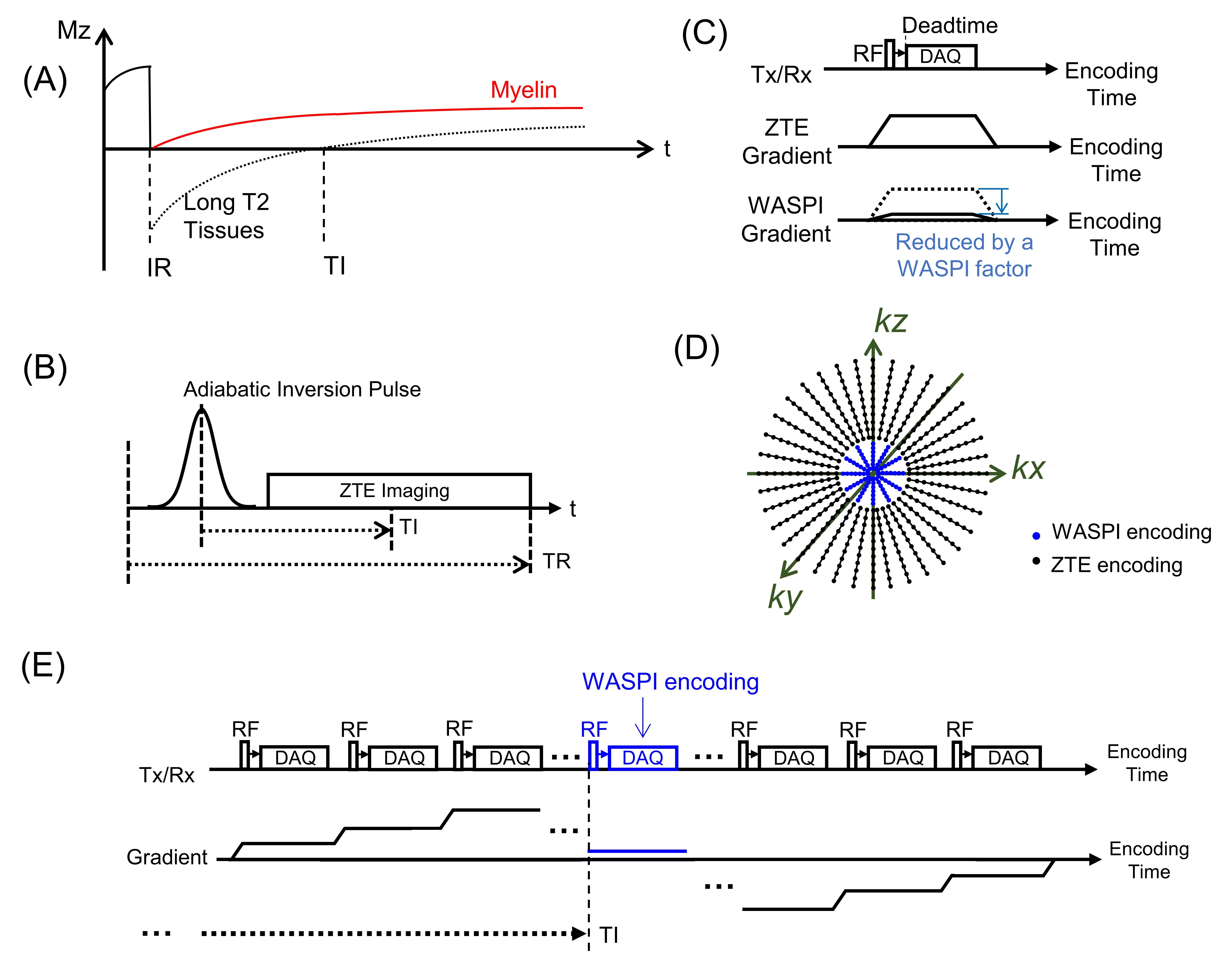
Figure 1A illustrates adiabatic inversion recovery (IR) of myelin and long T2 tissues (i.e., white matter (WM), gray matter (GM), and cerebrospinal fluid (CSF)). Due to the short T2* decay and relatively long duration of the adiabatic IR pulse (~8ms), the longitudinal magnetization of myelin is not inverted but partially saturated (red line in Figure 1A). The longitudinal magnetizations of long T2 tissues are inverted and undergo inversion recovery (black dotted line in Figure 1A). By acquiring UTE data at the nulling point of WM, we can selectively image myelin with long T2 WM signal suppressed. With a short TR, long T2 tissues with a wide range of T1s can be suppressed regardless of regional T1 variations4.We utilized a multi-spoke STAIR-ZTE sequence where several spokes are continuously acquired after each IR preparation (Figure 1B). ZTE imaging is typically performed using an excitation scheme where the RF pulse is applied with a fully ramped-up readout gradient to shorten the effective TE (Figure 1C)5. In this scheme, data during RF excitation and deadtime are missed, leaving a hole in the center of k-space (black dots in Figure 1D). WASPI encoding is used to fill the hole using a derated readout gradient (Figures 1C and 1D). Typically, the WASPI encoding is performed sequentially before or after ZTE encoding, but this scheme is not optimal for STAIR-ZTE since the WASPI data in the center of k-space can be largely contaminated by T1 variations. To address this issue, we proposed an interleaved encoding scheme where WASPI is interleaved at a desired inversion time (TI) (Figure 1E) in a manner similar to our previous development, interleaved hybrid encoding6. Another technical issue is unwanted slice selectivity caused by the RF excitation scheme of ZTE which could impact image quality in STAIR-ZTE imaging (e.g., blurring)7–9. To address this, we incorporated an amplitude- and phase-modulated hyperbolic secant (HSn) pulse as proposed by Schieban et al.10
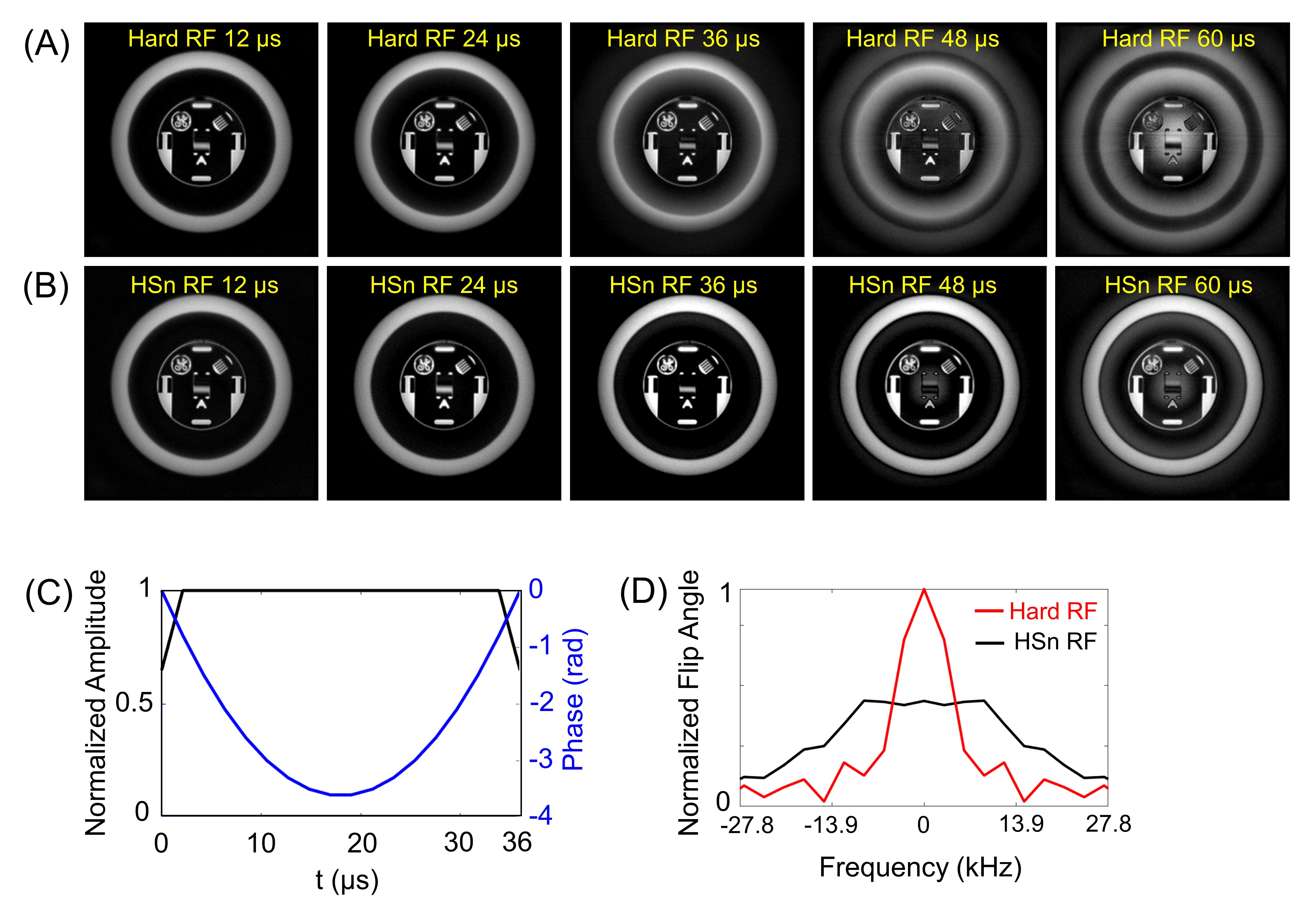
The STAIR-ZTE sequence was implemented on a clinical 3T MRI scanner (MR750, GE Healthcare). Five different HSn pulses with 12, 24, 36, 48, and 60µs duration were designed. Figures 2A and 2B show the resulting ZTE images with hard pulses and HSn pulses, respectively. The 12-36µs HSn pulse yielded reasonable image quality, and the 36µs HSn pulse was subsequently selected for in vivo study (Figures 2C, D). For in vivo investigation, three healthy volunteers (33-, 38-, and 47-year-old males) and two multiple sclerosis (MS) patients (22- and 54-year-old females) were recruited in compliance with the institutional review board and underwent an MRI exam using a GE 12-ch receive-only head-coil with the following parameters: 1) STAIR-ZTE: TR/TI/TE=142ms/65ms/12µs, spoke-to-spoke timing=1684µs, flip angle (FA)=7°, field of view (FOV)=300x300x300mm3, matrix=160x160x60, readout bandwidth (rBW)=62.5kHz, WASPI factor=8, #-of-spokes-per-IR=12, and scan time=9min 54sec, 2) MP-RAGE: TR/TI/TE=8.3/450/3.2ms, FA=12°, FOV=256x256x158mm3, matrix=256x256x132, rBW=83.4kHz, scan time=5min 51sec. 3) FLAIR: TR/TI/TE=6502/1925/127.8ms, FA=90°, FOV=256x256x317mm3, matrix=256x256x264, rBW=83.4kHz, scan time=5min 40sec. For healthy volunteers, STAIR-ZTE was repeated with four different configurations: 36µs HSn pulse or 24µs hard pulse paired with sequential or interleaved WASPI encoding. For MS patients, STAIR-ZTE was performed with a 36µs HSn pulse and interleaved WASPI encoding.
Results
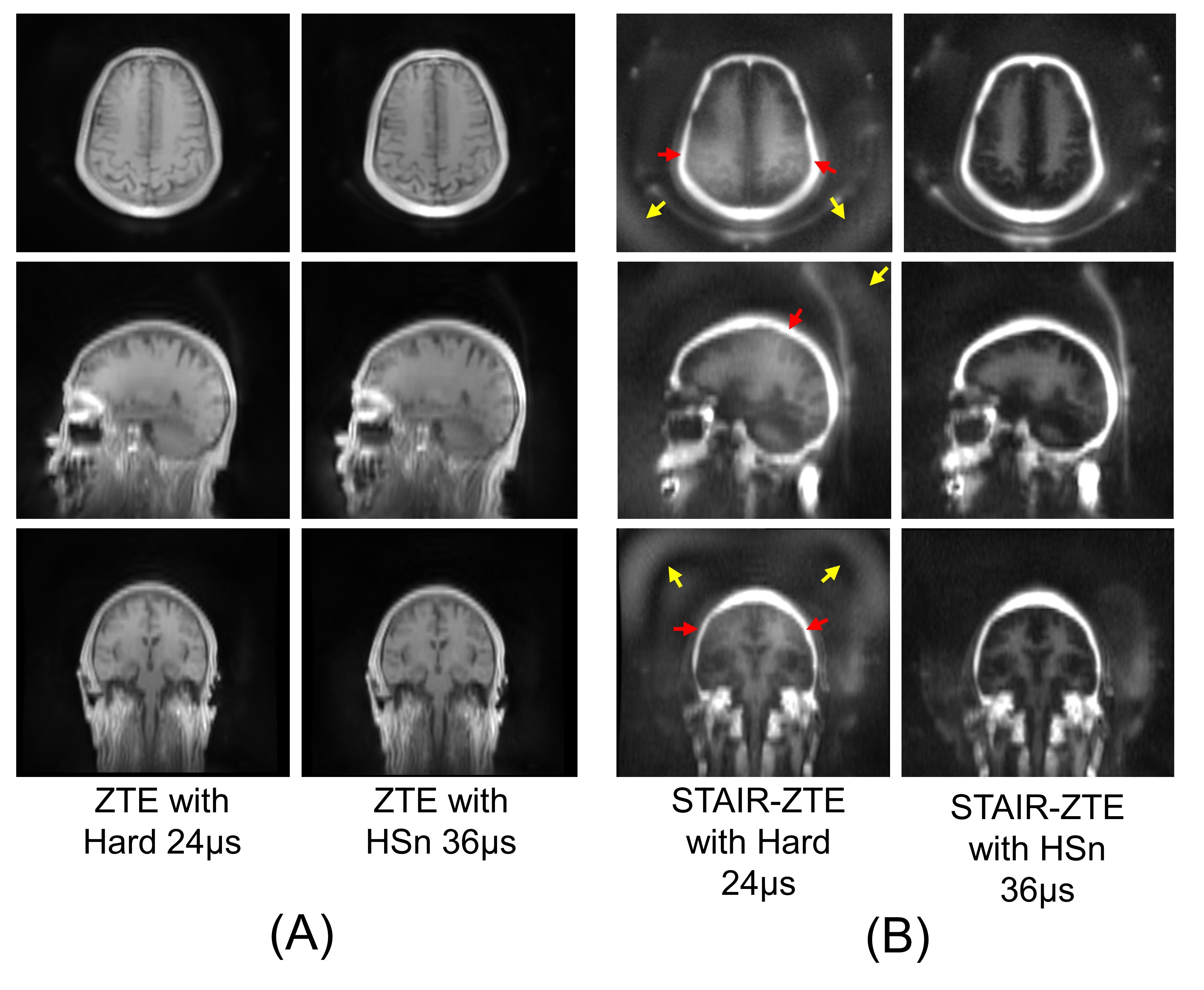
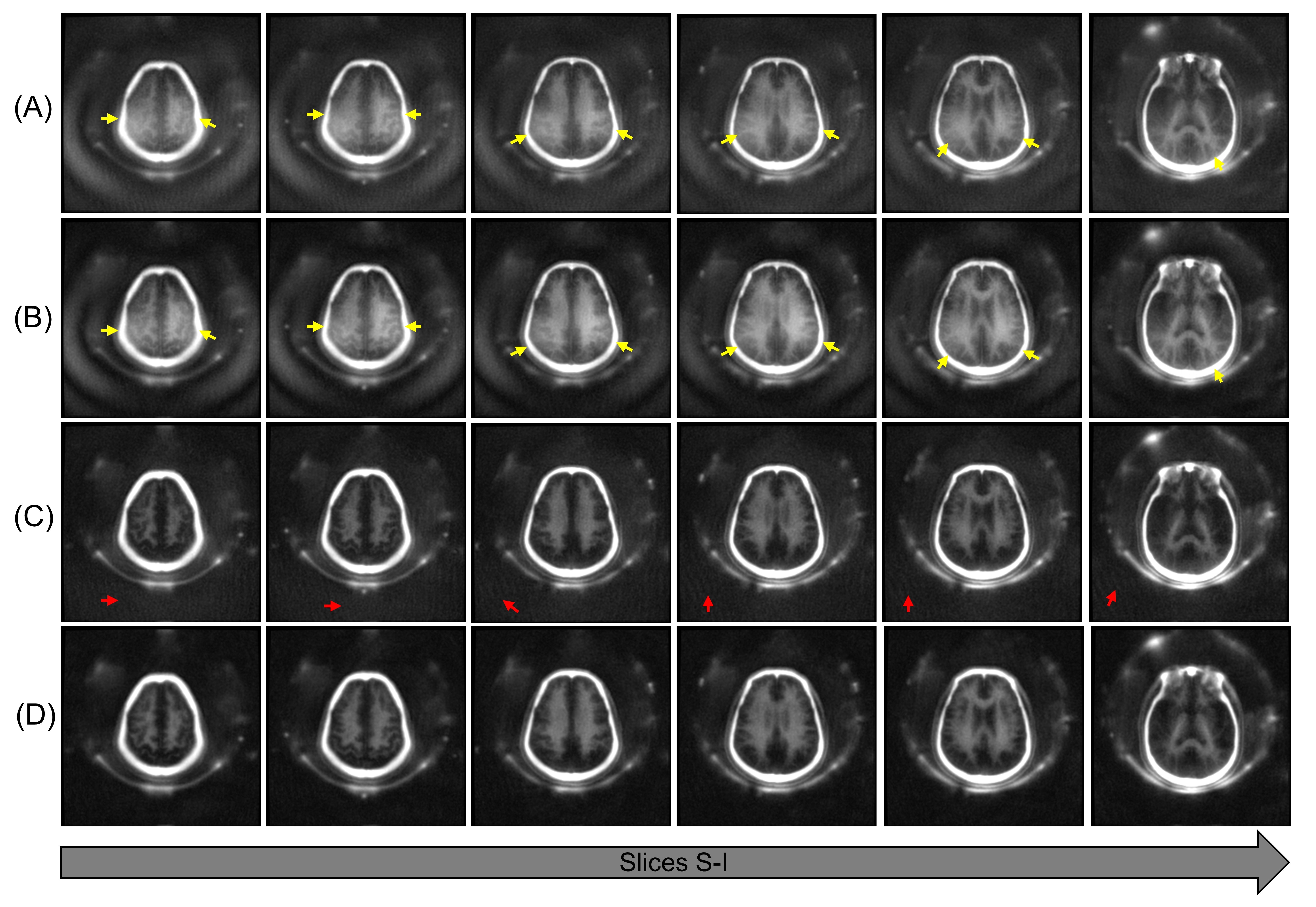
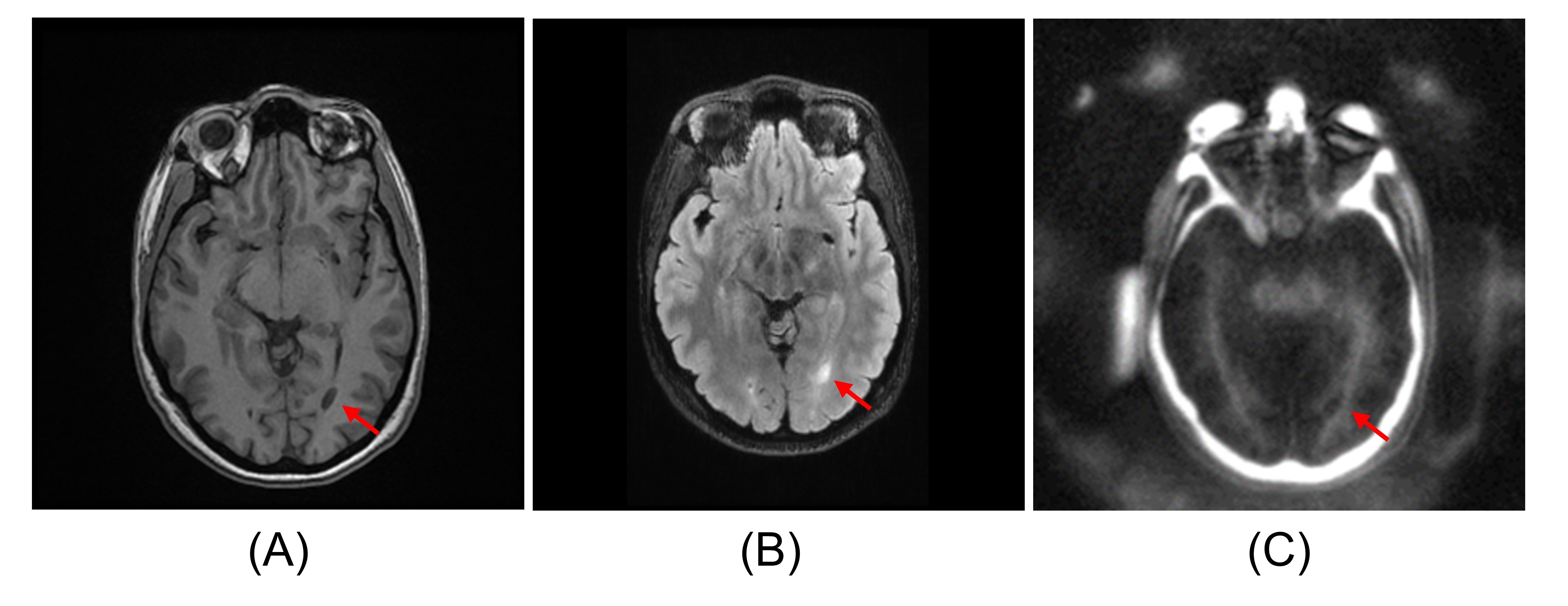
Regular ZTE imaging showed no dramatic improvement with an HSn pulse (Figure 3A). In contrast, STAIR-ZTE yielded significantly improved myelin contrast with an HSn pulse compared to with a hard pulse, where low-frequency bias (yellow arrows) impairing myelin contrast (red arrows) was well-suppressed. Figure 4 shows STAIR-ZTE with four configurations of RF types and encoding schemes. With a hard pulse, low frequency signal was propagated from outer FOV, causing strong signal bias regardless of the WASPI encoding schemes (yellow arrows in Figures 4A,B). STAIR-ZTE with an HSn pulse and sequential WASPI encoding showed improved myelin contrast but exhibited streaking artifacts across the image (red arrows in Figure 4C). STAIR-ZTE with an HSn pulse and interleaved WASPI encoding showed the best myelin contrast with suppression of both signal bias and streaking artifacts (Figure 4D). Figure 5 shows results with an MS patient (22F). The proposed STAIR-ZTE detected a demyelinated lesion and yielded myelin-specific images, corresponding well with clinical MRI.Discussion and Conclusion
As myelin signal intensity is very low compared to the surrounding long T2 tissues, even very subtle imaging artifacts (e.g., low-frequency bias or streaking/aliasing) may be critical factors, as shown in Figures 3 and 4. In this study, we showed efficacy of an HSn pulse and interleaved WASPI encoding for STAIR-ZTE in reducing such artifacts, improving myelin contrast. In future works, we will further optimize the protocol to achieve improved spatial resolution and SNR and evaluate it in a large number of subjects.Acknowledgements
The authors acknowledge grant support from the NIH (R01NS092650), Veterans Affairs (I01RX002604 and I01CX001388), and GE Healthcare.References
1. Sheth V, Shao H, Chen J, et al. Magnetic resonance imaging of myelin using ultrashort Echo time (UTE) pulse sequences: Phantom, specimen, volunteer and multiple sclerosis patient studies. Neuroimage 2016;136:37–44 doi: 10.1016/j.neuroimage.2016.05.012.
2. Ma Y, Jang H, Chang EY, et al. Ultrashort echo time (UTE) magnetic resonance imaging of myelin: Technical developments and challenges. Quant. Imaging Med. Surg. 2020;10:1186–1203 doi: 10.21037/QIMS-20-541.
3. Jang H, Wei Z, Wu M, et al. Improved volumetric myelin imaging in human brain using 3D dual echo inversion recovery‐prepared UTE with complex echo subtraction. Magn. Reson. Med. 2019:mrm.28082 doi: 10.1002/mrm.28082.
4. Ma Y, Jang H, Wei Z, et al. Myelin Imaging in Human Brain Using a Short Repetition Time Adiabatic Inversion Recovery Prepared Ultrashort Echo Time (STAIR-UTE) MRI Sequence in Multiple Sclerosis. Radiology 2020;297 doi: 10.1148/radiol.2020200425.
5. Jang H, Carl M, Ma Y, et al. Inversion recovery zero echo time (IR-ZTE) imaging for direct myelin detection in human brain: a feasibility study. Quant. Imaging Med. Surg. 2020;10:895–906 doi: 10.21037/qims.2020.04.13.
6. Jang H, Ma Y, Searleman AC, et al. Inversion recovery UTE based volumetric myelin imaging in human brain using interleaved hybrid encoding. Magn. Reson. Med. 2020;83:950–961 doi: 10.1002/mrm.27986.
7. Grodzki DM, Jakob PM, Heismann B. Correcting slice selectivity in hard pulse sequences. J. Magn. Reson. 2012;214:61–7 doi: 10.1016/j.jmr.2011.10.005.
8. Jang H, Wiens CN, McMillan AB. Ramped hybrid encoding for improved ultrashort echo time imaging. Magn. Reson. Med. 2016;76:814–825 doi: 10.1002/mrm.25977.
9. Cheng Li, Magland JF, Seifert AC, Wehrli FW. Correction of Excitation Profile in Zero Echo Time (ZTE) Imaging Using Quadratic Phase-Modulated RF Pulse Excitation and Iterative Reconstruction. IEEE Trans. Med. Imaging 2014;33:961–969 doi: 10.1109/TMI.2014.2300500.
10. Schieban K, Weiger M, Hennel F, Boss A, Pruessmann KP. ZTE imaging with enhanced flip angle using modulated excitation. Magn. Reson. Med. 2015;74:684–693 doi: 10.1002/mrm.25464.
Figures