4177
Simultaneous Measurements of Low-b Spin-Echo and High-b Stimulated-Echo DWI for Ultrahigh-b DWI1Utah Center for Advanced Imaging Research, University of Utah, Salt Lake City, UT, United States, 2NeuroImunology and NeuroVirology, University of Utah, Salt Lake City, UT, United States, 3Neurology, University of Utah, Salt Lake City, UT, United States, 4Radiology and Imaging Sciences, University of Utah, Salt Lake City, UT, United States
Synopsis
The signal-b curve of the Ultrahigh-b DWI (UHb-DWI) with bmax = ~ 10,000 s/mm2 is sensitive to the water exchange at the myelin sheath in white-matter. Ultrahigh-b DWI is obtained using DW-stimulated-echo (DW-STE) sequence, however, a half the prepared magnetization is discarded in conventional STE imaging. Therefore, the main objective of this work has been to measure low-b DW-spin-echo and DW-stimulated-echo in a single sequence, and to correct/combine DWSE and DWSTE for ultrahigh-b DWI (UHb-DWI) of cervical spinal cord (CSC).
Introduction
High-b diffusion coefficient DH of ultrahigh b radial DWI is sensitive to the water permeability at the myelin sheath and can be measured with increased diffusion time using diffusion-weighted stimulated-echo EPI (DWSTEPI) using three 90o RF pulses 1. To achieve UHb-DWI with b ~ 10,000 s/mm2 on clinical MRI system with limited gradient strength (Gmax = 40 mT/m), we varied the mixing time TM up to 450 ms, and permuted slice ordering within the long TM to improve the time-efficiency of the measurement 2,3. The method requires removing T1 decay effect on DWSTEPI data and it was unreliable at data with low SNR, such as in b > 5,000 s/mm2. On modern MRI system with increased gradient performance (Gmax = 80 mT/m), DWI of b ~ 10,000 s/mm2 can be obtained using DWSTEPI with a fixed TM (~ 100 ms). However, its time-efficiency is greatly reduced, because a half of the transverse magnetization is wasted, while being restored back to the longitudinal space by the second RF pulse. The fraction of the wasted magnetization is more than 50 % due to the imperfect 90o flipangle in real situation. Therefore, for successful combination of DWSE and DWSTE, the effect of imperfect 90o must be corrected, as in previous report 4,5. In this work, we present a method to obtain UHb-DWI for human CSC using simultaneously measured DWSEPI and DWSTEPI with simple RF correction.Methods
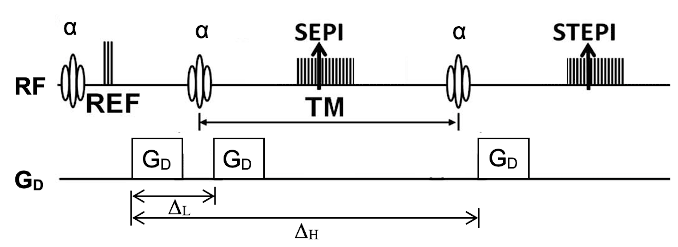
Pulse Sequence: Manufacturer’s 2D singleshot diffusion-weighted EPI sequence was modified to implement 2D singleshot DW-SESTEPI using a pulse sequence development environment (IDEA, Siemens Medical Solution, Erlangen, Germany). Three diffusion gradients are applied with the identical duration and amplitude. Diffusion-weightings for the low-b DWSE and high-b DWSTE are (γGDδ)2(ΔL-δ/3) and (γGDδ)2(ΔH-δ/3), respectively.Combining DWSE and DWSTE 4,5: The magnetizations for DWSE and DWSTE are derived as,
$M_{SE}(r; b_L) =M_+^o (r) \sin {\alpha} \sin^2 {\alpha/2} e^{-TE/T_2} e^{-bD_L}$. ... (1)
$M_{STE}(r; b_H) =M_+^o (r) \sin^3 {\alpha} e^{-TE/T_2} e^{-TM/T_1} e^{-bD_H}$. ... (2)
Therefore, low-b DWSE images can be combined to DWSTE data by multiplying a correction map $g(r)$ as in,
$M_{STE}(r; b_L) = M_{SE}(r; b_L} g(r)$. . . . (3)
Here, the correction map $g(r)$ can be estimated using eqns. (1, 2),
$g(r) = s \cos^2{\alpha/2} e^{-TM/T_1} = M_{STE}(r;b_o)/M_{SE}(r;0)$ . . . (4).
Eq. (4) indicates indicates that the DWSTE signal for the same b-value differs from DWSE by imperfect 90o and additional T1 decay.
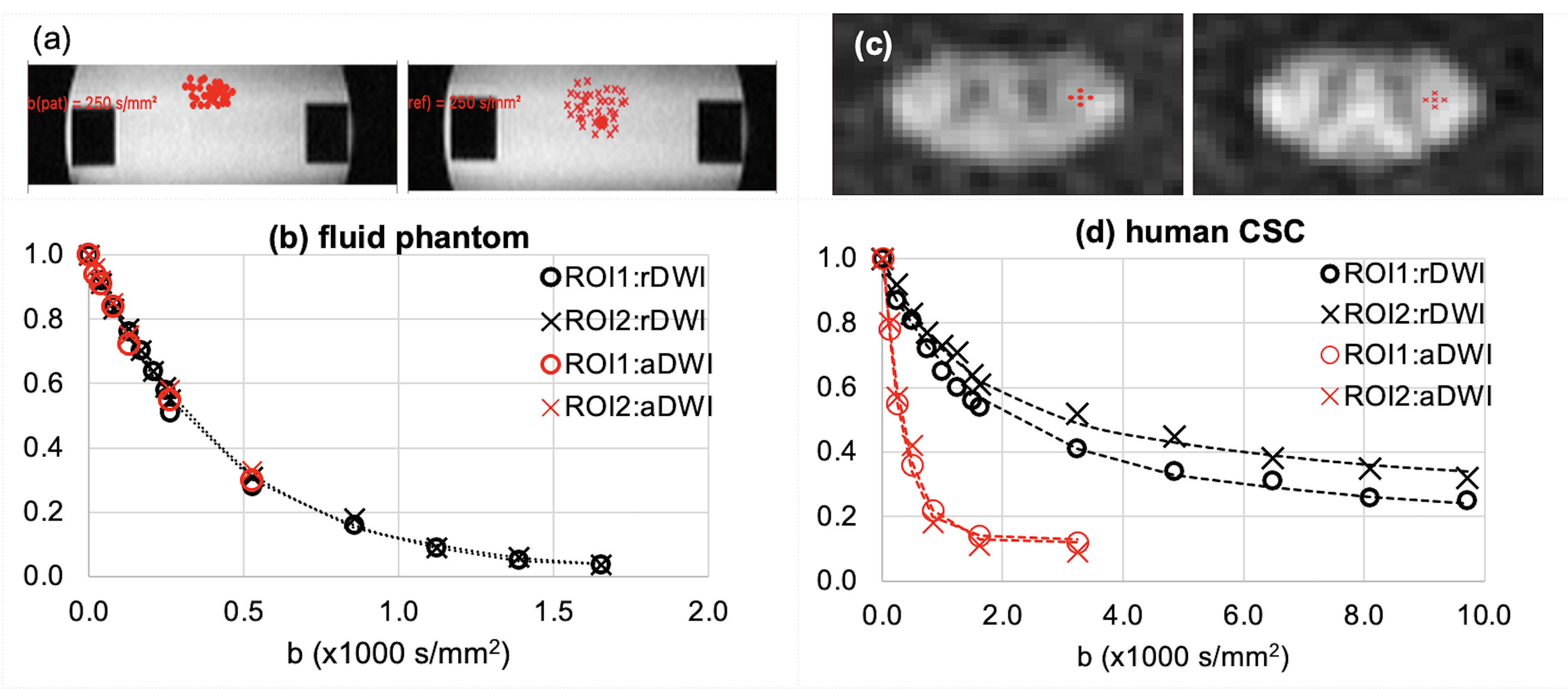
MR Imaging: UHb-DWI experiment was performed at 3T MRI system (Prisma, Siemens, Erlangen, Germany) using 64 channel head/neck array coil. 2D ss-DW-SESTEPI was applied to a fluid phantom, filled with 0.1 mMol MnCl2/water, with T1/T2 = 950/80 ms, 1x1 mm2 in-plane resolution and 4 mm slice thickness, TR 4900 ms, TE 76 ms, 8 averaging, 7 pairs of DWSE and DWSTE with b values ((40, 264), (80, 528), (130, 858), (170, 1123), (210, 1387), (250, 1651)) and ((20, 132), (40, 264), (80, 528)) s/mm2 along two orthogonal directions. Then, it was applied to obtain a set of UHb-DW images from CSC of a healthy subject, under an approved IRB, using the identical imaging parameters as those for phantom, except for 7 pairs of DWSE and DWSTE with b values ((250, 1620), (500, 3240), (750, 4860), (1000, 6480), (1250, 8100), (1500, 9721)) and ((130, 842), (250, 1620), (500, 3240)) s/mm2 along radial (rDWI) and axial (aDWI) to the cord direction, respectively. Data acquisition time for UHb-DWI was about 6 min for twenty-one slices, covering C1 ~ T2 vertebrae. The measured DWIs are processed, including RF/T1 correction and analyzing signal-b curve, at an offline UNIX computer using a home-developed software, written in Python 3.x.
Results and Discussions
DWSEPI and DWSTEPI images were successfully combined. From the combined images set, diffusion coefficient of the water at room temperature was estimated as D = 2.352 x 10⁻³ mm2/s, of which the signal-b curve is illustrated in Fig. 2b. The axial-DWI signal (red symbols in Fig. 2d) of the human CSC fits well to a single-exponential function with respect to b value with diffusivity 2.66x10⁻³ mm2/s. However, human radial-DWI signal fits to a double exponential function with average fast and slow decay constants (Df, Ds) = (0.700, 0.050) x 10-3 mm2/s and relative fractions (54 %, 46 %). This rDWI signal behavior is very similar to those using the variable TM for UHb-DWI 3. Current method utilizes the entire diffusion-prepared magnetization, unlike the conventional DWSTE. Because the method uses a fixed mixing time and does not require T1 correction, signal-b curve is significantly stable, particularly at the high-b region, say b > 5000 s/mm2, compared with the method presented in the reference 2,3.Synopsis
Ultrahigh-b DWI is obtained using DW-stimulated-echo (DW-STE) sequence, with a cost of a half the prepared magnetization. Therefore, the main objective of this work has been to measure low-b DW-spin-echo and DW-STE in a single sequence, and to correct/combine them for UHb-DWI of cervical spinal cord (CSC).Acknowledgements
This work was supported by NIH R56 R01 NS106097-01A1.References
1. Sapkota N, Yoon S, Thapa B, Bisson EF, Bowman BM, Miller SC, Shah LM, Rose JW, Jeong EK. Characterization of spinal cord white matter by suppressing signal from hindered space. A Monte Carlo simulation and an ex vivo ultrahigh-b diffusion-weighted imaging study, J. Magn. Reson. (2016): 272; 53~59.
2. Sapkota N, Shi X, Bisson EF, Shah LM, Rose JW, Jeong EK. 2D Single-Shot Diffusion-Weighted Stimulated EPI with Reduced FOV for Ultra-high-b Radial Diffusion-Weighted Imaging of Spinal Cord. Magn. Reson. Med. (2016), 77(6), 2167-2173.
3. Thapa B, Sapkota N, Lee YJ, Jeong KE, Bisson EF, Shah LM, Rose JW, Jeong EK, Ultra-High-b Radial Diffusion Weighted Imaging (UHb-rDWI) of Human Cervical Spinal Cord, (2019) J. Magn. Reson. Imag. 49(1): 204-211.
4. Franconi F, Sonier CB, Seguin F, Le Page A, Akoka A, Acquisition of spin echo and stimulated echo by a single sequence: Application to MRI of diffusion, Magn. Reson. Imag.1994, 12 (4): p605 – 611.
5. Shi XF, Jeong EK, Singleshot T1 Mapping using Simultaneous Acquisitions of Spin- and STimulated-EP Imaging (2D ss-SESTEPI), Magn Reson Med, 2010, 64, 734.
Figures