4176
Multi-Slice 2D pTx Readout-Segmented Diffusion-Weighted Imaging Using Slice-by-Slice B1+ Shimming1Imaging Centre of Excellence, University of Glasgow, Glasgow, United Kingdom, 2Siemens Healthcare Ltd., Frimley, United Kingdom, 3Siemens Healthineers, Erlangen, Germany
Synopsis
For ultra-high field, parallel transmit (pTx) plays a key role in the mitigation of B1+ field inhomogeneity. Slice-selective 2D imaging is particularly challenging, where common solutions such as spokes haven’t been robustly implemented in standard multi-slice acquisitions. In this abstract we use slice-by-slice shimming to combat B1+ homogeneity in a multi-slice readout-segmented diffusion-weighted sequence with preliminary results shown in a human subject. Slice-by-slice shimming is compared to single-transmit and volumetric-shimming, with promising improvements in image quality.
Introduction
Parallel transmit (pTx) MRI [1,2] is seen as a critical development for the clinical application of 7T MRI because of the capacity for pTx to mitigate B1+ inhomogeneity at ultra-high field strengths. Recently, robust methods to apply pTx in 3D sequences have been generated [3,4], often with minimal computation time. However, slice-selective pTx for 2D sequences faces larger challenges. The popular spokes approach [5] can be computationally prohibitive, can be highly sensitive to off-resonance [6], and can result in poor slice profiles [7], limiting multi-slice capability. An example of spokes Universal Pulses has been implemented, yet only for low-flip-angle gradient-echo acquisitions [8].In this abstract, we implement multi-slice 2D pTx with a single spoke, more commonly known as slice-by-slice B1+ shimming [9], for RF excitation and refocusing in multi-shot, diffusion-weighted imaging. This approach improves flip-angle homogeneity compared to more conventional volumetric-shimming, yet doesn’t face as many of the aforementioned challenges of full pTx spokes.
Methods
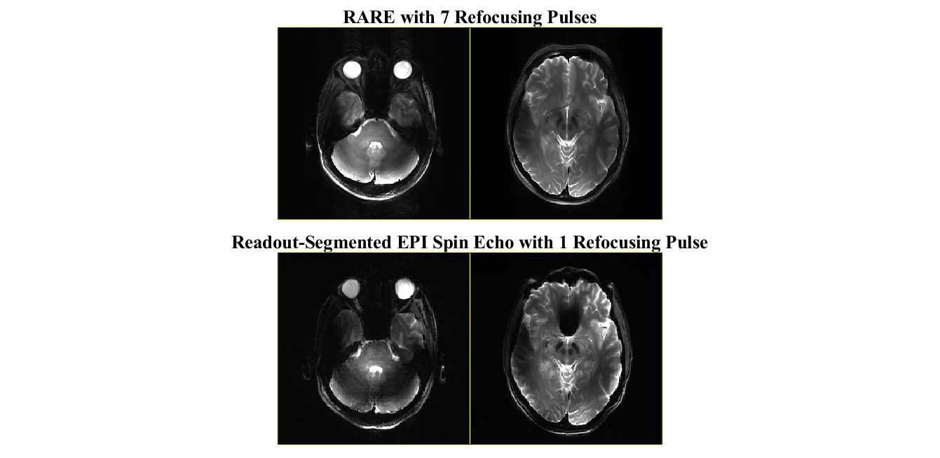
A 2D readout-segmented, diffusion-weighted sequence (Siemens’ RESOLVE [10]) was chosen for investigating slice-by-slice shimming. Compared to other spin-echo-based sequences, such as RARE [11] (Siemens’ TSE), readout-segmented EPI produces images with an increased sensitivity to B1+ homogeneity, despite only using one large-flip-angle refocusing pulse for imaging (example comparison shown in Figure 1). The readout-segmented, diffusion-weighted sequence was modified to include dynamic slice-selective pTx waveforms for a 7T scanner (MAGNETOM Terra, Siemens, Erlangen, Germany).A set of multi-channel B1+ maps was acquired in a healthy subject on the 7T scanner with a presaturation TurboFLASH sequence [12] on a self-built 8Tx/32Rx head coil [13]. Five axial slices were chosen for slice-by-slice shimming, each separated by 2.5 cm. First, a volumetric shim was performed on the five slices using a custom magnitude least-squares [5] optimization with a flip-angle standard-deviation constraint of 25° and constraints on local SAR. This volumetric shim generated relative magnitude and phase configurations for each transmit channel that could be broadly applied to all excitation and refocusing pulses in the sequence. Next, the same shimming optimization was applied on a slice-by-slice basis for all five slices. In this case, each excitation and refocusing pulse were shimmed individually for each slice (10 pTx waveforms in total). These waveforms were each inserted into the readout-segmented diffusion-weighted sequence for their respective slice location. The time to design and generate all 10 pTx waveforms was under one minute on a standard laptop computer (Intel(R)Core(TM)i5-8250U CPU) using MATLAB 2019b (Mathworks, MA, USA).
Three multi-slice 2D scans were performed in the healthy subject using the 8Tx/32Rx head coil (Siemens’ RESOLVE:TE/TR/FOV/matrix/slice thickness=58ms/4890ms/229cm2/224x224/5mm): one without any shimming (i.e., single-transmit equivalent), one with the volumetric B1+ shim, and one with the slice-by-slice B1+ shim waveforms. In all cases, the nominal/target flip angle for excitation was 90° and for refocusing, 180°. In these preliminary measurements, only the b=0 (non-diffusion-weighted) image was collected.
Results
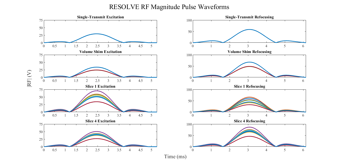
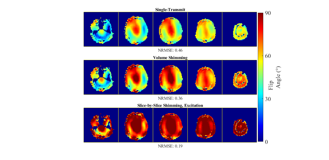
Figure 2 plots a representative sample of RF magnitude waveforms for the spin-echo excitation and refocusing pulses used in the three sets of acquisitions. The single-transmit and volumetric-shimming waveforms are scaled by the scanner for a nominal flip angle of 90° and 180° using a calibration reference voltage, while the slice-by-slice shimming waveforms are scaled during optimization directly with the subject-specific B1+ maps.Figure 3 shows the simulated excitation flip-angle maps for single-transmit, volumetric-shimming, and slice-by-slice shimming across all five slices used in the acquisition.
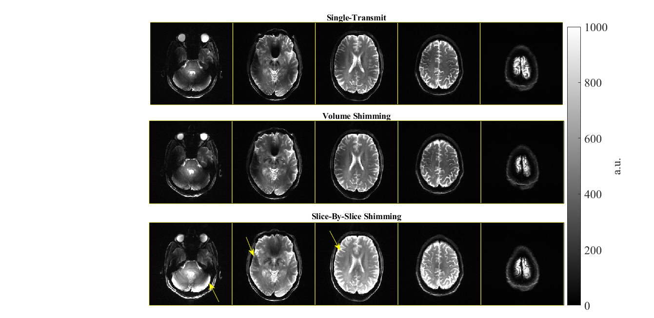
Figure 4 shows the experimental images for the healthy subject with single-transmit, volumetric-shimming, and slice-by-slice shimming, where the measured local SAR was 0.26, 0.25, 0.62 W/kg, respectively. Despite more than a two-fold increase in local SAR for slice-by-slice shimming, the overall estimated SAR is well below normal operating mode limits of 10 W/kg.
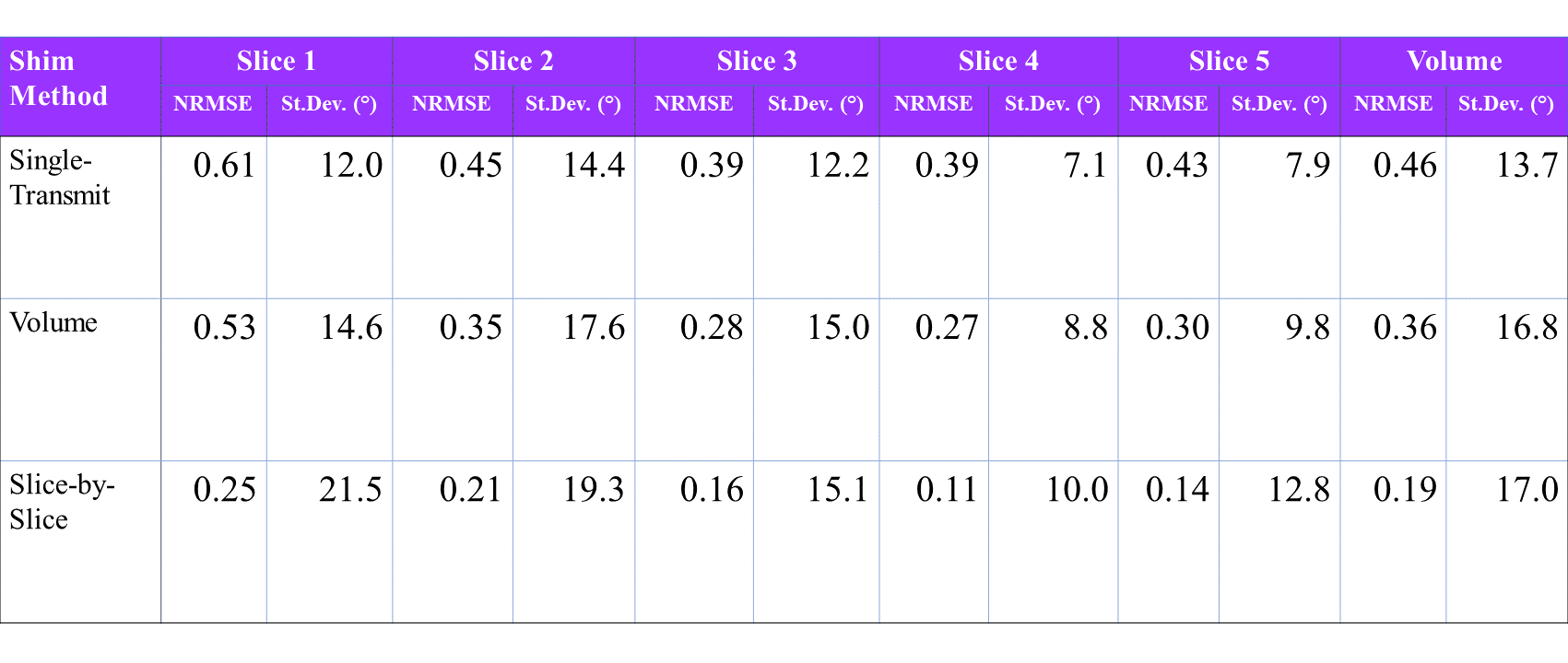
Figure 5 summarizes the performance using the normalized root mean squared error (NRMSE) and standard deviation of the flip-angle for the three scans on a slice-by-slice and full-volume basis.
Discussion & Conclusion
In this work we present an implementation of slice-by-slice shimming pTx in a 2D readout-segmented diffusion-weighted sequence, chosen for its notoriously poor B1+ homogeneity at 7T. This method is simple and efficient, which can lead to easy adoption in routine clinical MRI.The image-quality improvements shown in this preliminary study provide motivation for future work. First, a full simulation of the readout-segmented diffusion-weighted spin-echo sequence could help bridge the connection between the presented excitation simulations and spin-echo experimental data. Secondly, as slice-by-slice design time was minimal in this 5-slice experiment, more slices with thinner slice thickness could be implemented. Additionally, the slice-by-slice shimming optimizations could also be tailored to have slice-varying constraints on flip-angle standard-deviation based on the overall heterogeneity of the particular slice’s B1+ map. Finally, slice-by-slice shimming could be compared to full 2D pTx with spokes. Interestingly, it is possible that the best way to balance B1+ homogenization and the challenges of online 2D pTx pulse design is a hybrid of both techniques.
Acknowledgements
The authors would like to acknowledge Dr. Shajan Gunamony, Dr. Paul McElhinney, Dr. Sarah Allwood-Spiers for their work in designing, building, and validating the self-built pTx coil used in this investigation. We also thank Jürgen Herrler for his help with abstract revision.References
[1] T.S. Ibrahim et al., MRI, 2000. [2] U. Katscher et al., MRM, 2003.[3] V. Gras et al., MRM 2017.[4] J. Herrler et al., MRM 2020. [5] K. Setsompop et al., MRM 2008. [6] B. Guérin et al., MRM 2015.[7] B. Guérin et al., ISMRM 2020. [8] C. Le Ster et al., PLOS ONE 2019. [9] L. Hayes et al., MAGNETOM Flash 2014 [10] J. Hennig et al., MRM 1986. [11] A. Curtis et al., MRM, 2012. [12] S. Chung et al., MRM, 2010. [13] G. Shajan et al., ISMRM 2019.
Figures