4171
Can we achieve a better performance in metamaterial-assisted MRI combined to an SLR-based fast spin-echo sequence?1Department of Physics and Engineering, ITMO University, Saint Petersburg, Russian Federation, 2Aix-Marseille Universite, CNRS, Centre de Résonance Magnétique Biologique et Médicale, Marseille, France, 3Center of Diagnostics and Telemedicine Technologies, Department of Health Care of Moscow, Research and Practical Clinical, Moscow, Russian Federation
Synopsis
There has been previously proved that a metamaterial-based wireless coil (WLC) provided a 48-fold lower SAR in 1.5T wrist MRI in comparison to conventional RF setup. This can allow to extend the acceptable threshold of energy deposition for SAR-demanding pulse sequences. This study demonstrates that SLR-based FSE together with the WLC allowed to increase the slice selectivity while still being within the safe SAR limits. The actual energy deposition was decreased as compared to a conventional RF setup. The combination of this coil and SLR-based FSE offers an interesting alternative for investigations which require scanning in a “Low SAR” regim.
Introduction
Metamaterials-based radiofrequency (RF) devices have been reported to allow concentrating the RF magnetic field in an area of interest while shifting the electric field away from the patient’s body during the RF pulse transmission in magnetic resonance imaging (MRI)[1]. The specific absorption rate (SAR) of electromagnetic energy, has been proved to be 48 times lower for wrist imaging at 1.5T in comparison to conventional RF setups[2]. Even at the lowest conventional magnetic field, i.e 1.5T, the SAR aspect limits the utilization of high amplitude RF pulses, which may allow obtaining high precision of slice selection, robust saturation, efficient fat suppression, etc. Metamaterial-based devices may provide an opportunity to extend the acceptable threshold of RF energy deposition for such pulse sequences.The aim of the present study was to investigate the possibility of using highly-selective pulses, within the current RF safety limits thanks to the use of wireless metamaterial-based coil (WLC)[2]. A fast spin-echo (FSE) pulse sequence with clinical parameters and integrating highly-selective pulses computed using the Shinnar–Le Roux (SLR) algorithm[3] was numerically simulated. It was tested in a phantom experiment using the WLC.
Methods
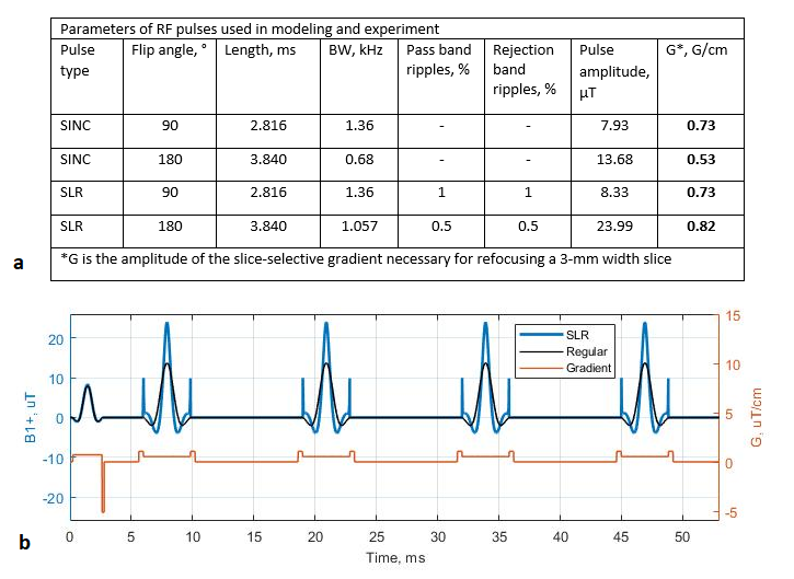
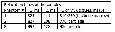
A “Bloch Equations Simulator”[4] was used for modeling. Spatial profiles of the transverse magnetization component Mxy, were considered as slice profiles. Apodized SINC pulses commonly used in clinical FSE sequences in “Low SAR” mode on Siemens Magnetom Avanto were used as a reference. The SLR pulses were constructed in the MATPULSE program[5]. The durations of the SLR pulses were similar to the ones of corresponding reference pulses in order to keep the echo times. Particular attention was paid to the optimization of the refocusing pulse profile. To increase the time-bandwidth product (TBW), i.e. the selectivity, the frequency bandwidth (BW) and ripples percentage of the 180° SLR pulse were optimized.The slice selection in T1-weighted FSE pulse sequence from Siemens wrist imaging protocol was modeled (parameters are listed in the capture to Fig.1). We assessed the signal evolution in three samples with different relaxation times (Fig.2). T1 times were close to the ones reported in musculoskeletal tissues at 1.5T[6]. The selectivity of the pulses was estimated for each echo through the ratio between the signal (S) accumulated within the desired slice thickness (TH) and the total signal (S1) accumulated within a full width of the main lobe together with the first side lobes of the slice profile (TH1).
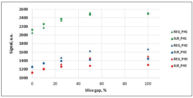
The SLR pulses were implemented in the software of the Siemens Magnetom Avanto scanner. The FSE pulse sequence was tested in "Low SAR" mode. Three calibrated phantoms (Eurospin II) with T1 times as in modeling were positioned in the center of the WLC. The images were obtained in a multi-slice mode (7 slices) with different gaps between slices of 100%, 50%, 25%, 10%, and 0% in order to investigate the “slice cross-talk” effect, which reduces the signal intensity when the slice profile is far from rectangular. The average signal intensity was measured in each phantom in a medial slice of each multi-slice set.
Results and Discussion
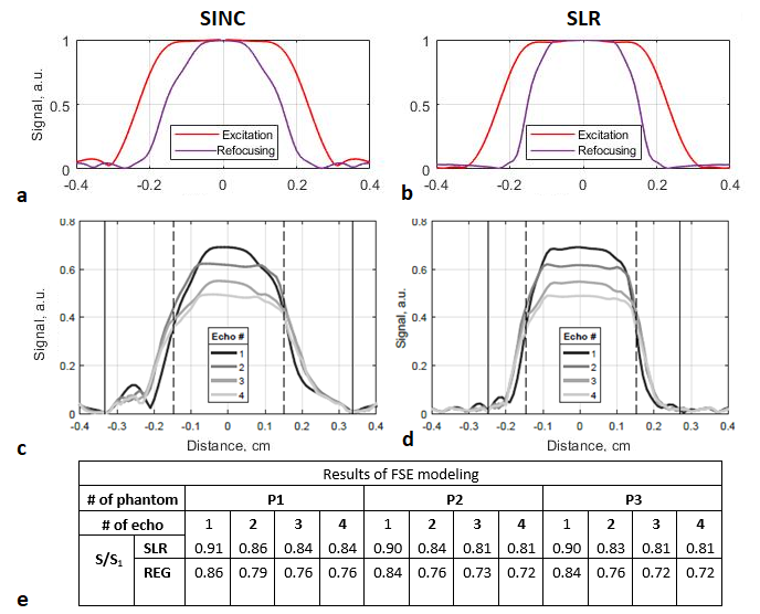
The RF and gradient pulses parameters used for the modeling are summarized in Fig.1(a). The BW of the refocusing SLR pulse was extended by a factor of 1.55 as compared to the regular pulse thereby allowing the TBW to be increased to 4. All excitation and refocusing profiles are illustrated in Fig.3(a,b).For the FSE, time-dependent RF magnetic field and slice selection gradient amplitudes that were generated are displayed in Fig.1(b). For a whole pulse sequence cycle, the energy deposition was 1.74-fold larger, using SLR pulses.
Let us consider the initial SAR generated in a duty cycle of a regular FSE with a conventional RF setup as SAR1. It was previously reported that with the introduction of the WLC to a conventional setup, a 48-fold SAR reduction can be observed[2] i.e. SAR2=SAR1/48. Thus, combining the WLC utilization with an SLR-based FSE provides a 1.74-fold increase of energy deposition (SAR2) i.e. SAR3=SAR2*1.74. On that basis, the latter setup still provides 28 times lower energy deposition i.e. SAR3=SAR1/27.59 as compared to the conventional situation.
Examples of the slice profiles (for sample P1) for the FSE sequence are displayed in Fig.3(c,d). The corresponding selectivity metrics for each echo were always higher for SLR pulses (Fig.3(e)). Signal intensities measured in phantoms are summarized in Fig.4. The “slice cross-talk” effect reduced the signal intensity by 13%-15% in the SLR-based sequence when the slice gap was decreased from 100% to 0% (Fig.4). At the same time, this signal reduction was significantly higher (19%-24%) for the regular pulse sequence. This illustrated that the “slice cross-talk” effect was reduced in SLR-based FSE and confirmed the increased RF pulses selectivity shown in the simulation.
Conclusions
The present study demonstrated that SLR-based FSE sequences combined with the use of a WLC allowed increasing the slice selectivity while still being within the safe SAR limits. In addition, the actual energy deposition was decreased as compared to a conventional RF setup. The combination of WLC and SLR-based FSE offers an interesting alternative for MR investigations that require scanning in a “Low SAR” regime such as those for children, pregnant women, and patients with implanted devices.Acknowledgements
This work was supported by the Russian Science Foundation (Grant No. 18-79-10167). E. Brui acknowledges the 'Ostrogradski' program of the Embassy of France in Russia.References
[1] Shchelokova A, et al. 2016 IEEE International Symposium on Antennas and Propagation (APSURSI), 2016:1397-1398
[2] Shchelokova A, et al. Magn Reson Med. 2018;80(4):1726-1737
[3] Shinnar M, et al. Magn. Reson. Med.1989;12:75-87
[4] Hargreaves B A, et al. Magn Reson Med. 2001;46(1):149-158
[5] Matson G B, Magn. Reson. Imaging.1994;12(8):1205-25
[6] Kato H, et al. Med Phys. 2005;32(10):3199-208
Figures