4164
Type and Time of Dialysis Are Independent Indicators for Carotid Atherosclerosis in End-stage Renal Disease Patients on Dialysis1Center for Biomedical Imaging Research, Medical School, Tsinghua University, Beijing, China, 2The Central Hospital of Zhejiang Lishui, Lishui, China
Synopsis
In this work, the vessel wall characteristics of carotid artery was measured on T1w, T2w and SNAP images in end-stage renal disease patients on dialysis. Totally, 94 patients were included. The time on dialysis was significantly and positively correlated with the mean wall area (p=0.012), normalized wall index (p=0.006), maximal wall thickness (p=0.005) and mean wall thickness (p=0.010). The presence of plaque was found to be significantly and independently associated with the dialysis type (p=0.047) and time on dialysis (p=0.032).
Synopsis
In this work, the vessel wallcharacteristics of carotid artery was measured on T1w, T2w and SNAP images in end-stage
renal disease patients on dialysis. Totally, 94 patients were included. The
time on dialysis was significantly and positively correlated with the mean wall
area (p=0.012), normalized wall index (p=0.006), maximal wall thickness
(p=0.005) and mean wall thickness (p=0.010). The presence of plaque was found
to be significantly and independently associated with the dialysis type (p=0.047)
and time on dialysis (p=0.032).
Introduction
Cardiovascular disease was the main causeof mortality in dialysis patients, and rapid progression of atherosclerosis was
found in end-stage renal patients. [1-3] In several previous studies, ultrasound
measurements of the intima-media thickness (IMT) of the carotid arteries were used
as an indicator of carotid atherosclerosis. [4-6] Recently, high resolution black-blood
MRI was used to evaluate carotid atherosclerosis which can provide more
characteristics of atherosclerotic plaque [7-9]. However, to the best of our
knowledge, there was no study using MRI to study vessel wall characteristics on
end-stage renal patients.
Therefore, this work aimed to investigate
the carotid vessel wall characteristics such as wall thickness, area, maximal
wall thickness and normalized wall index (NWI), which were measured by MRI on end-stage
renal patients on dialysis.
Methods
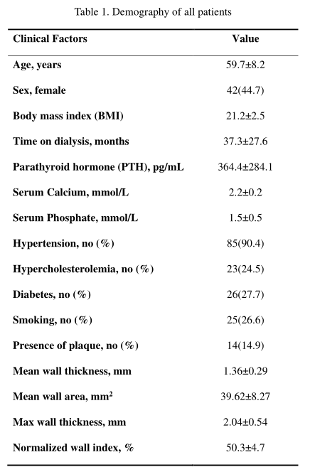
Totally, 94 patients (42 women; medianage: 59.5 years; range: 51-82 years) were recruited with institutional review
board approval and all the signed consent form acquired.
The inclusion criteria included: age greater than 50 years, with end-stage
renal disease, on dialysis, and without any contraindication for MRI scan. The patient
clinical parameters were collected, including age, gender, body mass index
(BMI), type of dialysis, time on dialysis, parathyroid hormone (PTH), serum
Calcium, serum Phosphate, hypertension, hypercholesterolemia, diabetes and
smoking.
All the patients were imaged on a Philips
3.0T MR scanner (Achieva; Philips, Best, the Netherlands)
with a customized carotid coil. The MR imaging protocol included: T1W-VISTA, TR/TE=600/30ms,
flip angle=90°, T2W-VISTA, TR/TE=1300/260ms, flip angle=90°; SNAP [10], TR/TE=10.4/5ms,
flip angle=11°. The following parameters are the same for these three
sequences: slice thickness=0.8mm, field of view=250x250mm2, reconstruction
resolution=0.39x0.39mm2, imaging direction: coronal.
MR images were re-sliced into 48 cross-sectional
slices (re-slicing slice thickness=1mm, in-plane resolution=0.4mmx0.4mm)
centered at carotid artery bifurcation. One experienced radiologist delineated
the carotid artery lumen and outer wall contours on T1W-VISTA images with SNAP
and T2W-VISTA images as references in CASCADE [11] software. Then the mean wall
thickness, mean wall area, maximal wall thickness and mean normalized wall
index (wall area / [lumen area +wall area] x 100%) were calculated based on
slice-wise results for each patient. The prevalence of atherosclerotic plaque
was defined as the minimal wall thickness larger than 2 mm.
Pearson correlation, t-test
and logistic regression were used to evaluate the association between the
clinical parameters and the carotid vessel wall imaging parameters.
Results
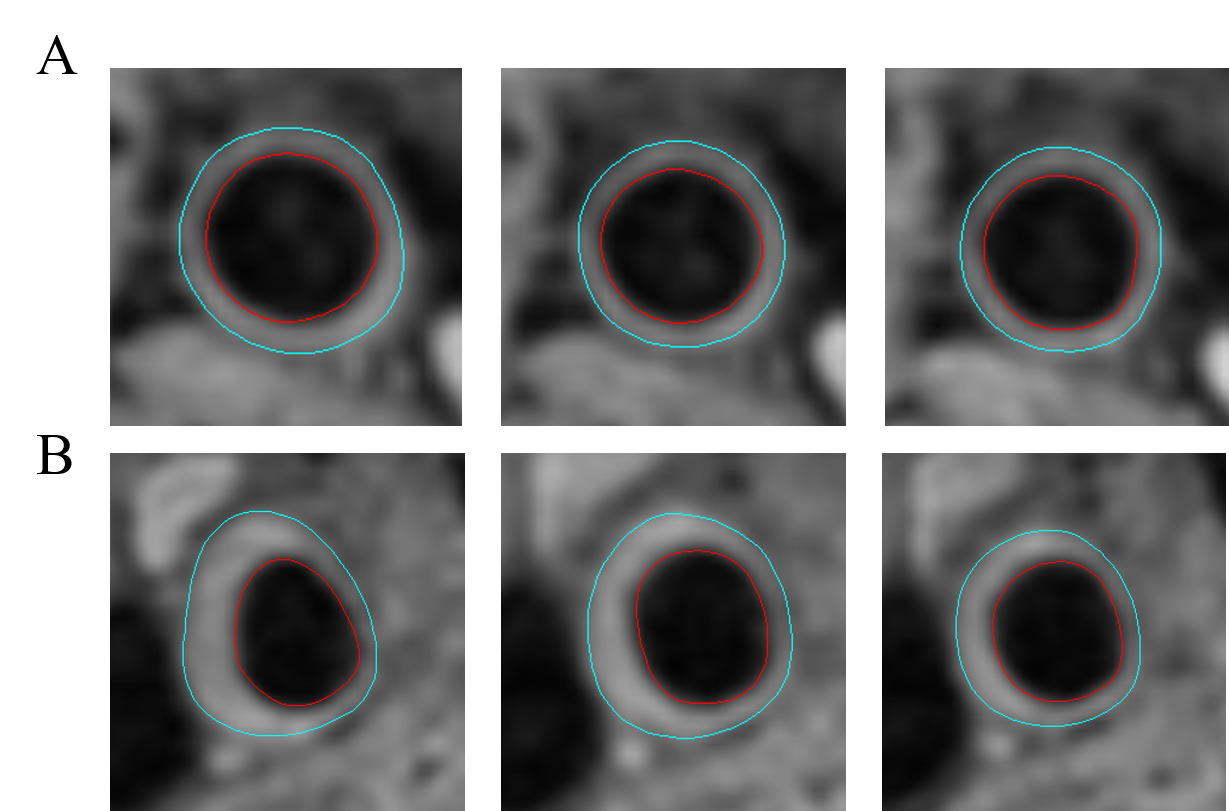
Figure 1A and B showed the carotid vessel wall images of an example patient (58-year-old, male) with long time on dialysis (62 months) and an example patient (62-year-old, male) with short time on dialysis (6 months). An atherosclerotic plaque can be clearly seen on the images of the patient with long time on dialysis, while the patient with shorter time on dialysis have relative thin carotid vessel wall.Table 1 showed the demography
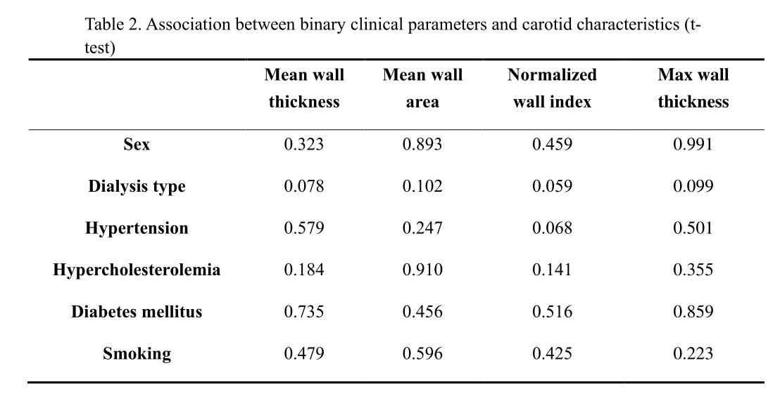
and the imaging parameters of patients. Table 2 shows the t-test between the clinical
parameters and carotid vessel wall characteristics. No statistical significance
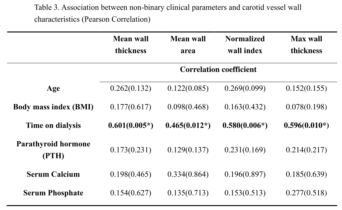
was found. Table 3 shows the result of Pearson correlation between non-binary
clinical parameters and carotid vessel wall characteristics. The time on
dialysis was significantly and positively correlated with the mean wall area
(p=0.012), NWI (p=0.006), maximal wall thickness (p=0.005), and mean wall
thickness (p=0.010).
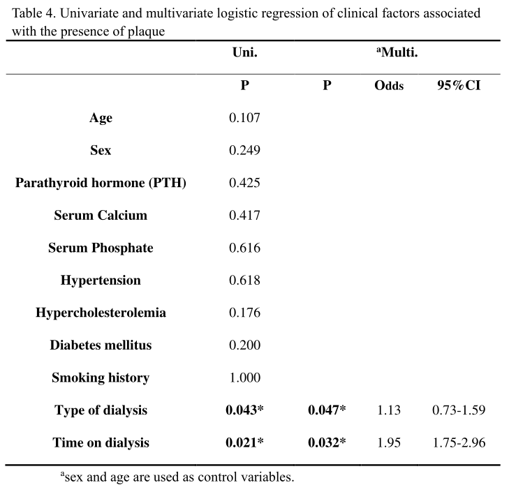
The univariate and multivariate analysis
of clinical factors associated with the presence of plaque were shown in Table
4. In univariate analysis, the type of dialysis (p=0.043) and time on dialysis
(p=0.021) were found to be significantly associated with the presence of
plaque. In multivariate analysis with age and sex as control variables (Model
1), the type of dialysis (p=0.047) and time on dialysis (p=0.032) were still
significantly associated with the presence of plaque, indicating they may be independent
risk factors of the atherosclerosis.
Discussion and Conclusion
This study found that the time on dialysiswas significantly and positively correlated with the mean wall area, normalized
wall index, maximal wall thickness and mean wall thickness. The presence of
plaque was also found to be significantly correlated with type of dialysis and
time on dialysis. After the control of age and sex the correlation remained
significant, which indicated the time and type of dialysis may be an independent
indicator of the atherosclerosis.
Acknowledgements
NoneReferences
1. Amann K, Tyralla K, Gross ML, Eifert T, Adamczak M, Ritz E. Spcial characteristics of atherosclerosis in chronic renal failure. Clin. Nephrol. 2003;60(1):13-21.
2. Stenvinkel P. Inflammation in end-stage renal disease: a fire that burns withn. Contrib Nephrol. 2005;149(5):185-199.
3. Kaysen G A. Association between Inflammation and Malnutrition as Risk Factors of Cardiovascular Disease. Blood Purif. 2006;24(1):51-55.
4. Fabris F , Zanocchi M , Bo M , et al. Carotid plaque, aging, and risk factors. A study of 457 subjects[J]. Stroke, 1994, 25(6):1133-1140.
5. Hojs R . Carotid Intima‐Media Thickness and Plaques in Hemodialysis Patients[J]. Artificial Organs, 2000, 24(9).
6. Sato M , Ogawa T , Sugimoto H , et al. Relation of Carotid Intima-Media Thickness and Silent Cerebral Infarction to Cardiovascular Events and All-Cause Mortality in Chronic Hemodialysis Patients[J]. Internal Medicine, 2012, 51(16):2111-2117.
7. Moody AR, Murphy RE, Morgan PS, et al. Characterization of complicated carotid plaque with magnetic resonance direct thrombus imaging in patients with cerebral ischemia. Circulation. 2003;107(24):3047–3052.
8. Zhu DC, Ferguson MS, DeMarco JK. An optimized 3D inversion recovery prepared fast spoiled gradient recalled sequence for carotid plaque hemorrhage imaging at 3.0 T. Magn Reson Imaging. 2008;26(10):1360–1366.
9. McNally JS, Kim SE, Yoon HC, et al. Carotid magnetization-prepared rapid acquisition with gradient-echo signal is associated with acute territorial cerebral ischemic events detected by diffusion-weighted MRI. Circ Cardiovasc Imaging. 2012; 5(3):376–382.
10. Takemoto K, Takano K, Abe H, Okawa M, Iwaasa M, Higashi T, et al. The new MRI modalities “BPAS and VISTA” for the diagnosis of VA dissection. Acta Neurochir Suppl. 2011;112:59–65.
11. D Xu, WS Kerwin, T Saam, M Ferguson, and C Yuan. Cascade: Computer aided system for cardiovascular disease evaluation. ISMRM. 2004:1922.
Figures