4161
Evaluation of registration accuracy for cerebral vessel on pre- and postcontrast T1 black blood images by Elastix1Center for Biomedical Image Research, Department of Medicine, Tsinghua University, Beijing, China
Synopsis
Image registration plays a prominent role in medical image processing pipeline1. It’s of interest to assess pre-post variation for registered vessels on black blood MR images. In this research, Elastix tool was applied to register pre-contrast T1 and post-contrast T1 black blood images, use our own methods to actually and accurately evaluate the accuracy of the registration, and achieved promising quantitative results. The study suggests that Elastix performs good registration accuracy of the two images, and can be directly used for automatic image processing, so as to more conveniently serve clinical applications.
Introduction
Recently, multi contrast magnetic resonance(MR) vessel wall imaging techniques are increasingly applied to identify and characterize cerebral atherosclerotic lesions2. Tedious manual operations are required to register the multi contrast data. However, the state-of-art tools(e.g., Elastix) are designed to register the whole brain tissue, which lacks the evaluation metric of registration accuracy of vessels3.It is of significance to investigate the registration performance of Elastix for major cerebral vessels on pre- and postcontrast MR images.Methods
MR Acquisition: MRI acquisitions were performed on a 3 T MRI scanner (Discovery 750, GE MEDICAL SYSTEMS). Pre- and post-contrast T1 TSE , were acquired in the coronal position with the same protocols: Matrix: 512512192 pixels, resolution of 0.4492mm*0.4492 mm*0.6mm. FA=90, TR/TE=800ms/15.2ms, echo train length=30.Image process: A total of 20 patients with unilateral symptomatic MCA infarction were included in the study. All analysis were conducted with Slicer(Elastix) 4.11.0 for registration and Mimics Medical 17.0 for segmentation. The processing procedures were divided into two related parts:
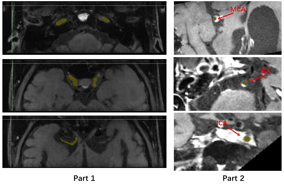
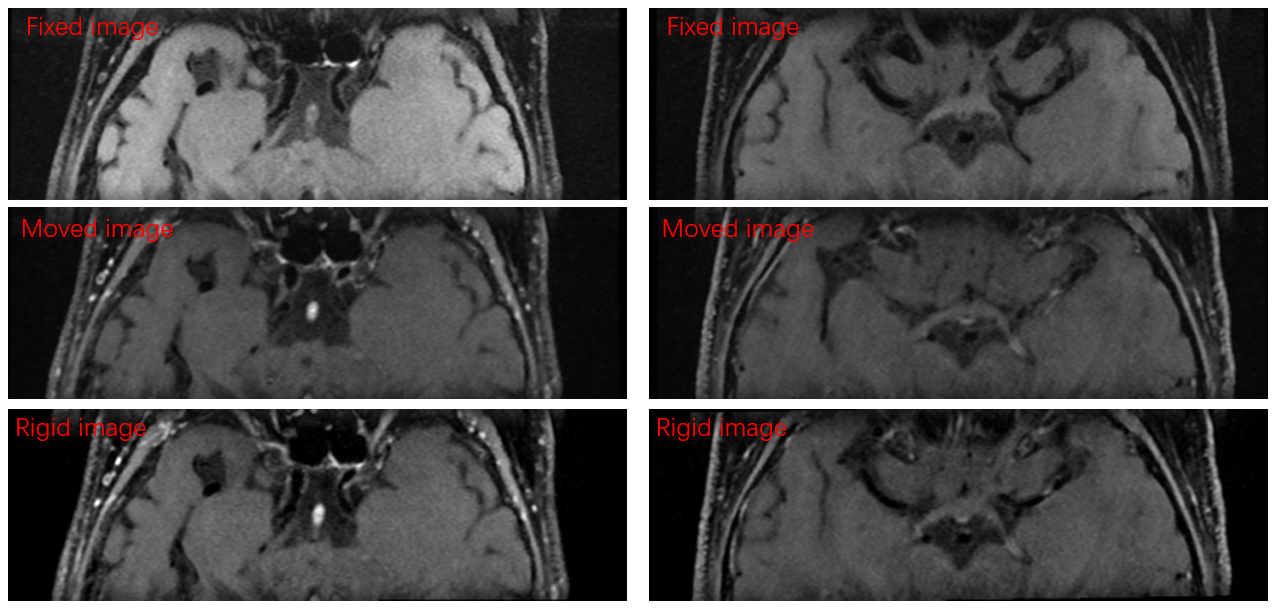
For part 1, “Generic” and “Rigid” registration between pre- and postcontrast T1 were performed. Pre- and postcontrast images are used as fixed image and moved image respectively. Four images dataset (original pre-T1, original post-T1, generically-registered post-T1, and rigidly-registered post-T1) were included in evaluation. For each patient, 3 slices from different positions were chosen to manually delineate lumen areas for all images.A example diagram of the selected areas is shown in Figure 1. Dice Similarity Coefficient of lumen mask was utilized to assess the registration accuracy. Intra-observer and inter-observer analysis were used to evaluate the reproducibility of the study.
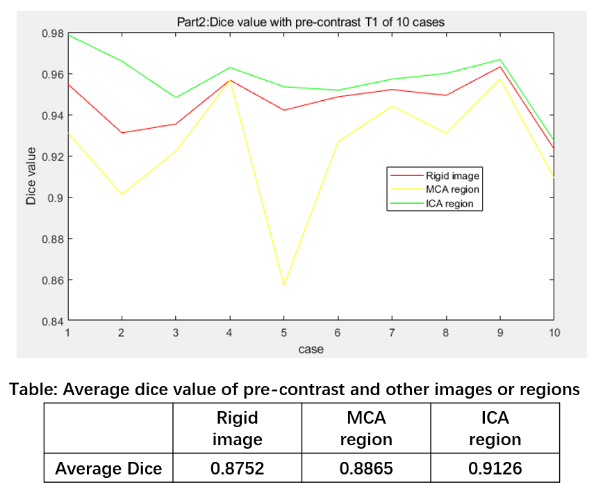
For part 2, Multiplanar reconstruction(MPR) method was used to select the axial sections of middle cerebral arteries (MCAs), internal carotid arteries (ICAs) and basilar artery (BA), 5 reconstructed planes were chosen for each patient. A example diagram of the selected areas is shown in Figure 1. 10 cases were included in part 2. Subsequent analysis were just the same as part 1.
Results
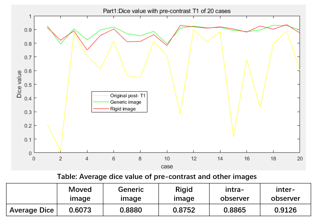
For part 1, we get the registration accuracy of blood vessel measured by the dice value after the pre-contrast T1 and post-contrast T1 brain images are registered by Elastix. The registration results are shown in figure 2. The moved images can be registered to fixed images. The quantitative results are shown in Figure 3, including the dice value with pre-contrast image of postcontrast image, Generic image and Rigid image. For part 2, the registration accuracy of Rigid image and the axial slices of the MCAs and ICAs is shown in Figure 4. The registration accuracy of MCAs is slightly worse than the accuracy of the whole and ICAs due to their small area.Discussion and Conclusion
According to the quantitative study results, we can draw these conclusions: This dice value of about 90% means that the Generic and Rigid registration both have good accuracy and there was no significant difference within intra-observer and inter-observer measurements. Good registration of MCAs, ICAs and BA is helpful for further automatic information mining from pre- and postcontrast images and related clinical research. Rigid registration requires significantly less time than Generic registration and the accuracy of the two is similar, so rigid registration of Elastix can meet our registration requirements for such data processing. However, the cases enrolled may not include very complex or poor-quality data, which may have a little impact on the generalization of study.Acknowledgements
No acknowledgement found.References
1. Alam F, Rahman SU, Din AU, Qayum F. Medical image registration: Classification, applications and issues. J Postgrad Med Inst 2018; 32(4): 330-7.
2. Kim JM, Jung KH, Sohn CH, Moon J, Shin JH, Park J, et al. Intracranial plaque enhancement from high resolution vessel wall magnetic resonance imaging predicts stroke recurrence. Int J Stroke. 2016;11:171–179.
3. Grant Haskins, Uwe Kruger, Pingkun Yan. Deep learning in medical image registration: a survey. Machine Vision and Applications 31, 13 (2020).
Figures