4152
Preliminary study on plaque characteristics of intracranial artery atherosclerotic stroke in young adults1Department of MRI, Shaanxi Provincial People's Hospital, Xi'an, China, 2Philips Healthcare, Xi'an, China
Synopsis
The aim of this study was to investigate plaque characteristics and clinical risk factors of intracranial atherosclerotic stroke in young adults. The vessel wall characteristics of plaque and clinical data were compared between young and old patients. Our results showed that maximum vessel wall thickness, narrowest vascular area and vessel wall area of young patients were significantly lower than those of old patients, while the number of young patients with plaque positive remodeling was higher. In addition, the number of young people suffering from hyperhomocysteinemia was significantly higher than that of old patients, while suffering from hypertension was significantly lower.
Introduction
Epidemiological evidence suggests that the incidence of intracranial artery atherosclerotic stroke in young adults has increased substantially [1]. MRI as a noninvasive, radiation free examination with high resolution and contrast plays a vital role in the qualitative and quantitative diagnosis of stroke [2]. Previous studies have analyzed the clinical risk factors and potential etiology of atherosclerotic stroke in young patients, but there are few studies on the characteristics of atherosclerotic plaque [3]. In this study, we intended to investigate the vascular wall characteristics of intracranial atherosclerotic stroke in young patients and to explore its underlying pathophysiological mechanisms.Methods
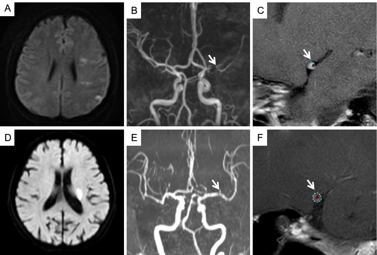
Sixty-two patients with intracranial large artery atherosclerotic stroke were included in this study. And they were divided into two groups: 26 young people (22 men; 38±8 years), 36 old people (29 men, 64±9 years). All MRI images were obtained by using a 3.0 T MR scanner (Ingenia CX, Philips Healthcare, the Netherlands) with a standard 32 channel head coil. The black blood imaging, T1WI Vista imaging were acquired as the following parameters: for black blood sequence, high resolution (HR) MRI scan 2 layers per sequence, TR/TE =2500/67 ms (T2WI), for T1WI, 700/14 ms; FOV 80 mm×80 mm, matrix 256×256, slice thickness =2 mm. The differences of vessel wall properties of plaque (maximum wall thickness (mm), vessel area(mm2), vessel fluid area and vessel wall area of narrowest level (mm2), plaque load, degree of stenosis, positive remodeling) and clinically relevant factors (smoking, hypertension, Coronary disease, diabetes mellitus, hyperlipidemia, hyperhomocysteinemia) in young and old atherosclerotic stroke patients were analyzed. All HRMRI images were measure by two radiologists blinded to the clinical data (Figure 1), both with over 10 years of experience. And the mean values of the above parameters measured by two radiologists for final statistical analysis. The qualitative data between groups were compared using the chi-square test or Fisher exact test, whereas quantitative data were compared using independent sample t test if continuous variables satisfy normal distribution, or Mann-Whitney U test if not. The intraclass correlation coefficient was used to find the and inter-observer reproducibility for the measurements of plaque characteristics.Results
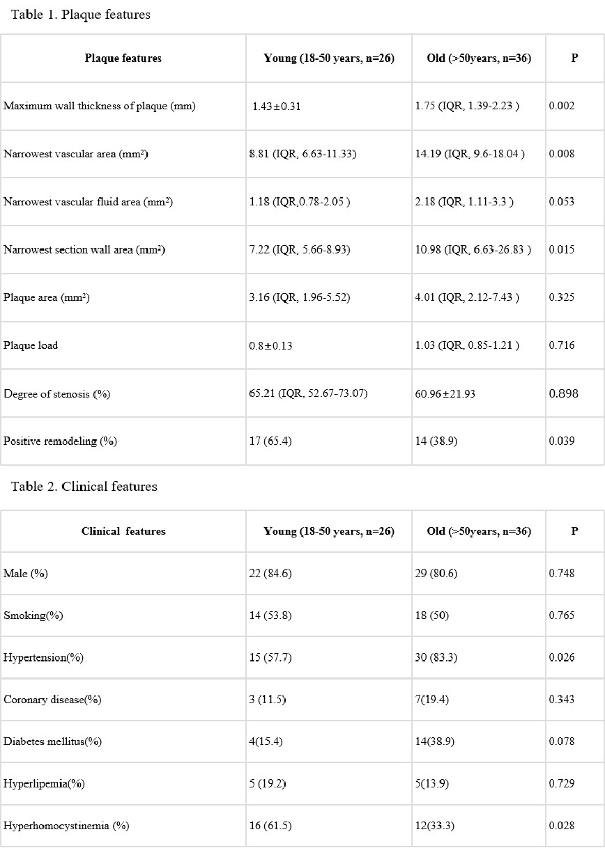
Measurement consistency between the two observers was good (r = 0.899, 95% confidence interval (0.859-0.988) ). The maximum wall thickness (mm), vessel area and vessel wall area of narrowest level (mm2) of young patients were significantly lower than those of old patients (1.43mm vs 1.75 mm, 8.81 mm2 vs 14.19 mm2, 7.22 mm2 vs 10.98 mm2, p < 0.05, Table 1). while the number of patients with positive remodeling in young patients was significantly higher than that in the old patients (65.4% vs 38.9%, p < 0.05, Table 1). In addition, the number of young people suffering from hyperhomocysteinemia was significantly higher than that of old patients (61.5% vs 33.3%, p < 0.05, Table 2), while suffering from hypertension was significantly lower.(57.7% vs 83.3%, p < 0.05, Table 2).Discussion
We concluded that there were differences in plaque characteristics between young and old atherosclerotic stroke patients through intracranial high resolution magnetic resonance imaging. And there were significant differences in some clinical factors between young and old patients. We found that maximum vessel wall thickness, vessel area and vessel wall area of narrowest level of young patients were significantly lower than those of old patients. In addition, there were more cases of hyperhomocysteinemia in young patients and more cases of hypertension in old patients. These results may indicate that plaque formation in young people may be closely related to hyperhomocysteinemia. The time of atherosclerotic plaque formation for young patients is relatively short, the development of plaque is still in the initial stage, more tend to positive remodeling. While atherosclerotic plaque formation in old patients may be closely related to the long-term chronic development of hypertension, and plaques take longer to form and develop slowly, which makes the plaque morphological characteristics more significant than that of the young patients. With the prolongation of time, the development of plaques was tends to negative remodeling when stroke occurred, which was consistent with the development process of plaque remodeling previously studied. In addition,these results also may suggest that young people are at greater risk of ischemic stroke in patients with hyperhomocysteine.Conclusion
This study is helpful to reveal the plaque characteristics and clinical susceptibility factors of young atherosclerotic stroke and to prevent the occurrence of intracranial atherosclerotic stroke in young people.Acknowledgements
No acknowledgement foundReferences
1. Ekker M S , Boot E M , Singhal A B , et al. Epidemiology, aetiology, and management of ischaemic stroke in young adults. The Lancet Neurology, 2018, 17(9):790-801.
2. Ahn S H , Lee J , Kim Y J , et al. Isolated MCA disease in patients without significant atherosclerotic risk factors: a high-resolution magnetic resonance imaging study. Stroke; a journal of cerebral circulation, 2015, 46(3):697-703.
3. Xu Y Y , Li M L , Gao S , et al. Etiology of intracranial stenosis in young patients: A high-resolution magnetic resonance imaging study. Annals of Translational Medicine, 2017, 5(16):319-319.
Figures