4143
Frequency Dependent Altered Functional Connectivity of Declarative Memory Networks in Patients with Dural Arterio-Venous Fistula1Imaging Sciences and Interventional Radiology, Sree Chitra Tirunal Institute for Medical Sciences and Technology, Thiruvananthapuram, India, 2Neurology, Sree Chitra Tirunal Institute for Medical Sciences and Technology, Thiruvananthapuram, India
Synopsis
Dural Arteriovenous fistula (dAVF) causes cognitive deficits and associated memory impairment. The objective of this study was to understand the frequency dependent correlates of resting state functional connectivity of memory deficit presentations in dAVF. Our results reveal reduced functional connectivity, in a frequency dependent fashion, among hippocampus, parahippocampal cortex and dorsolateral prefrontal cortex, i.e.: regions involved in declarative memory functional processing.
Introduction
Analysing the resting state functional connectivity (rsFC) of brain helps to understand the altered functionality of brain structures and is being widely used to understand the neural activity for a variety of pathological conditions. Analysis of rsFC among medial temporal lobe structures to understand memory impairment has never been investigated in dAVF patients, to the best of our knowledge1. The purpose of this study is to investigate the rsFC among medial temporal lobe regions for understanding memory impairments in dAVF by utilising frequency decomposition of BOLD signal to explore potential frequency dependency of neural activity of memory related structures in dAVF.Methods
Resting state Functional Magnetic Resonance Imaging (rsfMRI) analysis was performed in a total of 60 subjects. The data used in this study is from a cohort of 30 prospectively recruited dAVF patients (DP) from the outpatient clinic of department of Imaging Sciences and Interventional Radiology (IS&IR), Sree Chitra Tirunal Institute for Medical Sciences and Technology, Trivandrum, Kerala, India and 30 age matched healthy controls (HC).The Blood oxygen level dependent (BOLD) spectrum was band pass filtered in the range of 0.008-0.09 Hz (Full band – FB). It was decomposed to four narrow frequency bands FB1(0.008-0.028)Hz, FB2(0.028-0.049)Hz, FB3(0.049-0.069)Hz, FB4(0.069-0.09)Hz to investigate any frequency modulations of rsFC in DP compared to HC. The fMRI Region of Interest ROI-ROI analysis was performed using CONN toolbox2. The smaller frequency bands were individually band pass filtered. Medial temporal lobe structures - Hippocampus and Parahippocampal cortex and dorsolateral prefrontal regions were chosen for analysis based on their memory functional role reported in literature studies3, 4. Neuropsychological assessments: Rey Auditory Verbal Learning test, Wechsler Memory Scale (WMS) verbal visual memory subsets and WMS paired associate learning test has been done in both groups to assess their memory functions. A two sample t test gave significant group difference (p<0.01) for all these tests with lower scores in DP.Results
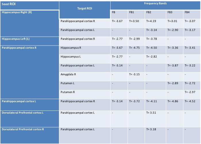
The rsFC among right hippocampus, right posterior parahippocampal cortex, left posterior parahippocampal cortex , left and right dorsolateral prefrontal areas were significantly altered in DP than HC (pFDR <0.01).We observed that altered functional connectivity was frequency dependent. A summary of significantly altered (pFDR <0.01) rsFC and its frequency dependency for DP>HC are shown in Table1.Discussion
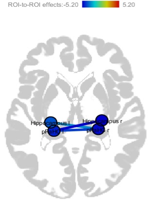
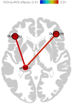
Several Frequency dependent fMRI analysis works have been conducted in recent years in various pathological conditions, also inspired by contributions of electrophysiological studies that reported the frequency specific neural oscillations in the brain and the altered functional connectivity patterns could be distinct to specific frequency bands5,6. Our work gave insights on the reduced functional connectivity of hippocampus and posterior parahippocampal cortex (pFDR<0.001) shown in Fig 1, suggesting impaired memory encoding and retrieval functions7.The increased connectivity of left (pFDR = 0.0008) and right (pFDR=0.0023) dorsolateral prefrontal cortex with left parahippocampal cortex shown in fig 2, found exclusively in FB 2 could be due to impairment in working memory8 contributing to maladaptive functioning of dAVF brain. Our findings with frequency decomposition based rsfMRI analysis demonstrate that frequency analysis enables to capture frequency dependent minute BOLD signal changes which in turn help understand the altered functioning of brain regions in dAVF. This work reports the frequency dependent functional connectivity of memory related structures in DP which are altered and some contributing to exclusive frequency ranges. Right hippocampus- right parahippocampal cortex altered connectivity is visible at all the frequency bands.Conclusion
Results of this study suggest that neural activity visualized through resting state BOLD signal could function at several frequencies and some functions could even be distinct to particular frequency bands. Furthermore, it is beneficial to incorporate frequency decomposition of BOLD signal of rsfMRI, along with the full frequency band analysis to fathom the pathologically altered brain neural function estimation better.Acknowledgements
The authors would like to acknowledge the financial support for this study by the following projects:
TDF project 6107/ 2018, SCTIMST, Department of Science and Technology, Govt: of India
DST/CSRI/2018/11, Department of Science and Technology, Govt: of India
References
1. Brito A, Tsang ACO, Hilditch C, et al. Intracranial Dural Arteriovenous Fistula as a Reversible Cause of Dementia: Case Series and Literature Review. World Neurosurg. 2019;121:e543-e553.
2. Whitfield-Gabrieli S, Nieto-Castanon A. Conn: a functional connectivity toolbox for correlated and anticorrelated brain networks. Brain Connect. 2012;2(3):125-41.
3. Thompson RF, Kim JJ. Memory systems in the brain and localization of a memory. Proc Natl Acad Sci U S A. 1996;93(24):13438-44.
4. Funahashi S. Working Memory in the Prefrontal Cortex. Brain Sci. 2017;7(5):49.
5. Penttonen M, Buzsa´ki G. Natural logarithmic relationship between brain oscillators. Thalamus & Related Systems. 2003;2: 145–152
6. Zuo XN, Di Martino A, Kelly C, et al. The oscillating brain: complex and reliable. Neuroimage. 2010;49(2):1432-45.
7. van Strien NM, Cappaert NL, Witter MP. The anatomy of memory: an interactive overview of the parahippocampal-hippocampal network. Nat Rev Neurosci. 2009;10(4):272-82.
8. Braver TS, Barch DM, Kelley WM,et.al. Direct comparison of prefrontal cortex regions engaged by working and long-term memory tasks. Neuroimage. 2001;14(1):48-59.
Figures