4130
Comparing venous diameter from SWI with ground-truth venous diameter in straight sinus
Mehdi Zoghinia1, Mohammed Ayoub Alaoui Mhamdi1, and Russell Butler1,2
1Bishops university, sherbrooke, QC, Canada, 2Diagnostic Radiology, University of Sherbrooke, sherbrooke, QC, Canada
1Bishops university, sherbrooke, QC, Canada, 2Diagnostic Radiology, University of Sherbrooke, sherbrooke, QC, Canada
Synopsis
Paramagnetic deoxyhemoglobin introduces magnetic susceptibility to venous vessels in the brain, causing intra-voxel dephasing of surrounding tissue, appearing dark on a susceptibility-weighted image (SWI). However, empirical studies quantifying the extent of this dephasing is lacking. Using time-of-flight (TOF) angiogram as ground truth, and focusing on the straight sinus, we quantified the extent to which SWI vessel diameters deviated from true (TOF) vessel diameters. We find straight sinus diameter based on SWI segmentation to be nearly twice the diameter of TOF segmentation, and the difference between SWI and TOF straight sinus diameter was dependent on vessel location in the y-axis.
Introduction
As early as 1990 Ogawa et al demonstrated blood-oxygenation sensitive contrast in gradient-echo images, showing that when blood oxygenation level is lowered, venous vessels appear more prominently (Ogawa et al. 1990). This is the origin of susceptibility-weighted imaging (SWI) a popular technique for quantifying brain venous vasculature (Haacke et al. 2004). Recent results (Halefoglu and Yousem 2018) indicate that venous vasculature may be implicated in disorders such as multiple sclerosis and Alzheimer’s, and venous diameter in large vessels (such as straight sinus) may emerge as key biomarkers for these (and other disorders). One problem with using SWI images to quantify venous diameter is that the dephasing effect depends on both orientation and diameter of the vessel (Buxton 2002). However, due to lack of ground truth for venous vessel diameter, no study has yet examined the extent to which SWI vessel diameters deviate from the true vessel diameter. Time-of-flight (TOF) contrast, which is based on blood flow velocity, allows for a more accurate quantification of vessel diameter because the effect does not extend beyond the vessel. Here, we compare TOF diameter with SWI diameter in straight sinus, to quantify the extent to which SWI diameters deviate from true vessel diameter.Materials and methods
We used the Forrest dataset from openneuro, which contains both SWI (3-Tesla) and TOF (7-Tesla) brain images. An example from one subject is shown (Figure 1) where the straight sinus is visible on both SWI and TOF and a large dephasing artifact presents in the SWI as blurred image intensity.Registration: SWI and TOF images were brought into the same space using an intermediate image (T2-weighted) which was also part of the same dataset. Skull-stripped T2 was registered to both TOF and SWI using FLIRT FSL (Jenkinson et al. 2012). Then, the T2 to SWI matrix was inverted and applied to SWI, bringing SWI into T2 space. Finally, the T2 to TOF matrix was applied to SWI_in_T2, bringing SWI into TOF space.
Segmentation of straight sinus: straight sinus is a prominent vessel with well-defined morphology. Segmentation was achieved by using a simple intensity threshold on the SWI and TOF images (Figure 2), the threshold was based on visual inspection of raw datasets.
Diameter extraction: straight sinus diameters were obtained from output of Kimimaro skeletonization python package. The input to kimimaro was the binary image of segmented vessels (see segmentation).
Results
Figure 2, the SWI segmentation and the TOF segmentation side by side. The threshold was obtained from visual inspection of raw image intensity. Figure 3, kimimaro skeletonization of straight sinus segmentation yielded a set of skeleton points along the centerline of the straight sinus vessel. Each skeleton point is associated with a diameter, which is the radius of the straight sinus at that point in the vessel. In the SWI, the straight sinus decreases in diameter along the y-axis. SWI vs TOF diameter: performing a T-test between SWI and TOF diameters in the straight sinus revealed that SWI diameters were on average nearly double that of TOF diameters (Figure 4A). In Figure 5, we plot the SWI minus TOF diameter as a function of straight sinus location along the y-axis. The SWI minus TOF vessel diameter in straight sinus is dependent on y-position.Discussion
We compared the diameter of venous vessels derived from SWI and TOF images along the anterior-posterio extent of the straight sinus. On average, we find that the SWI segmentation over-estimates the true vessel diameter by nearly double. We provide the following possible interpretations:1) the TOF diameter (ground truth) is not the true diameter. TOF depends on flow-related enhancement to generate contrast between vessels and surrounding tissue. This works well for arteries, and as can be seen from figure 1, also highlights veins and sinuses. However, blood moves more slowly in the veins, and the TOF segmentation which depends on a simple threshold of TOF image intensity may be missing some voxels which comprise the vein, in which case we would be systematically under-estimating TOF vessel diameter.
2) TOF diameter is the true diameter, and venous diameters derived from SWI images are exaggerated. We believe this to be the more likely scenario. Little work has been done to understand the extent to which SWI images over-estimate vessel diameter. If SWI diameters are double the true diameter in straight sinus as these results seem to indicate, a more systematic investigation is needed to better understand how SWI vessel diameter depends on deoxyhemoglobin content, vessel radius, and vessel orientation.
Acknowledgements
NSERC - Natural Science and Engineering Research Council of CanadaReferences
- Buxton RB. 2002. Introduction to functional magnetic resonance imaging : principles and techniques. 1st ed. Cambridge University Press.
- Haacke EM, Xu Y, Cheng YCN, Reichenbach JR. 2004. Susceptibility weighted imaging (SWI). Magn Reson Med. 52:612–618.
- Halefoglu AM, Yousem DM. 2018. Susceptibility weighted imaging: Clinical applications and future directions. World J Radiol. 10:30–45.
- Jenkinson M, Beckmann CF, Behrens TEJ, Woolrich MW, Smith SM. 2012. FSL. Neuroimage. 62:782–790.
- Ogawa S, Lee TM, Kay AR, Tank DW. 1990. Brain magnetic resonance imaging with contrast dependent on blood oxygenation. Proc Natl Acad Sci U S A. 87:9868–987
Figures
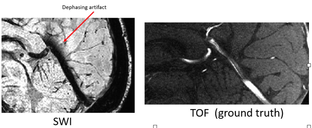
The straight sinus on both SWI and TOF
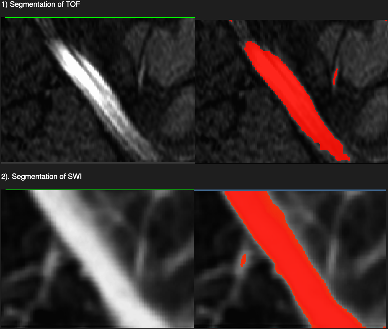
Segmentation results
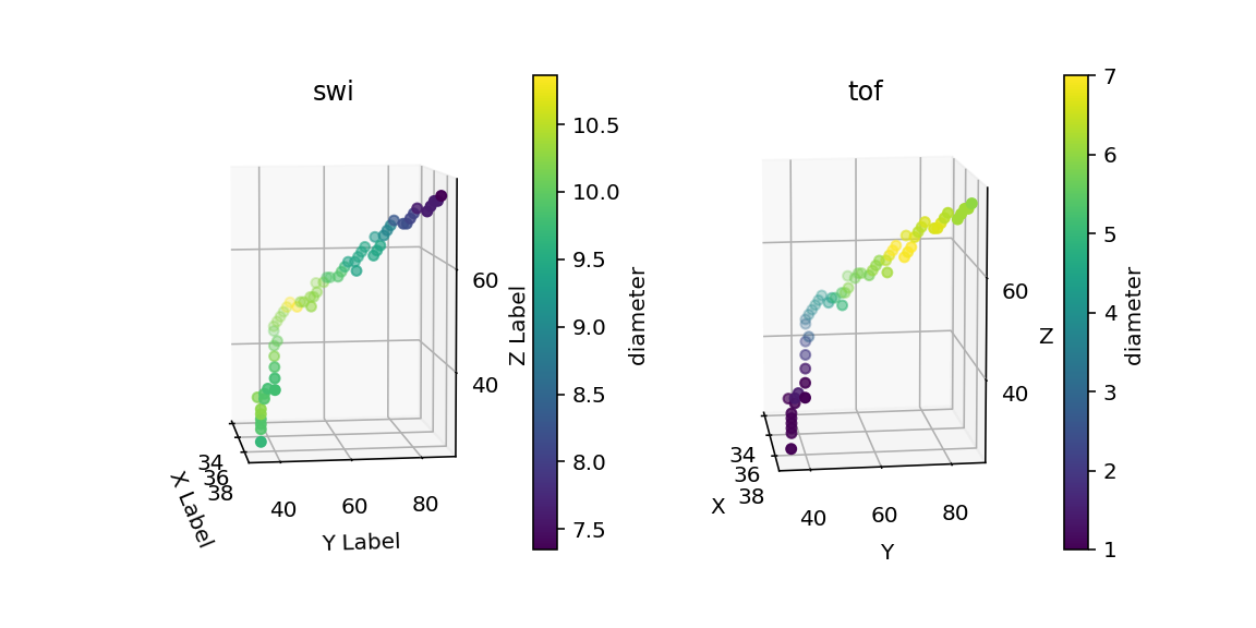
diameters
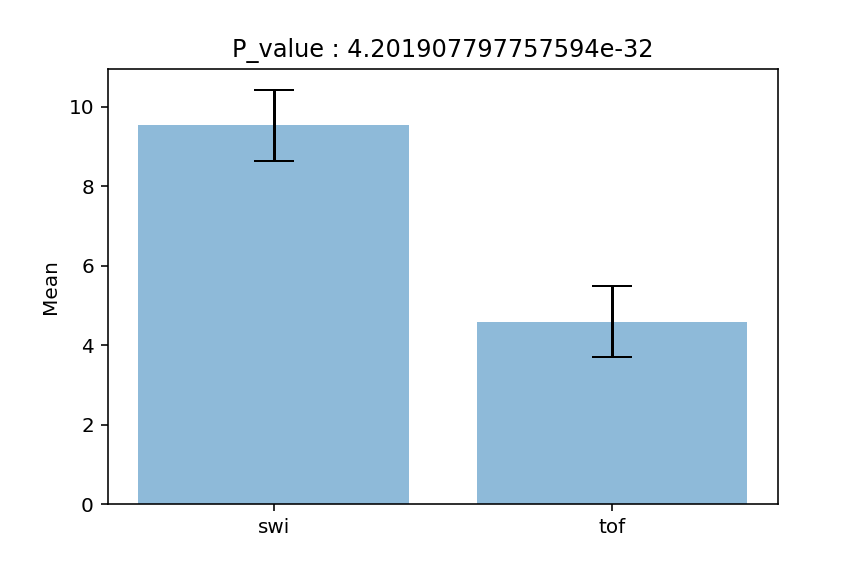
SWI vs TOF diameters
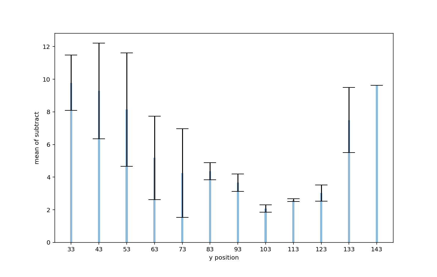
SWI minus TOF diameter