4116
Repeatability of Radiomic Features in T2-Weighted Prostate MRI: Impact of Pre-processing Configurations1Department of Circulation and Medical Imaging, Norwegian University of Science and Technology, Trondheim, Norway, 2Department of Radiology and Nuclear Medicine, St. Olavs Hospital, Trondheim University Hospital, Trondheim, Norway, 3Department of Cancer Research and Molecular Medicine, Norwegian University of Science and Technology, Trondheim, Norway, 4Department of Urology, St. Olavs Hospital, Trondheim University Hospital, Trondheim, Norway
Synopsis
Identifying repeatability of radiomic features in T2-Weighted prostate MRI is important to develop consistent imaging biomarkers and evaluate prostate cancer. This study aims to investigate the impact of pre-processing configurations on radiomic features repeatability in two short-term measurements. Repeatability was analyzed through pre-processing combinations of quantization, bin number, normalization, and filtering, and PyRadiomics feature extraction tool. The results show that repeatability of texture features varies depending on anatomical zones and lesions, pre-processing, and feature itself. Four texture features provide good repeatability independently of pre-processing configuration. Moreover, different feature groups have the highest repeatability in whole prostate, prostate zones and lesions.
Introduction
T2-Weighted (T2W) MRI is important in the evaluation of prostate cancer (PCa), and identifying radiomic features can be valuable to develop consistent imaging biomarkers and facilitate clinical decision making1-2. However, repeatability of the extracted radiomic features is a challenge, as it may be influenced by various factors3-5. Pre-processing is needed for an optimal quantitative analysis, and a number of pre-processing techniques have been recommended by the Imaging Biomarkers Standardization Initiatives6. The aim of the current study was to investigate the influence of pre-processing configurations on the repeatability of radiomic features in T2W-MRI of the prostate.Methods
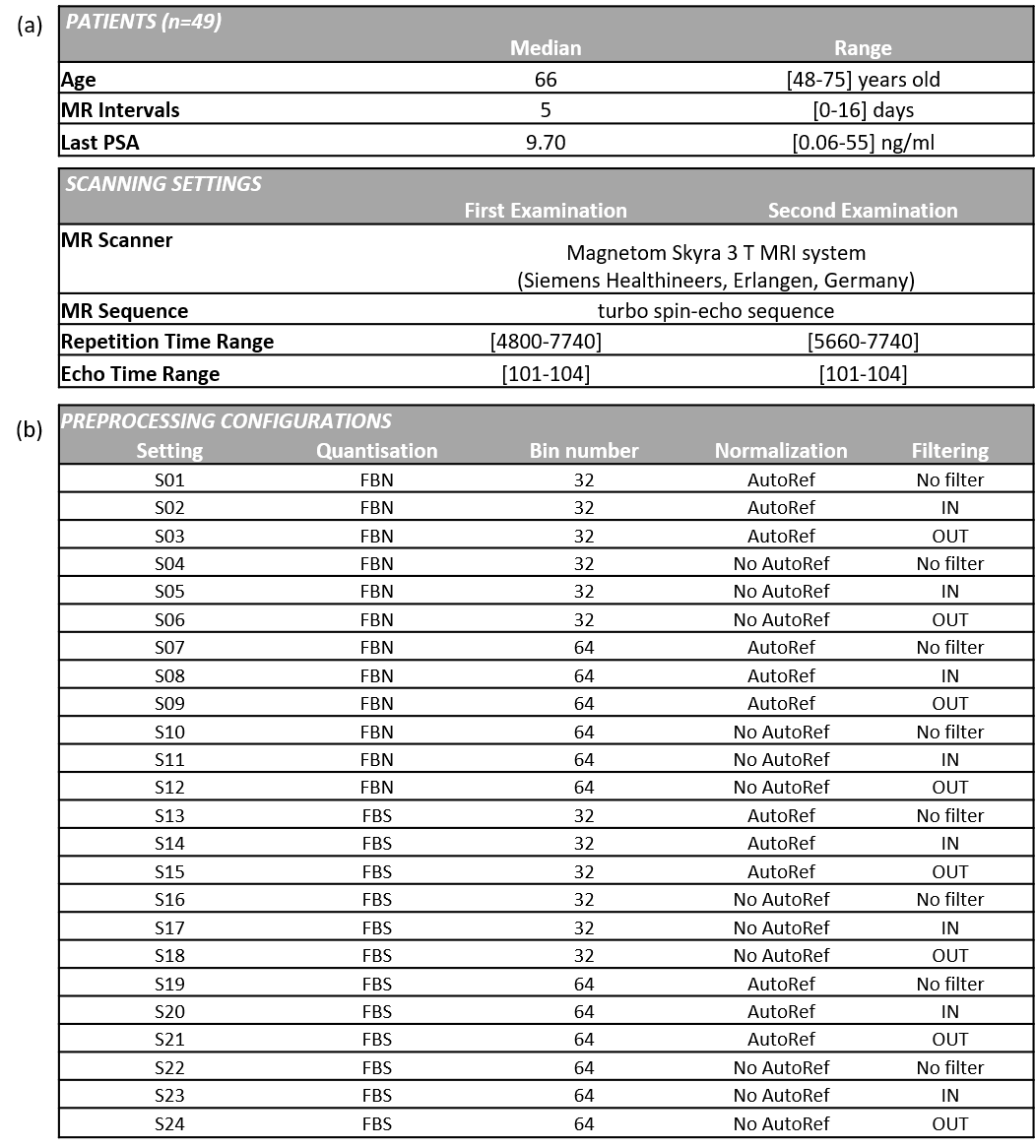
T2W-MRI datasets from two consecutive mpMRI examinations (median interval 5 days, range 0-16 days) were collected from 49 patients. The patients were referred for prostate mpMRI examination at St. Olavs Hospital, Trondheim University Hospital, Norway, between March 2015 and December 2017, due to suspicion of PCa. The first examination was a detection scan according to the PI-RADS recommendations7, and the second was to guide in-bore biopsies. All patients were scored with PI-RADS≥3 and presented with at least one lesion. The study was approved by the regional ethics committee. Scan parameters of both T2W-MRI datasets are provided in Figure 1(a). The T2W-MRI datasets were manually segmented into Whole Prostate Gland (WP, n=49), Peripheral Zone (PZ, n=49), Transitional Zone (TZ, n=49) and lesions (L, n=73) by a radiology resident using ITK-SNAP8 under supervision of a senior radiologist.All T2W-MRI datasets were corrected using a set of pre-processing steps, resulting in 24 combinations; Quantization (Fixed-Bin Number (FBW) and Fixed-Bin Size (FBS) methods)6, Bin number (32 and 64)6, Normalization (with and without AutoRef)9, and Filtering (No filter, IN (set voxels with intensities inside µ±3s outlier), and OUT (set voxels with intensities outside µ±3s outlier))10, see Figure 1(b).
Radiomic features (n=107) were extracted using PyRadiomics v3.011. The extracted features were derived from six texture feature groups; First-Order (FO, 18 features), Gray-Level-Co-occurrence Matrix (GLCM, 24 features), Gray-Level-Run-Length-Matrix (GLRLM, 16 features), Gray-Level-Size-Zone-Matrix (GLSZM, 16 features), Neighboring-Gray-Tone-Difference-Matrix (NGTDM, 5 features), Gray-Level-Dependence-Matrix (GLDM, 14 features), and one morphologic feature group; Shape (14 features).
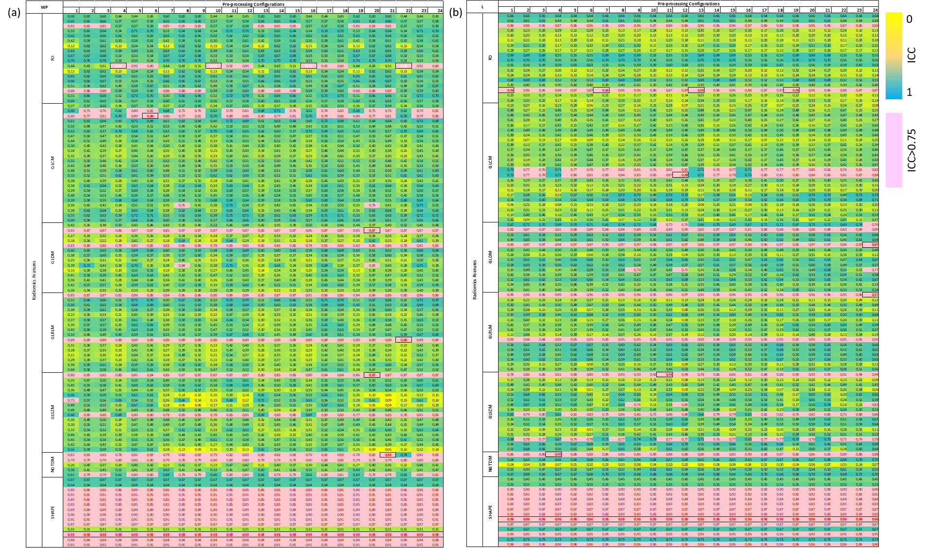
Feature repeatability of paired T2W-MRI datasets from all 24 pre-processing configurations was assessed by calculating two-way random effects Intra-class Correlation Coefficient (ICC2, absolute agreement, single rater)12 using Matlab R2020a (The MathWorks, Inc., USA). A good repeatability was defined as ICC>0.7512.
Results
Figure 2 shows the repeatability results in heatmap of ICCs with selected good features in WP and L datasets as examples. Figure 3 summarizes the distribution of good repeatability based on pre-processing configurations and prostate regions and lesions. Figure 4 describes the highest repeatability results according to radiomic features and pre-processing configurations.Discussions
Repeatability of radiomics features from T2W MRI in an actual clinical setting has not yet been reported. This study addresses the impact of pre-processing configurations on repeatability of radiomic features derived from two consecutive T2W-MRI scans of the prostate acquired within a short-term interval. Based on ICCs, we show that the repeatability of texture features varies depending on the ROI they are derived from (WP, PZ, TZ, or L), pre-processing configurations, and the feature itself. Four texture features were identified to have good repeatability in all configurations for all prostate regions and lesions (GLDM-DependenceNonUniformity, GLDM-GrayLevelNonUniformity, GLRLM- GrayLevelNonUniformity, GLRLM-RunLengthNonUniformity). Importantly, more than 14% of texture features have good repeatability in more than one pre-processing configuration, and more than 12% texture features have good repeatability in more than twelve pre-processing configurations in all prostate regions and lesions. For shape features, as this study only utilized manual segmentation, no pre-processing configuration affects the repeatability, but the manual segmentation itself has affected the repeatability. Yet, seven shape features have been found to have good repeatability in all prostate regions and lesions (MajorAxisLength, Maximum2DDiameterColumn, Maximum2DDiameterSlice, Maximum3DDiameter, MeshVolume, MinorAxisLength, VoxelVolume). This shows that some texture features provide good repeatability for routine scanning of clinical patients. Furthermore, the best combination has been found to differ depending on prostate regions and lesions. FO features seem to provide highest repeatability in WP, GLRLM features produce best results in PZ and TZ, while GLDM and GLRLM features are the best options for L. AutoRef also appears to consistently provide better repeatability, mostly in FO features in all prostate regions and slightly in GLCM and GLDM in WP and TZ, and GLRLM in PZ, and GLSZM in L.Conclusions
Repeatability of radiomic texture features is known to be susceptible to pre-processing settings. This study visualizes the repeatability of radiomic features in T2W-MRI of the prostate with verified and delineated anatomical zones and lesions. The results show that four texture features provide good repeatability independently of the pre-processing configuration. Moreover, different feature groups have the highest repeatability in the whole prostate, the prostate zones and lesions. This knowledge provides basic information necessary for studies implementing radiomic features in clinical applications, such as a supplement for PCa characterization or detection of progression in patients on Active Surveillance.Acknowledgements
No acknowledgement found.References
1. Cattell R, Chen S, Huang C. Robustness of radiomic features in magnetic resonance imaging: review and a phantom study. Visual Computing for Industry, Biomedicine, and Art. 2019 Dec 1;2(1):19.
2. Fedorov A, Vangel MG, Tempany CM, Fennessy FM. Multiparametric magnetic resonance imaging of the prostate: repeatability of volume and apparent diffusion coefficient quantification. Investigative radiology. 2017 Sep;52(9):538.
3. Traverso A, Wee L, Dekker A, Gillies R. Repeatability and reproducibility of radiomic features: a systematic review. International Journal of Radiation Oncology* Biology* Physics. 2018 Nov 15;102(4):1143-58.
4. Fornacon-Wood I, Mistry H, Ackermann CJ, Blackhall F, McPartlin A, Faivre-Finn C, Price GJ, O’Connor JP. Reliability and prognostic value of radiomic features are highly dependent on choice of feature extraction platform. Eur. Radiol.. 2020 Jun 1.
5. Schwier M, van Griethuysen J, Vangel MG, Pieper S, Peled S, Tempany C, Aerts HJ, Kikinis R, Fennessy FM, Fedorov A. Repeatability of multiparametric prostate MRI radiomics features. Scientific reports. 2019 Jul 1;9(1):1-6.
6. Zwanenburg A, Vallières M, Abdalah MA, Aerts HJ, Andrearczyk V, Apte A, Ashrafinia S, Bakas S, Beukinga RJ, Boellaard R, Bogowicz M. The image biomarker standardization initiative: standardized quantitative radiomics for high-throughput image-based phenotyping. Radiology. 2020 May;295(2):328-38.
7. Padhani AR, Barentsz J, Villeirs G, Rosenkrantz AB, Margolis DJ, Turkbey B, Thoeny HC, Cornud F, Haider MA, Macura KJ, Tempany CM. PI-RADS Steering Committee: the PI-RADS multiparametric MRI and MRI-directed biopsy pathway. Radiology. 2019 Aug;292(2):464-74.
8. Yushkevich PA, Piven J, Hazlett HC, Smith RG, Ho S, Gee JC, Gerig G. User-guided 3D active contour segmentation of anatomical structures: significantly improved efficiency and reliability. Neuroimage. 2006 Jul 1;31(3):1116-28.
9. Sunoqrot MR, Nketiah GA, Selnæs KM, Bathen TF, Elschot M. Automated reference tissue normalization of T2-weighted MR images of the prostate using object recognition. Magnetic Resonance Materials in Physics, Biology and Medicine. 2020 Jul 31:1-3.
10. Collewet G, Strzelecki M, Mariette F. Influence of MRI acquisition protocols and image intensity normalization methods on texture classification. Magnetic resonance imaging. 2004 Jan 1;22(1):81-91.
11. Van Griethuysen JJ, Fedorov A, Parmar C, Hosny A, Aucoin N, Narayan V, Beets-Tan RG, Fillion-Robin JC, Pieper S, Aerts HJ. Computational radiomics system to decode the radiographic phenotype. Cancer research. 2017 Nov 1;77(21):e104-7.
12. Robert Matthew (2020). f_ICC (https://github.com/robertpetermatthew/f_ICC), GitHub. Retrieved December 11, 2020. Shrout PE, Fleiss JL. Intraclass correlations: uses in assessing rater reliability. Psychological bulletin. 1979 Mar;86(2):420.
Figures