4102
Development of Distortion-Free MR Elastography Methods for Slip Interface Imaging of the Prostate1Radiology, Mayo Clinic, Rochester, MN, United States, 2Urology, Mayo Clinic, Rochester, MN, United States
Synopsis
Slip interface imaging is a MR elastography based technique that enables assessing the mechanical connectivity between the tumor and its sourronding tissues. In this study, we have implemented a distortion-free MRE method (DIADEM-MRE) for detecting slip boundary of the prostate capsule. This technique has the great potential to be used to locate the margin of tumor adherent to the prostatic capsule, thus predicting the present of extraprostatice extension of prostate cancer.
Introduction
A key factor affecting the prognosis and choice of treatment in patients with prostate cancer is the presence of extraprostatic extension (EPE) [1, 2]. Reliable tools for predicting EPE are desired to avoid unnecessary therapy-induced morbidity or treatment failure. It is our hypothesis that the longer the margin of tumor adherence to the prostatic capsule, the higher the risk of EPE. MR elastography (MRE) slip-interface imaging (SII) can evaluate tumor adherence and may be useful for evaluating the degree of EPE noninvasively [3, 4]. Despite recent progress, geometric distortion is still a well-known challenge for prostate MRE based on EPI imaging methods due to its proximity to the often air-filled colon[5, 6]. These artifacts are of particular importance for prostate cancer EPE detection because the artifacts typically occur at the border of the rectum and the peripheral zone of the prostate, where 70% of prostate cancers are located [7]. To address the EPI distortion problem, DIADEM-EPI (Distortion-free Imaging: A Double Encoding Method)[8] has been adapted to MRE and demonstrated in brain MRE [9]. In this study, we implemented this approach for prostate MRE to substantially reduced the geometric distortion and demonstrated its feasibility for prostate SII in healthy volunteers.Methods
The distortion-free DIADEM MRE technique is based on a multiband, spin-echo, EPI-MRE sequence [10] with an additional spin-warp, phase-encoding gradient immediately before the EPI acquisition [11, 12]. With IRB approval, three healthy volunteers were scanned on a 3T scanner (Signa Premier, GE Healthcare, Waukesha, WI) using a 30-channel flexible anterior array coil and 60-channel embedded posterior array coil (GE Healthcare, Waukesha, WI). Shear waves at 60 Hz were introduced into the prostate using a 10-cm diameter, rigid, high-density polyethylene driver that was strapped to the lower abdominal wall over the symphysis pubis. The DIADEM-MRE data were acquired with the following imaging parameters: TR/TE=1417/47.2 ms; FOV=38.4 cm; 128×128 matrix; 34 contiguous 3-mm-thick axial slices; 2x phase acceleration; 2x multiband (MB) acceleration, 544-μs echo spacing, 60-Hz mechanical vibrations; 6 motion-encoding directions, 3 phase offsets; 8 shots for each DIADEM dataset and total acquisition time of 3:27 minutes. SII-derived octahedral shear strain (OSS) maps were calculated as described in a previous study [13].Results
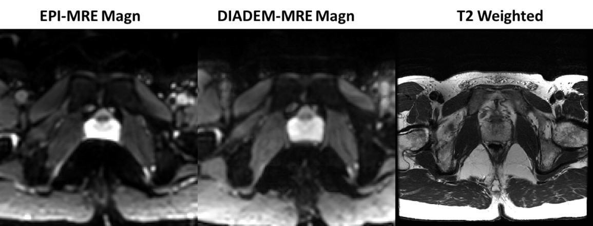
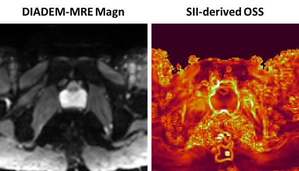
Figure 1 compares the standard EPI-MRE and DIADEM-MRE magnitude images for one volunteer. The prostate in EPI MRE is heavily distorted where it is adjacent to the rectum. In contrast, DIADEM MRE shows no distortion when compared with the T2-weighted anatomical image. Figure 2 compares the DIADEM-MRE magnitude image and the SII-derived OSS map for the same volunteer as in Figure 1. A high-intensity boundary (i.e., a slip interface) can be seen around the healthy prostate.Discussion and Conclusion
We have shown in this preliminary result that it is feasible to acquire OSS map of the prostate using DIADEM MRE, where the geometric distortion near the prostate boundary was substantially reduced. For a healthy prostate, the existence of a complete prostate capsule creates a slip boundary between different tissue layers (i.e., the prostate moves relative to other organs at a submicron level under vibration). The displacement discontinuity results in a high-valued OSS contour at the tumor boundary. Additional studies involving prostate cancer patients with varying degrees of EPE are still required to determine if this technique may be useful for predicting prostate cancer EPE.Acknowledgements
This work was supported by grants from the NIH (R01 EB001981 and R01 NS113760) and Mayo Clinic imaging awards CIM-92541650.References
1. Epstein, J.I., et al., Prediction of progression following radical prostatectomy. A multivariate analysis of 721 men with long-term follow-up. Am J Surg Pathol, 1996. 20(3): p. 286-92.
2. Mikel Hubanks, J., et al., The presence of extracapsular extension is associated with an increased risk of death from prostate cancer after radical prostatectomy for patients with seminal vesicle invasion and negative lymph nodes. Urol Oncol, 2014. 32(1): p. 26.e1-7.
3. Yin, Z., et al., Slip interface imaging predicts tumor-brain adhesion in vestibular schwannomas. Radiology, 2015. 277(2): p. 507-517.
4. Hu, B., et al. Slip-interface imaging preoperatively predicts hepatocellular carcinoma microvascular invasion. in ISMRM 27th Annual Meeting & Exhibition. 2019. Montréal, QC, Canada.
5. Glaser, K.J., A. Manduca, and R.L. Ehman, Review of MR elastography applications and recent developments. J Magn Reson Imaging, 2012. 36(4): p. 757-74.
6. Pepin, K.M., R.L. Ehman, and K.P. McGee, Magnetic resonance elastography (MRE) in cancer: Technique, analysis, and applications. Prog Nucl Magn Reson Spectrosc, 2015. 90-91: p. 32-48.
7. Rickards, D., Transrectal ultrasound 1992. Br J Urol, 1992. 69(5): p. 449-55.
8. In, M.H., et al., Distortion-free imaging: A double encoding method (DIADEM) combined with multiband imaging for rapid distortion-free high-resolution diffusion imaging on a compact 3T with high-performance gradients. J Magn Reson Imaging, 2019.
9. Sui, Y., et al. High-Resolution Distortion-Free Whole-Brain MR Elastography using Multiband DIADEM (DIADEM-MRE). in 2020 ISMRM 2020.
10. Sui, Y., et al., Fast Brain MR Elastography Using a Simultaneous Multislice EPI Acquisition on a Compact 3T Scanner. ISMRM Proceeding, 2019: p. 3971.
11. Robson, M.D., J.C. Gore, and R.T. Constable, Measurement of the point spread function in MRI using constant time imaging. Magnetic resonance in medicine, 1997. 38(5): p. 733-740.
12. Zeng, H. and R.T. Constable, Image distortion correction in EPI: comparison of field mapping with point spread function mapping. Magn Reson Med, 2002. 48(1): p. 137-46.
13. Yin, Z., et al., Slip interface imaging based on MR-elastography preoperatively predicts meningioma-brain adhesion. J Magn Reson Imaging, 2017. 46(4): p. 1007-1016.
Figures