4100
POST-ACQUISITION WATER SIGNAL REMOVAL IN 3D WATER-UNSUPPRESSED 1H-MR SPECTROSCOPIC IMAGING DATA OF THE PROSTATE1Radiology, Radboud UMC, Nijmegen, Netherlands, 2Icometrix, Leuven, Belgium, 3KU Leuven, Leuven, Belgium, 4Radboud UMC, Nijmegen, Netherlands
Synopsis
Water-unsuppressed MRSI acquisitions could obliviate the need for additional reference data sampling for signal quantification. We evaluated two methods for post-acquisition water signal removal in 1H-MRSI of the prostate. The method using Löwner Blind Source Separation for filtering the water signal outperformed the matrix-based Hankel Lanczos Singular Value Decomposition method. The two techniques were evaluated and compared against conventional water suppressed data acquisitions in 4 volunteers. The results demonstrate that post-acquisition water removal was successfully implemented in water-unsuppressed prostate MRS(I) data, and that the Löwner filter showed the best performance.
INTRODUCTION
Proton MR Spectroscopic Imaging (MRSI) is commonly performed with suppression of the dominating water signal during acquisition. This requires additional suppression pulses that often do not completely remove the water signal and can cause artefacts in the data such as signal attenuation of resonances with a chemical shift close to that of water. When not removed, or acquired separately, the water signal can be very useful as a reference to determine the tissue concentration of metabolites and to correct for artefacts such as line shape or phase distortions. For these purposes a separate data set without water suppression is often acquired in single voxel MRS, but this requires too much additional examination time in MRSI1. Alternatively, MRSI can be performed without suppression of the water signal, which then requires full removal of this water signal for metabolite quantification. The aim of this work is to develop a post-acquisition protocol to remove the water signal and its side bands from non-water suppressed MRSI of the prostate. We explored Löwner Blind Source Separation (BSS)2 and Hankel Lanczos singular value decomposition (HLSVD)3 as filters for water signal removal. The two techniques were evaluated and compared against conventional MRSI with water suppression1,2 and the additional value of the approach is illustrated with the determination of absolute tissue metabolite concentrations.METHOD
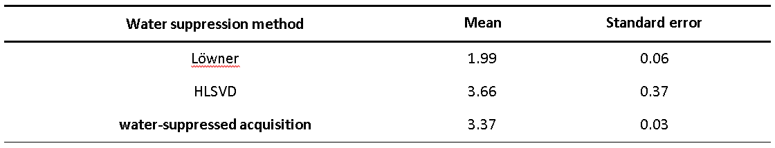
Four volunteers (27-55 years, mean age 47 years) were examined on a 3T MR system (MAGNETOM Prisma-Fit, Siemens, Erlangen) using a body phased-array coil (no endorectal coil) for reception and a GOIA semi-LASER pulse sequence for volume of interest (VOI) selection of the prostate4. For each volunteer, an MRSI dataset with and without suppression of the water signal was acquired. From water-unsuppressed data, first the sideband artefacts originating from the huge water peak were eliminated using the modulus of the signal1,5. Then, a Hilbert transformation was applied to recreate complex data to be able to apply the filtering algorithms. The filtering techniques tested were (a) the Löwner-BSS, applied to all voxels of interest at once, versus (b) HLSVD, applied voxel-by-voxel. The processed spectra were fitted and quantified with LC-model software (Version 6.3-1L) for signals of citrate, choline, spermine, creatine. Absolute quantification of metabolites was performed using the water unsuppressed MRSI spectra before water removal and the water filtered spectra or water signal suppressed spectra as input for LC-model with corrections for water and metabolite proton T1 and T2 relaxation times6. A repeated measures ANOVA statistical analysis was used to compare the effect of the filters on the metabolites of interest from 21 voxels in 4 volunteers from different locations. The performance of the filters was evaluated by the ratio of the variance of residual signal and noise in the range of the water resonance (4.2 to 5.4 ppm) to the variance of noise (11 to 12 ppm). The filtering techniques were compared to each other and to the result of the conventional MRSI acquisition with water suppression to identify possible bias of post-processed water removal on metabolite signal quantification. Finally, the water removal technique was applied to the MRSI data of a prostate cancer patient acquired with an endorectal coil.RESULTS AND DISCUSSION
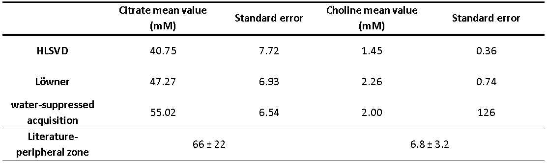
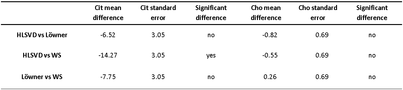
The effect of the filters is illustrated with MR spectra in the frequency range 1 to 6 ppm of one voxel from a 3D dataset of a volunteer (Figure 1). Both filters suppressed the water signal with a performance equivalent or better than in spectra from the same voxel using conventional MRSI with water suppression. Table 1 presents the calculated mean values of the ratios of the variance in the water area to the noise area. The results show that Löwner filtering provides the best suppression of the water peak with respect to the noise level with the mean variance ratio at 1.99 ± 0.06. Table 2 presents the mean values and standard deviations of the absolute tissue concentrations of citrate and choline, within the range of those found in the literature for healthy individuals7. The absolute concentration values of citrate do not differ significantly in the case of Löwner filtering versus the values obtained from spectra recorded with water signal suppression, while HLSVD filtered quantification caused a significant difference with conventional water-suppressed acquisition (Table 3). Regarding choline, the absolute concentration values do not differ significantly between any of the three water suppression methods (Table 3). Finally, the metabolite map of citrate for Löwner water suppression technique is presented in Figure 2. In the area of a tumor, in the left peripheral zone close to the coil, the citrate concentration is lower in relation to the rest of the peripheral zone.CONCLUSIONS
We demonstrate that non-endorectal coil MRSI of the prostate without water signal suppression performs equally well as MRSI with water signal suppression, using a post-acquisition protocol for water signal removal. The Löwner filter showed the best performance in water removal. Altogether, our approach of post-acquisition water signal removal in MRSI of the prostate is a robust method that allows to use the water signal for referencing purposes such as absolute quantification of metabolites. No additional reference data is needed as the water signal is obtained from the same acquisition as the metabolite signals are.Acknowledgements
This work was funded by the European Union's Horizon 2020 research and innovation program INSPiRE-MED under the Marie Sklodowska-Curie grant agreement No 813120.References
1. Serrai H. et al. “Localized proton spectroscopy without water suppression: Removal of gradient induced frequency modulations by modulus signal selection”. Journal of Magnetic Resonance. 2002; 154.
2. Bharath H.N. et al. “Tensor-Based Method for Residual Water Suppression in 1H Magnetic Resonance Spectroscopic Imaging”. IEEE Transactions on Biomedical Engineering. 2019; 66: 584-594.
3. Laudadio T. et al. “Improved Lanczos algorithms for black box MRS data quantitation''. Journal of Magnetic Resonance. 2002; 157: 292-297.
4. Steinseifer I. K. et al. “Improved Volume Selective 1 H MR Spectroscopic Imaging of the Prostate with Gradient Offset Independent Adiabaticity Pulses at 3 Tesla”. Magnetic Resonance in Medicine. 2015; 74: 915–924.
5. Dong Z. “Proton MRS and MRSI of the brain without water suppression”. Progress in Nuclear Magnetic Resonance Spectroscopy. 2014.
6. Tayari N. et al. “In vivo MR spectroscopic imaging of the prostate, from application to interpretation”. Analytical Biochemistry. 2017; 529: 158-170.
7. Basharat M. et al. “Evaluation of short‐TE 1H MRSI for quantification of metabolites in the prostate”. NMR in Biomedicine. 2014; 27: 459-467.
Figures
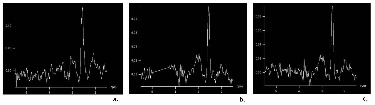
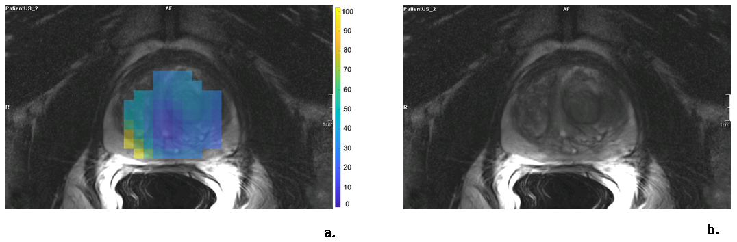
Figure 2. Patient with prostate cancer, acquired with an endorectal coil. a. metabolite map of absolute citrate concentration (mM) after Löwner filtering for water removal. The patient has a tumor in the center-left peripheral zone close to the coil, in the area with a lower citrate concentration. b. anatomy of the area of the metabolite map.