4094
Comparison of multiplexed sensitivity encoding (MUSE) and single-shot echo-planar imaging (ssEPI) for diffusion-weighted imaging of prostate
Chun-Ying Shen1, Chia-Wei Li2, Chien-Yuan Lin2, and Ching-Hua Yang1
1Department of Radiology, Taovuan General Hospital, Ministry of Health and Welfare, Taovuan, Taiwan, 2GE Healthcare, Taipei, Taiwan
1Department of Radiology, Taovuan General Hospital, Ministry of Health and Welfare, Taovuan, Taiwan, 2GE Healthcare, Taipei, Taiwan
Synopsis
This study aimed to evaluate and compare quantitatively the effect of image distortion of multiplexed sensitivity encoding (MUSE) and conventional single-shot echo-planar imaging (ssEPI) for diffusion-weighted imaging (DWI) of the prostate. Our result showed that prostate images by MUSE-DWI can provide significant less distortions and higher SNR than that by conventional ssEPI-DWI at the similar acquisition time.
INTRODUCTION
Previous experimental report has shown that diffusion-weighted imaging (DWI) using multiplexed sensitivity encoding(MUSE), a multi-shot segmented EPI technique, can reliably produce high-resolution DWI of the brain [1]. With the interleaved k-space acquisition in each shot and parallel imaging reconstruction, MUSE can provide higher resolution, lower distortion, as well as a better SNR compared with conventional ssEPI. However, shot-to-shot phase variations caused by subject motion can severely degrade the image quality in segmented EPI, which is a major hurdle in pelvis DWI. Indeed, segmented EPI has not mostly been implemented for non-neural DWI [1-3]. Therefore, the purpose of this study was to compare MUSE and conventional DWI techniques in prostate MRI. Using quantitative comparison, we will validate whether MUSE technique could provide prostate DWI with less noise and distortions in comparison with conventional DWI or not, and this validating could provide the possibly leading to better lesion detectability in prostate.METHODS
Five patients (mean age, 72.2 years) were enrolled in this study. Data were acquired using a 3T MR scanner (SIGNA Pioneer; GE Healthcare, Milwaukee, WI, USA) using a geometry-embracing method (GEM) 16-elements flexible array-coil and a 32-elements spine array-coil for signal detection, and a whole-body coil was used for radio-frequency excitation. MUSE-DWI sequence was acquired with the following parameters: TR/TE=7692/87.8 ms; FOV: 256 x 256 cm; matrix size: 128 x 128; slice thickness: 4 mm; total scan time: 4 min 30 sec. For quantitative comparing, the conventional ssEPI-DWI was acquired with the following parameters: TR/TE=11130/83.8 ms; FOV: 256 x 256 cm; matrix size: 118 x 118; slice thickness: 4 mm; total scan time: 4 min 26 sec. Three b-values (0, 800, and 1500) was acquired by those two DWI sequences. For spatial segmentation, high-resolution T2-weighted propeller images was acquired with the following parameters: TR/TE=7289/97.3 ms; FOV: 256 x 256; 320 x 320; slice thickness: 4 mm. All data processing steps were performed using MATLAB software. First, all the images were co-registered to eliminate the motion issue. Second, the manual spatial segmentation was applied in the high-resolution T2-weighted propeller images. Third, the segmented volume of prostate was applied into the B-null images by MUSE-DWI and conventional ss-DWI for signal-to-noise ratio (SNR) and distortion detection based on the overlapping between the segmented volume and the prostate regions by DWI images.RESULTS and DISCUSSION
It’s obviously that images by MUSE-DWI showed less distortion than that by conventional ssEPI-DWI (Figure 1). In the prostate diffusion-weighted images with zero b-value, the images by MUSE-DWI showed significant less (p<0.023; paired t-test) distortion than that by conventional ssEPI-DWI (Figure 2). Agreed with our hypothesis, images by MUSE-DWI showed significant higher SNR than the images by conventional ssEPI-DWI (p<0.049; paired t-test; demonstrated on the Table1). MUSE-DWI can provide multi-shot prostate images with fewer distortions and higher SNR, and it could provide better image quality than conventional DWI images.CONCLUSION
Compared with diffusion images in prostate by conventional ssEPI-DWI, MUSE-DWI can provide not only better spatial resolution but also better image quality that showed less distortions and higher SNR. Despite the relative longer acquisition time, MUSE-DWI could be useful for the detection and characterization of prostate lesion.Acknowledgements
References
1. Chen N-K, Guidon A, Chang H-C, Song AW. A robust multi-shot scan strategy for high-resolution diffusion weighted MRI enabled by multiplexed sensitivity-encoding (MUSE). Neuroimage. 2013;72:41–7. 2. Chen X, Zhang Y, Cao Y, Sun R, Huang P, Xu Y, et al. A feasible study on using multiplexed sensitivity-encoding to reduce geometric distortion in diffusion-weighted echo planar imaging. Magn Reson Imaging. 2018;54:153–9. 3. Herbst M, Deng W, Ernst T, Stenger VA. Segmented simultaneous multi-slice diffusion weighted imaging with generalized trajectories. Magn Reson Med. 2017;78:1476–81.Figures
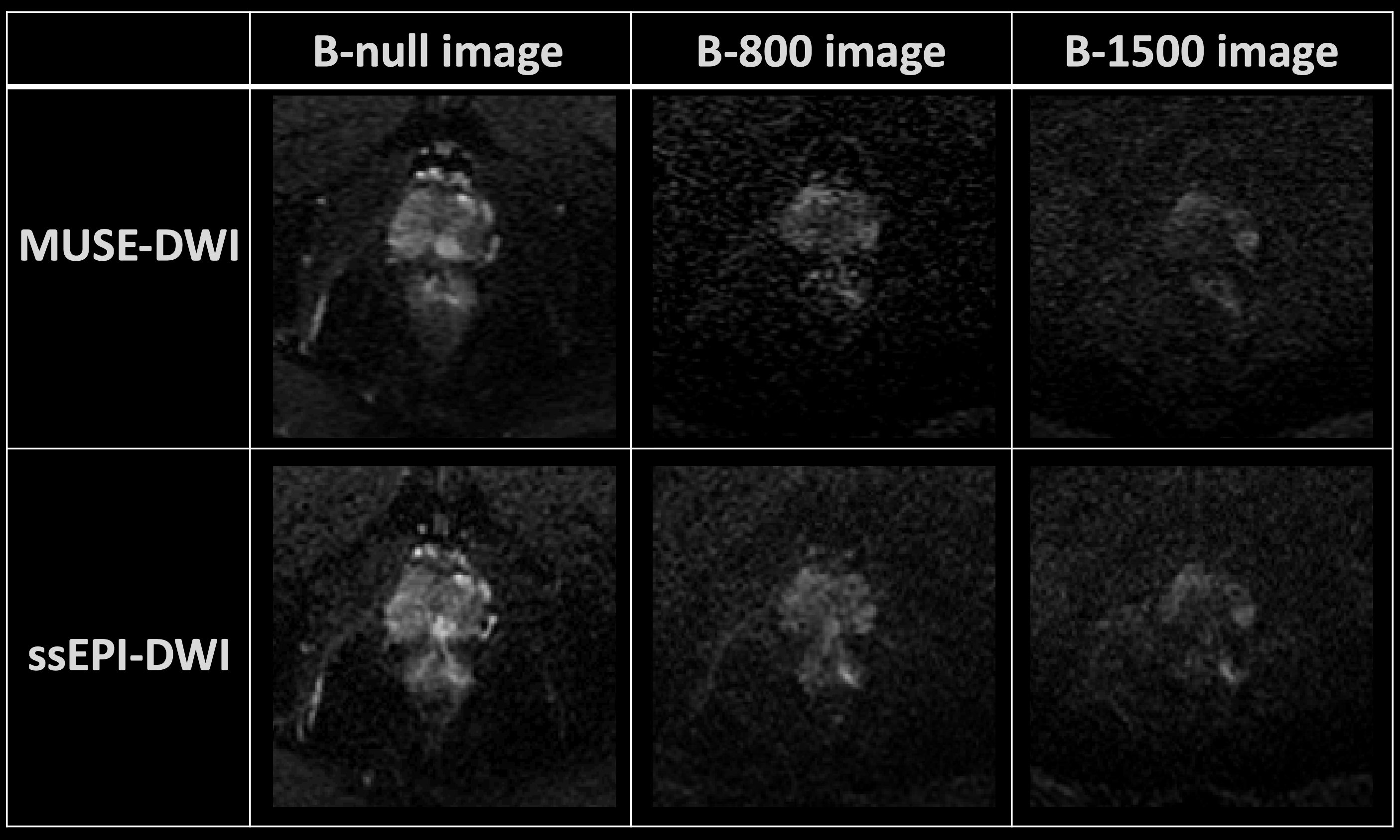
Figure 1. The diffusion-weighted images of prostate by ssEPI-DWI and MUSE-DWI.
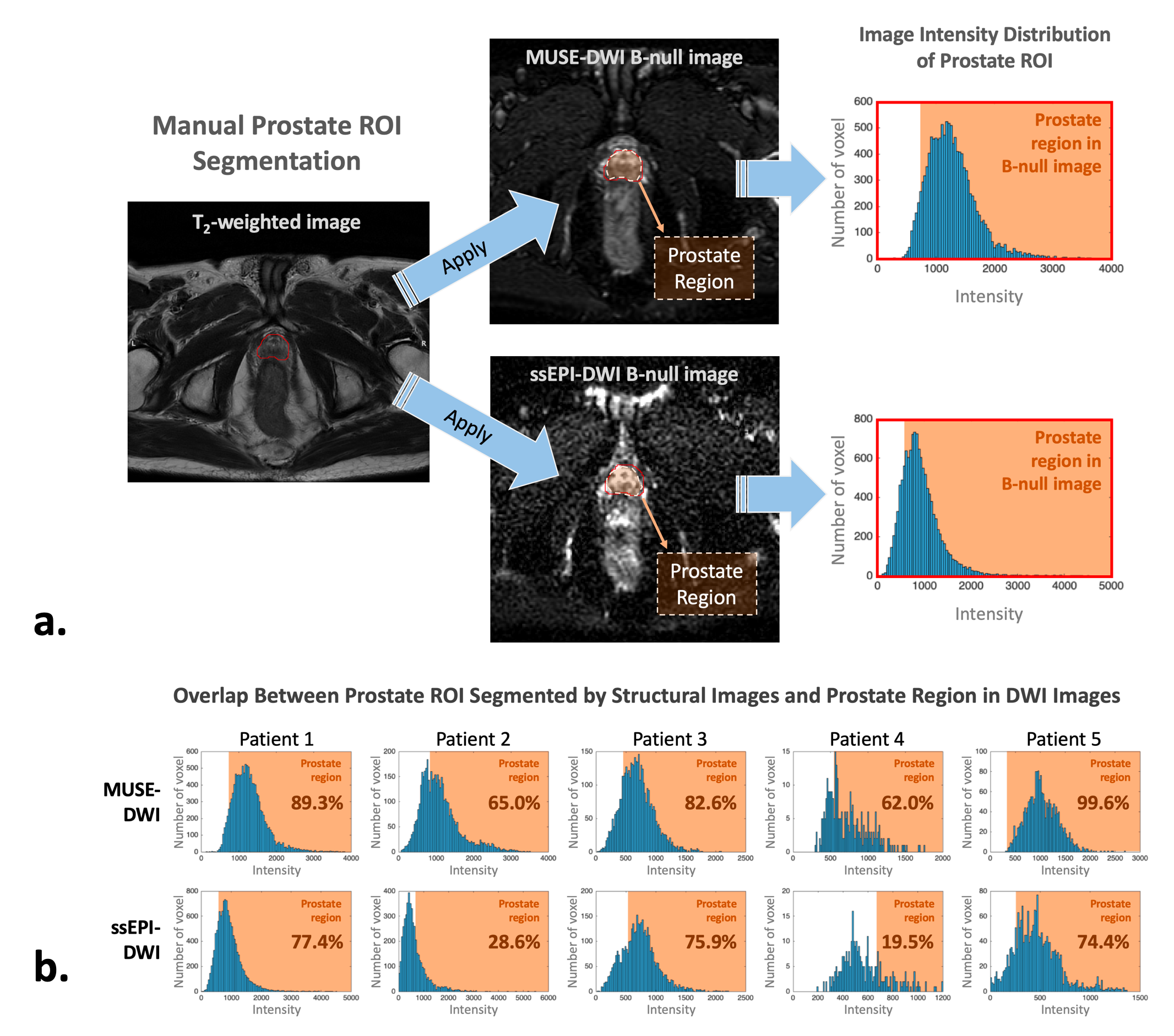
Figure 2. The data analysis procedure. (a) The prostate ROI was segmented manually from high-resolution structural images, and (b) the overlapping between the segmented prostate ROI and prostate range by DWI images was calculated.
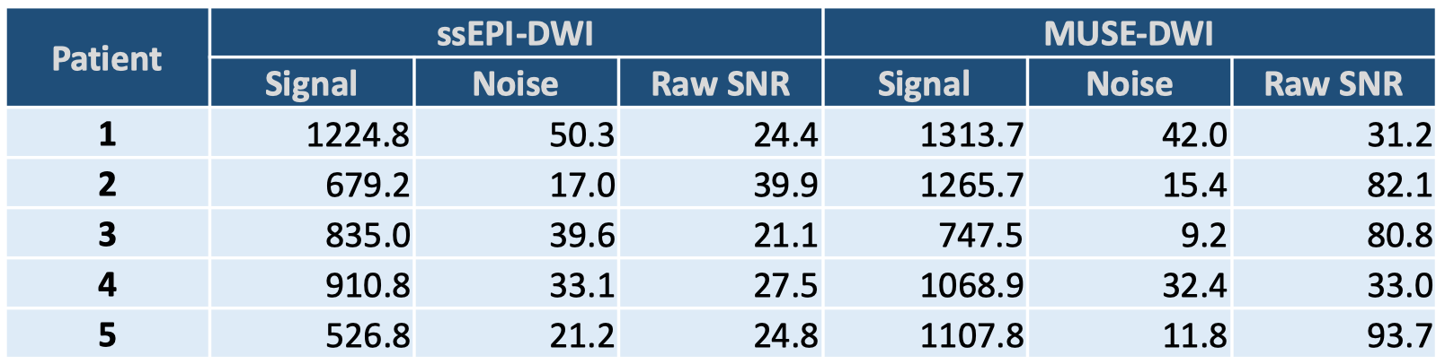
Table 1. The SNR of prostate images by ssEPI-DWI and MUSE-DWI.