4077
Associations between Bone Turnover Biomarkers and MR-based Knee composition and WORMS scores:cross-sectional
WANG BIN1, TAN HUI1, HE Taiping1, YU Nan1, and WANG Shaoyu2
1Shaanxi University of Chinese Medicine, SHAANXI, China, 2Siemens Healthineers, shanghai, China
1Shaanxi University of Chinese Medicine, SHAANXI, China, 2Siemens Healthineers, shanghai, China
Synopsis
We attempted to explore the relationship between bone markers (including bone formation and bone absorption) and cartilage, meniscus morphology, and WORMS socres in patients with osteoarthritis.
Objective
The aim of this study was to assess the cross-sectional association between bone turnover biomarkers of bone resorption and formation and MR-based cartilage, meniscus composition and Whole-Organ Magnetic Resonance Imaging Score (WORMS) of the knee osteoarthritis.Methods
30 subjects with Kellgren Lawrence(K-L) grades 1–3 in the knee with osteoarthritis were enrolled in this study and the date about serum bone turnover biomarkers were also recorded.The Western Ontario and McMaster Universities Osteoarthritis Index (WOMAC) scores were used to assess patients’ pain, function and quality of life. All the MRI pulse sequences were performed on a 3T MR scanner(MAGNETOM SKYRA, Siemens Healthineers, Erlangen, Germany), included sagittal 2D fat-saturated proton density (PD)-weighted fast spin-echo sequence, sagittal 3D dual-echo-steady-state (DESS) sequence and T2 mapping sequence. Cartilage T2 value measurements were performed in the patella, medial femur, medial tibia, lateral femur, and lateral tibia compartments. Meniscus T2 measurements were performed in the medial anterior, medial body, medial posterior, lateral anterior, lateral body and lateral posterior compartments. MRI-based cartilage, meniscus and osteophyte morphological scores were assessed in five regions (patella, medial femur, lateral femur, medial tibia and lateral tibia) using WORMS scores. Bone turnover biomarkers about serum osteocalcin, total procollagen type-1 intact N-terminal propeptide(tP1NP), β C-termianl telopeptide of type Ⅰ collagen(β-CTX), bone alkaline phosphatase(BAP), parathyroid hormone(PTH) were recorded and the correlation with MRI parameters(T2 and WORMS) were analysised using partial correlations adjusted for age, gender, BMI, and K-L grade in knees.Results
All the subjects had a mean age of 60.23±8.25 years, a mean BMI of 25.07±3.03 kg/m2, and the majority were famale(19, 63.3%).There was no association between bone turnover biomarkers and WOMAC scores(p>0.05).Among the cartilage T2, only PTH was correlated with T2 of the lateral tibia (r=0.482, P=0.013).Among the meniscus T2, osteocalcin, tP1NP and β-CTX was correlated with T2 of the lateral anterior(p<0.05).Among the morphologic measures, β-CTX was positively correlated with lateral tibia cartilage damage (r=0.453, p=0.020).BAP was positively correlated with lateral anterior meniscus damage(r=0.420, p=0.033).PTH was negatively correlated with sum and lateral body of meniscus worms damage(r= -0.402 and -0.442, p=0.042 and 0.024 respectively). And we also found that BAP was correlated with osteophyte worms of the lateral femur (r=0.394, p=0.046) and lateral tibia (r=0.395, p=0.046), and with sum scores of all knee regions (r=0.421, p=0.032).Conclusions
Our study suggested that there was significantlly correlations between osteocalcin, tP1NP and β-CTX and MRI T2 value of meniscus extra-cellular matrix degeneration. The elevated BAP represents a higher bone formation activity, which could be associated with bone formation of osteophytes.Acknowledgements
No acknowledgement found.References
No reference found.Figures
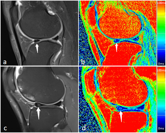
Figure 1 a and b: A 51-year-old famale with OA, right knee K-L 3, PD-weighted image(a) and T2 Mapping image(b), Meniscus lateral anterior is normal and the values of T2 is 24.72 ms, osteocalcin: 19.66 ng/ml, tP1NP: 64.94 ng/ml, β-CTX: 691 pg/ml. c and d: A 71-year-old famale with OA, right knee K-L 2, PD-weighted image(c) and T2 Mapping image(d), Meniscus lateral anterior is degeneration and the values of T2 is 28.44 ms, osteocalcin: 13.51 ng/ml, tP1NP: 46.25 ng/ml, β-CTX: 224 pg/ml.
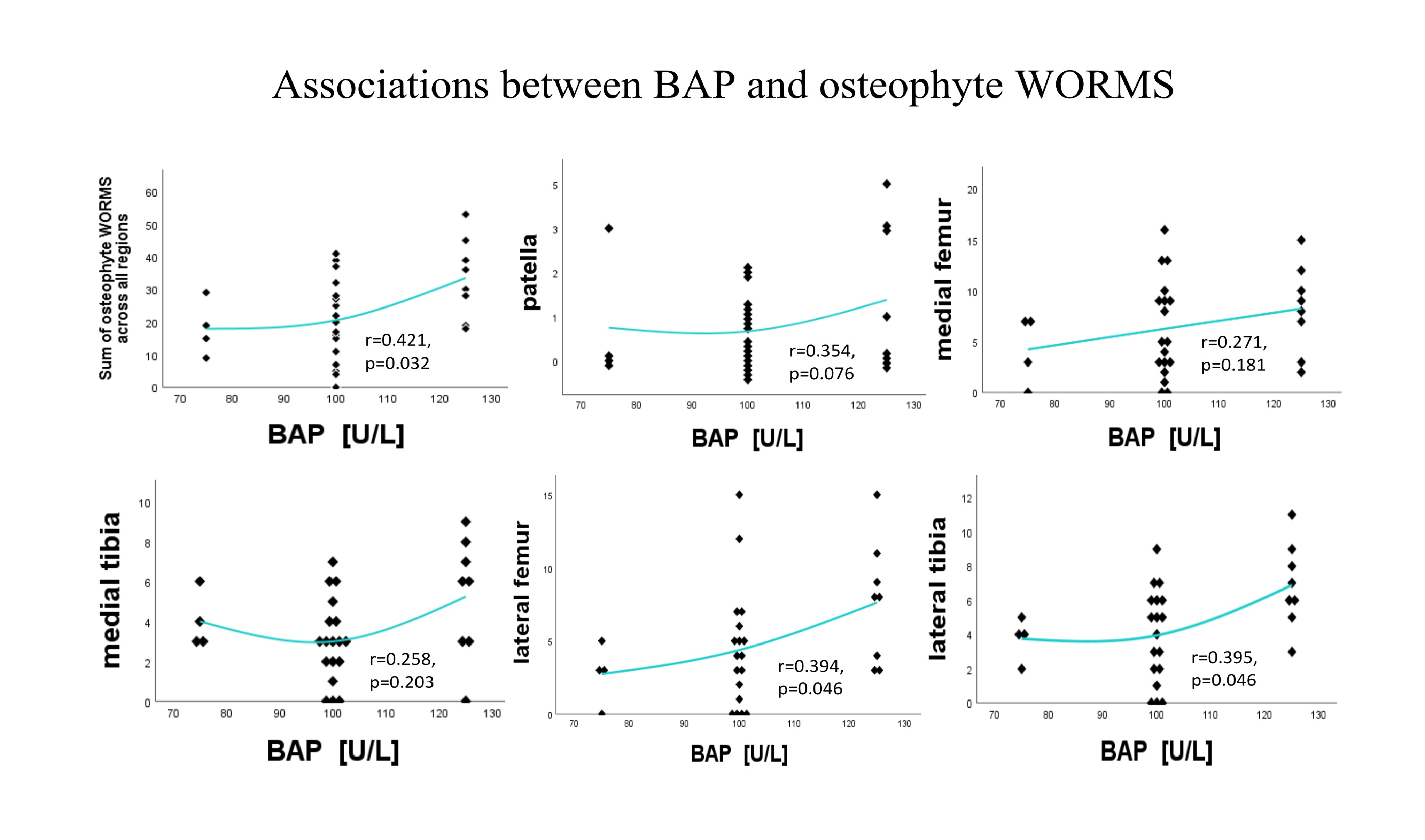
Figure 2. The correlation between BAP and Osteophyte WORMS scores was illustrated by correlation fitting scatter plots. Partial correlations are listed with adjustments for age, gender, BMI, KL grade.
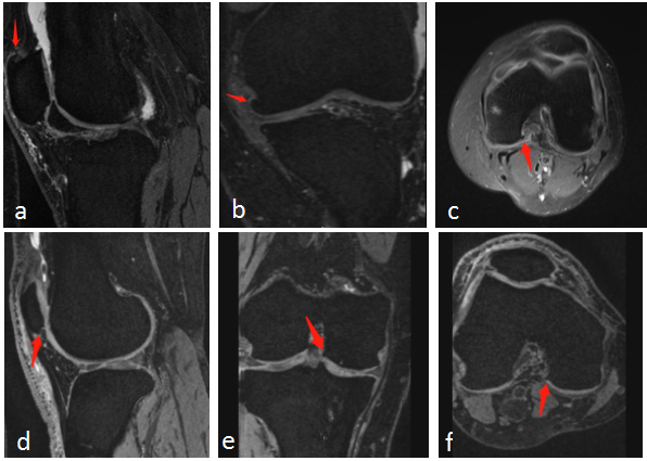
Figure 3 Sagittal(a), Coronal(b), Axial(c) of 3D DESS sequence: A 45-year-old male with OA, left knee K-L 2, summation of osteophyte WORMS scores was 36, patella score:7, medial femur score:10, medial tibia score:3, lateral femur score:11, lateral tibia score:5, the BAP leves is 125U/L.Sagittal(d), Coronal(e), Axial(f) of 3D DESS sequence: A 52-year-old male with OA, right knee K-L 1, summation of osteophyte WORMS scores was 6, patella score:1, medial femur score:3, medial tibia score:1, lateral femur score:0, lateral tibia score:1, the BAP leves is 75U/L.