4076
MRI and CT in an Ancient Child Mummy: Contrast Combination to Increase Tissue Differentiation1Dept. of Radiology, Medical Physics, Medical Center – University of Freiburg, Faculty of Medicine, University of Freiburg, Freiburg, Germany, 2German Consortium for Translational Cancer Research Partner Site Freiburg, German Cancer Research Center (DKFZ), Heidelberg, Germany, 3Institute of Evolutionary Medicine, Faculty of Medicine, University of Zurich, Zurich, Switzerland
Synopsis
MR images of a child mummy were acquired on a clinical 3T system using a dedicated RF coil with optimized RF switching hardware and 3D UTE sequence. In addition, dual-energy CT images were sampled and co-registered to compare MRI signal intensities and T2* relaxation times with CT Hounsfield Units and effective atomic numbers in bone, soft tissue and embalming material.
Introduction
Imaging of ancient remains dates back as early as 1896 when König applied X-ray imaging to study an ancient child mummy [1]. Today, X-ray imaging with CT is the gold standard for time-efficient imaging of ancient remains as it provides high resolution and excellent bone contrast [2]; however, recently the feasibility of MRI for mummy imaging was demonstrated at 1.5 and 3T clinical systems [3,4]. MRI offers additional contrasts that can supplement CT images - especially the visualization of soft tissue components with longer transverse relaxation times than bones. This is potentially valuable for pathological studies and could give improved insight of used materials or procedures of ancient mummification.This study aims to revisit the original child mummy investigated by König and adds proton density and T2* relaxation time information to compare and correlate signal intensities of selected tissue components.
Materials and Methods
An ancient Egyptian child mummy (Figure 1, 378-235 BC, Senckenberg Museum of Natural History, Frankfurt, Germany) was imaged at a clinical 3T MRI system (Prisma Fit, Siemens, Erlangen, Germany) using a dedicated quadrature Tx/Rx high pass birdcage coil (diameter: 25 cm, 8 legs, Q-factor: 128) [5]. The FID signal of the mummy was measured at different flip angles to determine the average T1, which was used to optimize TR and flip angle in the subsequent ultra-short echo time (UTE) sequence. The calibration resulted in the following UTE parameters: TE = 70, 90, 110, 200, 800 µs, TR = 2.5 ms, a = 14°, FOV = 250 mm and a matrix size of 2563. For each TE 230.000 spokes were acquired with a bandwidth of 1775 Hz/pixel. A T2* map was calculated from 5 different TEs. For comparison, CT (Somatom Definition Flash, Siemens) images were acquired at cathode voltages of 100 kV and 140 kV. From the dual-energy CT images effective atomic numbers were calculated [6]. MRI and CT data were then co-registered, and local tissue parameters were compared in a selected slice and in 3 different regions of interest (ROI) (Figure 2).Results
MR images of the child mummy show high SNR of 286±50 at the posterior cranial fossa location for shrunken brain remnants (with embalming resin) as marked in Figure 2. Cranial bone tissue has an SNR of 65±44, and the bandage material around the face has the lowest SNR with 26±15. The T2* map shows a mean 139±38 µs for cranial bone, 214±17 µs for the remnant tissue and 118±81 µs for the face bandage. Calculated effective atomic numbers Z show the same mean of 7.3 for bandage and remnant tissue and 11.5 for cranial bone. Scatter plots of MRI and T2* vs CT show weak correlations (R² ≤ 0.30) for all ROIs (Figure 3).Discussion
Conventional CT offers high resolution and high contrast but is limited in the dynamic range of selected tissues like the bandage and the remnant tissue (blue and red ROI). Here, MRI offers a strong contrast between the two tissues. CT provides a good differentiation between cranial bone and bandage ROI compared to MRI with much lower signal and higher overlap of scatter points (blue and yellow, Fig. 3). In the scatter plots with MR data against the calculated effective atomic number Z the different tissues can be further discriminated, which might be used to resolve elemental composition in a further analysis [7]. Cranial bone is clearly separated (Fig. 4a) with Z(cranial)=11.55 from the bandage and remnant ROI ranging around Z(bandage, remnant)=7.3, while bandage and remnant tissue is only discernable via the MR signal and T2* values (Fig. 4b,c and d). Here, quantitative MRI might be a good supplement to dual-energy CT data and can complement CT with more soft tissue contrast for additional material differentiation.Conclusion
Co-registration of CT and MR data from an ancient child mummy allows for visual correlation using scatter plots. Weak correlation shows high contrast differences and signifies the benefit of MRI as a supplementary imaging modality to distinguish different materials in addition to CT and derived DECT data.Acknowledgements
We thank Prof. Dr. Friedemann Schrenk (Senckenberg Research Institute, Frankfurt am Main) and Christine Hemm for the possibility to examine the child mummy with MRI and dual-energy CT.
Grant support from the Deutsche Forschungsgemeinschaft (DFG) under grant numbers BO 3025/8-1 and UL 1187/6-1 is gratefully acknowledged.
References
1. König W. Johann Ambrosius Barth; Leipzig: 1896. 14 Photographien mit Röntgen-Strahlen aufgenommen im Physikalischen Verein zu Frankfurt a. M.
2. Zesch S, et al. "From first to latest imaging technology: Revisiting the first mummy investigated with X-ray in 1896 by using dual-source computed tomography." European journal of radiology open 3 (2016): 172-181.
3. Özen AC, et al. "Comparison of ultrashort echo time sequences for MRI of an ancient mummified human hand." Magnetic resonance in medicine 75.2 (2016): 701-708.
4. Öhrström LM, et al. "Scenes from the past: MR imaging versus CT of ancient Peruvian and Egyptian mummified tissues." Radiographics 33.1 (2013): 291-296.
5. Tesfai AS, et al. "Multi-parameter Analytical Method for B1 and SNR Analysis (MAMBA): An open source RF coil design tool." Journal of Magnetic Resonance 319 (2020): 106825.
6. McCollough CH, et al. "Dual-and multi-energy CT: principles, technical approaches, and clinical applications." Radiology 276.3 (2015): 637-653.
7. Bewes JM, et al. "Imaging ancient and mummified specimens: Dual-energy CT with effective atomic number imaging of two ancient Egyptian cat mummies." Journal of Archaeological Science: Reports 8 (2016): 173-177.
Figures
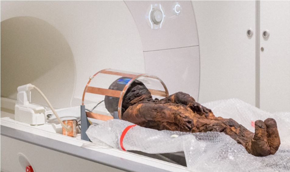
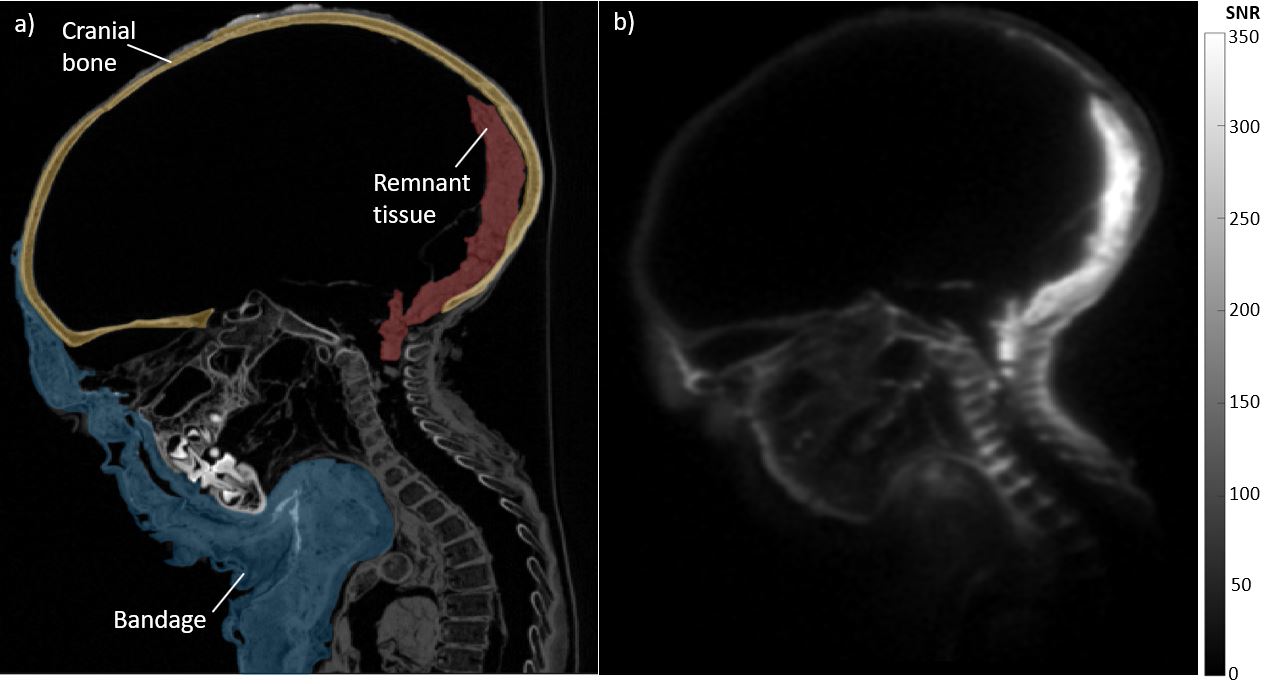
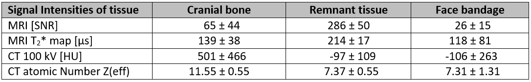
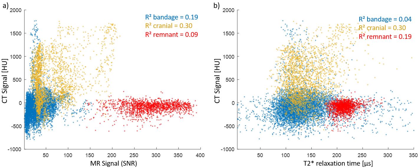
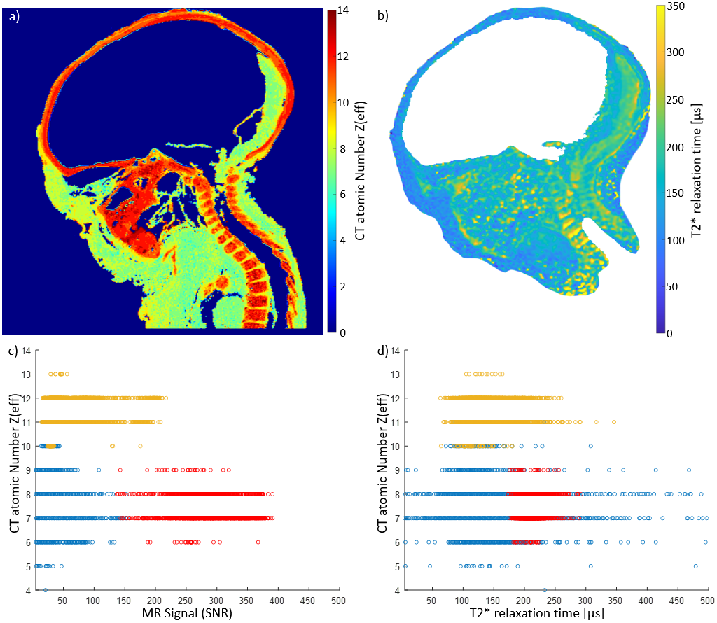
Figure 4: a) Calculated effective atomic number Z from dual energy CT measurement and b) acquired T2* map of the corresponding slice of child mummy head. Scatter plots show c) MR signal and d) T2* in comparison to CT effective atomic number.