4063
Retrospective Contrast Tuning from a Single T1-weighted Image Using Deep Learning1Stanford University, Palo Alto, CA, United States, 2University of California San Diego, San Diego, CA, United States
Synopsis
While versatile soft tissue contrasts are achievable in MRI, contrast attainable from each scan is predetermined by the imaging protocol. A retrospective tuning of contrast will provide an opportunity to normalize MRI data for radiomics analysis. In this study, we present a new paradigm to obtain a spectrum of contrasts from a single T1-weighted image. Using deep learning, T1 map, proton density map, and B1 map are predicted from every T1-weighted image, and new contrasts can be obtained with the application of Bloch equations. The method has been validated in knee MRI with high accuracy achieved.
Introduction
While versatile soft tissue contrasts are achievable in MRI, contrast attainable from each scan is predetermined by the imaging protocol. A retrospective tuning of contrast will provide an opportunity to normalize MRI data for radiomics analysis. In this study, we present a new paradigm to obtain a spectrum of tissue contrasts from a single T1-weighted image obtained in clinical practice. In this way, no additional scans are required for generating quantitative T1 maps.Methods
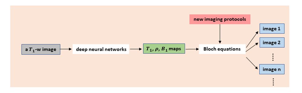
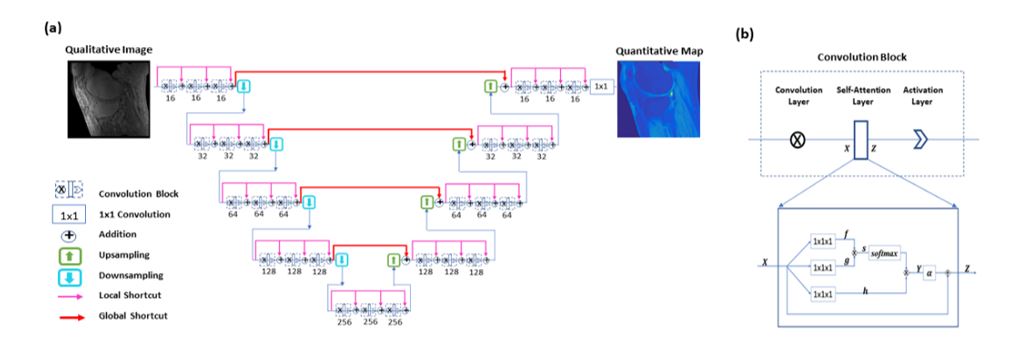
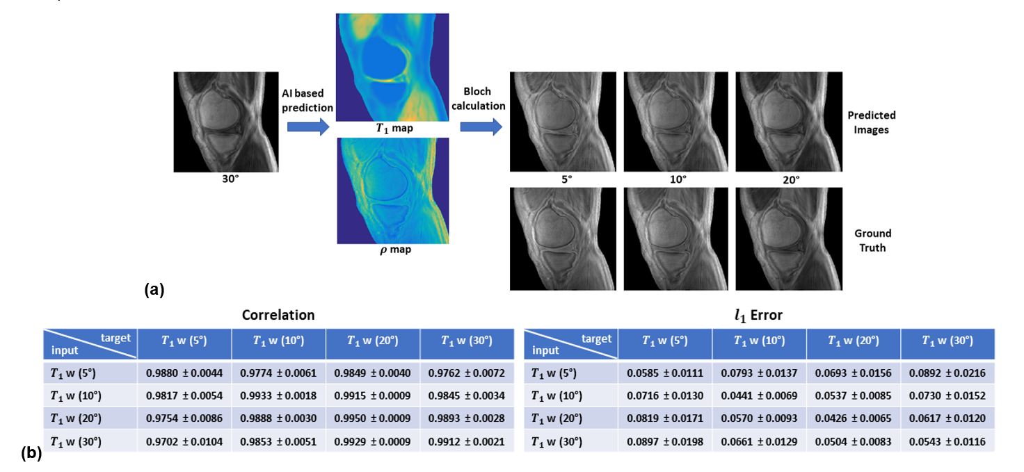
We propose a novel framework for retrospective tuning of MRI contrast, where deep learning based quantitative MRI is combined with Bloch equations. In theory, retrospective change of tissue contrast in MRI can be achieved by applying Bloch equations on tissue relaxation parametric maps, which however are hard to obtain due to the long scan time. Leveraging from the capability of deep learning, we propose a quantitative MRI approach to extract parametric maps from single MR images without conducting extra data acquisition. Using deep convolutional neural networks, T1 map, proton density map, and B1 map are predicted from every single T1-weighted image. Based on the predicted parametric/field maps, a spectrum of soft tissue contrasts can be obtained, where Bloch equations are applied with various imaging parameter values. The principle is illustrated in Figure 1.In the quantitative parametric mapping step, deep convolutional neural networks are used to provide direct mapping from single T1 weighted images to corresponding parametric/field maps. For ground truth, every T1 map is obtained from four T1-weighted images acquired with variable flip angle (5°, 10°, 20°, and 30°), proton density map is calculated from T1-weighted image and the corresponding T1 map, and B1 map is measured using the actual flip angle method [1]. In these tasks, self-attention convolutional neural network framework [2] is employed as shown in Figure 2, where the hierarchical network architecture is adopted (enabling feature extraction at various scales), global shortcuts and relatively dense local shortcuts are equipped (leading to an improved network performance), and the attention mechanism is integrated (to make efficient use of non-local information). A total of 1,224 slice images from 51 subjects are utilized for model training, and 120 images of 5 different subjects are employed for testing. In training, the Adam algorithm is used to update the network parameters, and the iterative procedure continues until convergence is reached. For a test image acquired using the same imaging protocol, quantitative T1 map, proton density map and B1 map are automatically generated from a single T1-weighted image by the established network models. The predicted T1 maps are compared with ground truth maps with L1 error and correlation coefficient calculated. In addition, every T1 map is also predicted from two T1-weighted images and gets compared with the maps predicted from single input images.
After parametric/field maps are estimated from a T1-weighted image, Bloch equations are used to calculate the signal intensity of MR images with the adoption of different imaging parameter values (flip angle, TR, etc). While a wide spectrum of contrasts can be obtained, the proposed method is only validated at certain contrasts (corresponding to the flip angles specified in training data) due to the availability of ground truth images.
Results
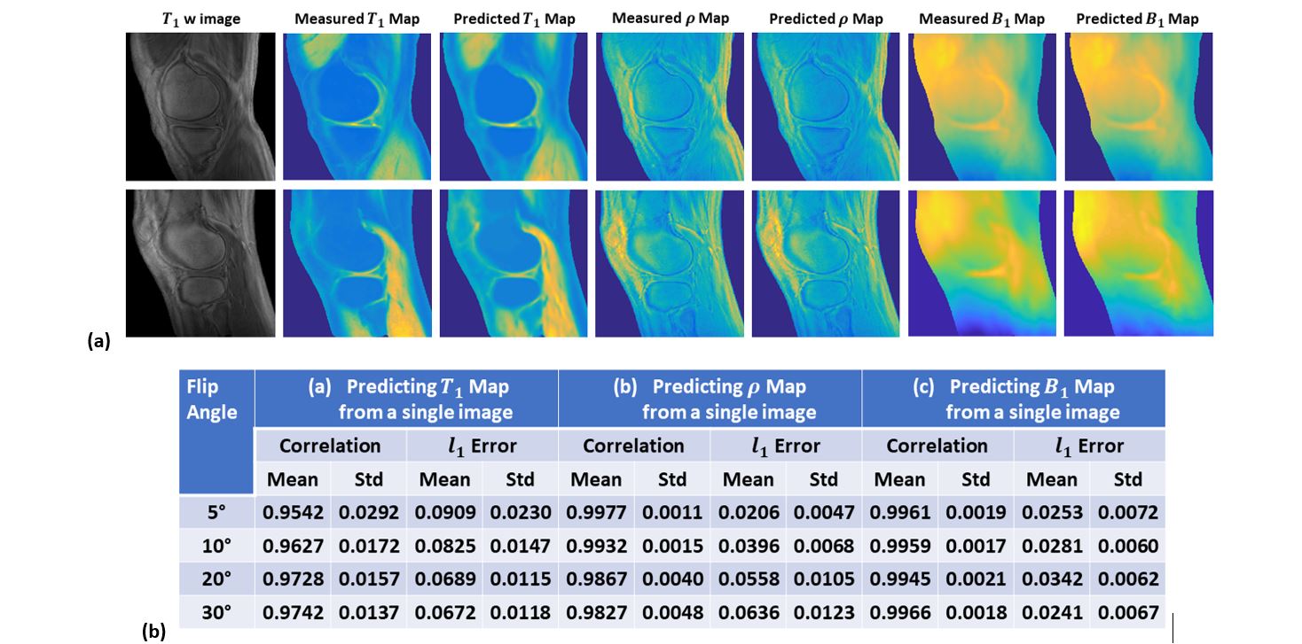
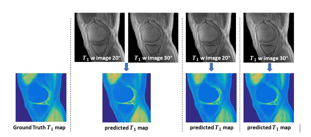
Using a large data set, deep learning models have been trained to predict quantitative T1 map, proton density map and B1 map from a single T1-weighted image. Images in two representative cases are shown in Figure 3(a), and the quantitative results for all test sets are given in Figure 3(b). The predicted maps show high image fidelity to the ground truth maps.In addition to the single image prediction, map is predicted from two input -weighted images acquired using 20° and 30° respectively (Figure 4). The difference between the resultant map and the corresponding maps derived from single -weighted images (acquired using 20° or 30°) is negligible. Quantitatively, the correlation coefficient and L1 error for the multi-image prediction are 0.9836 and 0.0567 respectively, as compared to 0.9728/0.9742 and 0.0689/0.0672 for the single image predictions.
After tissue relaxation parametric maps are obtained from a single image, the signal intensity of MR images presumably acquired using different flip angles are obtained with the application of Bloch equations. From a specific T1-weighted image (acquired using a flip angle of 30°), other T1-weighted images (corresponding to flip angle of 5°, 10° and 20°) are predicted and compared to the ground truth images as illustrated in Figure 5. High image fidelity is consistently achieved in the predicted images with low L1 error (between 0.04 and 0.09) and high correlation coefficients (ranging from 0.97 to 0.99).
Discussion
In the proposed method, quantitative maps are extracted from a single T1-weighted image with the aid of a priori knowledge. In the two-step contrast tuning strategy, deep neural networks are responsible for extracting inherent tissue relaxation property, and the use of Bloch equations imposes an explicit control over the imaging protocol, gaining unlimited possibilities of tissue contrast.Conclusion
A new data-driven strategy is proposed for retrospective MRI contrast tuning. Using deep learning models, a spectrum of tissue contrast is obtained from a single T1-weighted image without additional data acquisition, providing an opportunity to normalize MRI data for radiomics analysis.Acknowledgements
The research was supported by NIH/NCI (1R01 CA176553), NIH/NIAMS (1R01 AR068987), NIH/NINDS (1R01 NS092650).References
1. Y. J. Ma, W. Zhao, L. Wan, T. Guo, A. Searleman, H. Jang, et al., "Whole knee joint T1 values measured in vivo at 3T by combined 3D ultrashort echo time cones actual flip angle and variable flip angle methods," Magnetic resonance in medicine, vol. 81, pp. 1634-1644, 2019.
2. Wu, Y., Y. Ma, J. Liu, W. Zhao, J. Du et al., Self-attention convolutional neural network for improved MR image reconstruction. Information Sciences, 2019. 490: p. 317-328.
Figures