4043
Triple-ring permanent magnet design for ultra-low-field MRI systems1Laboratory of Biomedical Imaging and Signal Processing, The University of Hong Kong, Hong Kong SAR, China, 2Department of Electrical and Electronic Engineering, The University of Hong Kong, Hong Kong SAR, China
Synopsis
MRI has impacted modern healthcare tremendously. However, MRI accessibility is low and extremely inhomogeneous globally. The recent shift in prioritizing MRI’s value has seen the emergence in developing point-of-care systems at ultra-low-field (<0.1T), particularly the use of permanent magnets for its light-weight, low power and low cost benefits. However, fundamentally, a relatively strong and uniform magnetic field is still desired for ultra-low-field MRI systems. Here, we propose a triple-ring permanent magnet design that aims to preserve and improve upon the fundamental field homogeneity requirements of MR imaging at ultra-low-field, without sacrificing the flexibility to perform both body and head imaging.
Introduction
The past 40 years have witnessed a remarkable development of clinical MRI from a mere experimental device to being perhaps the most powerful human imaging technology with superb image quality for both clinical diagnosis and advanced biomedical research1. The advent of the superconducting magnets that can produce strong and highly homogenous magnetic fields >1.0T has been the primary driver in MRI’s value. Despite the clear advantages and clinical impact of MRI, its accessibility is low and extremely inhomogeneous worldwide2,3. The recent emergence of ultra-low-field and ultra-low-power MRI systems4,5 (<0.1T) has shown that there is a fundamental paradigm shift in the next wave of MRI technologies that are focused on designing accessible point-of-care systems for wider integration across various healthcare sectors and in developing countries. Permanent magnet designs remain extremely desirable when developing such ultra-low-field systems to achieve light-weight, low power and low costs benefits, which are not possible with superconducting and resistive electromagnet designs. The typical permanent magnet design possesses two pronounced pieces (i.e., pole piece stacked on top of magnet) attached to a yoke that is connected by two to four columns6,7. As ultra-low-field systems inherently suffer from low SNR, it is paramount that the magnetic field in ultra-low-field systems is highly homogenous to preserve the MRI imaging capabilities. In this study, we propose a compact triple-ring permanent magnet design that aims to improve upon the field homogeneity and to achieve an elliptic field-of-view for both whole-body and head imaging.Methods
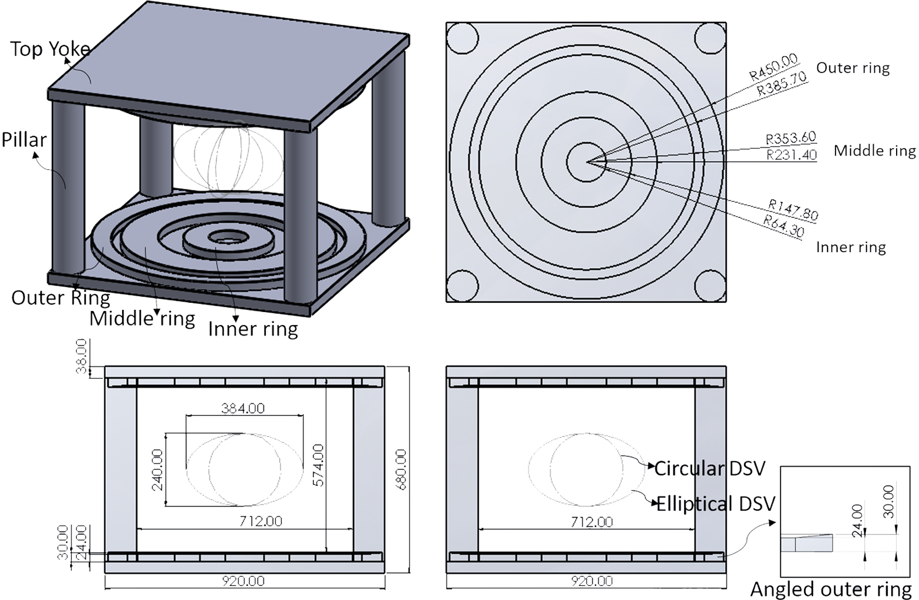
Numerical optimization of permanent magnet design: The magnet conceptually consists of two primary inner rings and one outer shimming ring for fabrication simplicity (Figure 1). Using ANSYS, numerical iterations were performed to optimize the triple-ring parameters, including (1) diameters of three rings; (2) gaps between the three primary rings; and (3) introduction of an angled/recessed thin outer shim ring to divert the field lines towards the imaging volume to improve homogeneity without adverse effects on fringe field. Given the requirements of our design for use in whole-body and head imaging, we defined two homogeneity DSVs (Figure 1), namely an elliptical DSV (240mm in the short axis and 384mm in the long axis) and a spherical DSV (250mm diameter). The targeted field inhomogeneity is 5000 ppm peak-to-peak within the DSV volume because such inhomogeneity level range can be easily shimmed by passive shimming according to our experience. To achieve a reasonable strong and uniform magnetic field, we chose to use neodymium magnets (NdFeB; remanence, Br=0.94T).Results
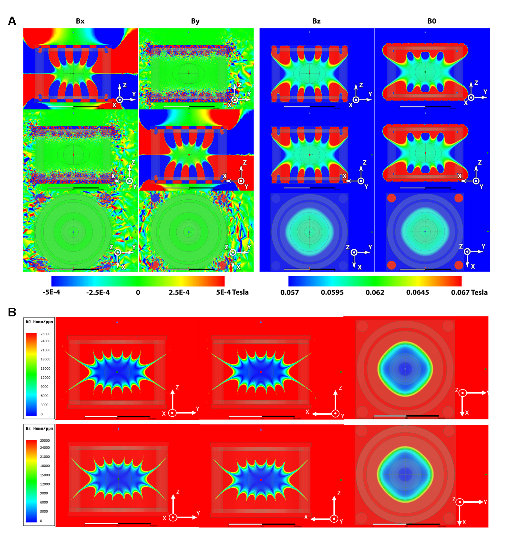
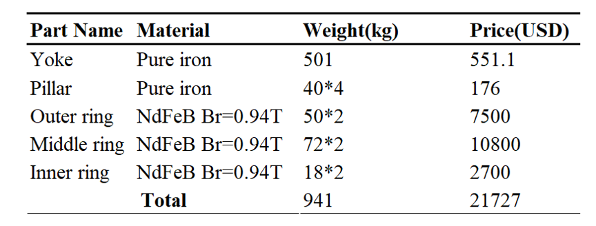
Main magnetic field strength and field homogeneity: Figure 2A shows the simulated main magnetic field vectors and their corresponding strength generated by the numerically optimized triple-ring magnet. With such design, the magnetic field, B0, is predominated by field vectors in the Bz direction, while those in the Bx and By direction are negligible as desired. This finding demonstrates that the design meets the fundamental criterion for MR imaging (i.e., a unidirectional static magnetic field able to polarize spins). The strength of the B0 field is ~60mT.Figure 2B shows the corresponding field homogeneity maps. Notably, our design is relatively homogeneous throughout the desired body and head imaging volume (i.e., within 5000ppm peak-to-peak over the 240x384mm elliptical and 250mm spherical DSV, respectively). Note that a B0 inhomogeneity of <10,000ppm peak-to-peak within the DSV can be shimmed through passive shimming as per our experience. Our findings indicate that our design is able to produce a uniform static magnetic field efficiently, which is crucial for encoding MRI signals with linear gradient fields, without sacrificing the flexibility to perform both whole-body and head imaging. Table 1 lists the resulting NdFeB magnet material weight, total magnet weight, and other magnet parameters of our design.
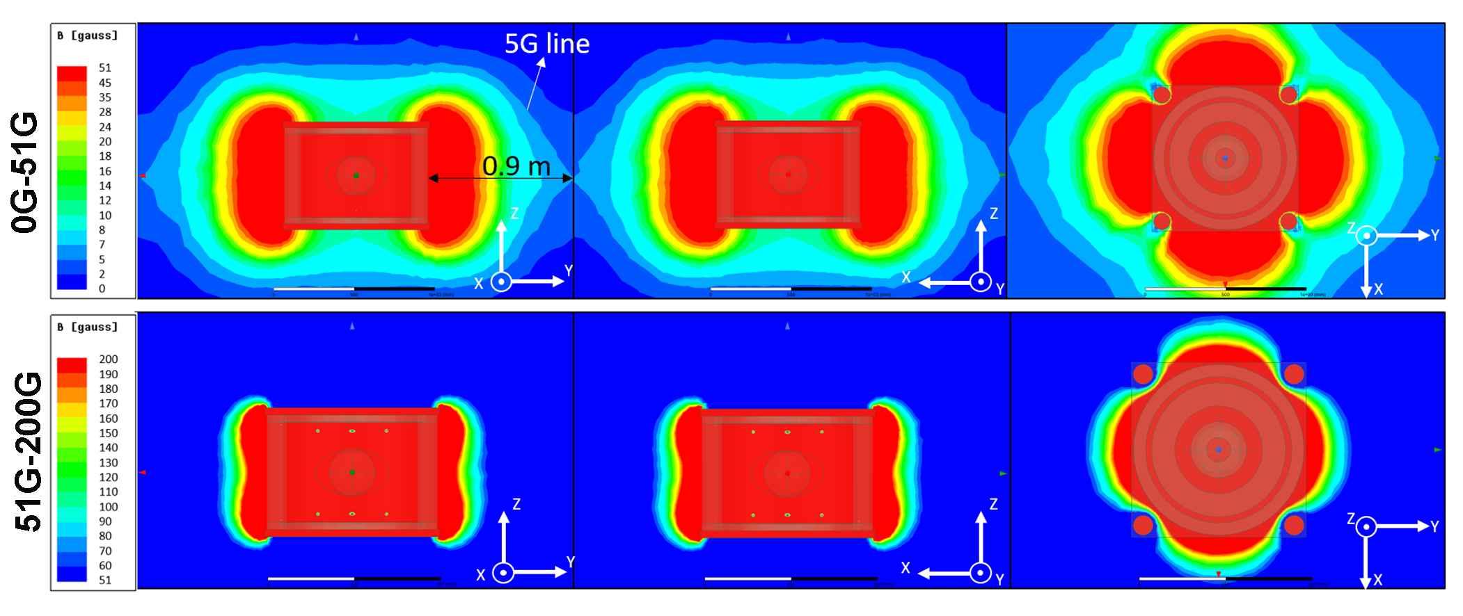
Fringe field: In addition to the main magnetic field strength and its homogeneity, we also considered the fringe field performance, as it is another important factor affecting the compactness and usability of our permanent magnet design. Figure 3 shows that the 5G line is located ~90cm from the magnet’s periphery.
Discussion and Conclusion
Our proposed triple-ring design presents an alternative approach to developing ultra-low-field permanent magnet subsystems. It improves B0 homogeneity without compromising its strength and the ability to perform whole-body imaging, including the head. Further, our current design with the additional angled thin shimming ring is highly manufacturable at low costs as it does not deviate significantly from the commonly used permanent MRI magnet shapes (e.g., circular). Note that the homogeneity reported in this study, which is within 5000ppm in both the elliptical and spherical DSVs, can be shimmed passively to 100-200ppm according to our experimental experience. Despite the demonstrated improvements in field homogeneity with our proposed design, the fringe field performance (i.e., defined by the 5G line) is not fully optimized in the present study, which is the goal of our future study. Additionally, we will further optimize certain components of the permanent magnet system to ensure that it is as light as possible. For example, the magnet yoke design, which at present is a cubic iron slab, can be recessed at specific locations with low magnetic flux density without compromising B0 strength and homogeneity, fringe field, and structural integrity.Acknowledgements
This study was supported by Hong Kong Research Grant Council (R7003-19, C7048-16G, HKU17112120, HKU17103819 and HKU17104020), Guangdong Key Technologies for Treatment of Brain Disorders (2018B030332001), and Guangdong Key Technologies for Alzheimer’s Disease Diagnosis and Treatment (2018B030336001).References
1. Fuchs, V.R. & Sox, H.C., Jr. Physicians' views of the relative importance of thirty medical innovations. Health Aff (Millwood) 20, 30-42 (2001).
2. Magnetic resonance imaging (MRI) units (indicator). 2019. (OECD). https://data.oecd.org/healtheqt/magnetic-resonance-imaging-mri-units.htm.
3. Geethanath, S. & Vaughan, J.T., Jr. Accessible magnetic resonance imaging: A review. J Magn Reson Imaging 49, e65-e77 (2019).
4. Marques, J.P., Simonis, F.F.J. & Webb, A.G. Low-field MRI: An MR physics perspective. J Magn Reson Imaging 49, 1528-1542 (2019).
5. Cooley, C.Z., McDaniel, P.C., Stockmann, J.P., Srinivas, S.A., Cauley, S.F., Sliwiak, M., Sappo, C.R., Vaughn, C.F., Guerin, B., Rosen, M.S., Lev, M.H. & Wald, L.L. A portable scanner for magnetic resonance imaging of the brain. Nat Biomed Eng (2020).
6. Miyamoto, T., Sakurai, H., Takabayashi, H. & Aoki, M. A Development of a Permanent-Magnet Assembly for Mri Devices Using Nd-Fe-B Material. IEEE Transactions on Magnetics 25, 3907-3909 (1989).
7. Webb, A.G. Magnetic resonance technology: hardware and system component design, (Royal society of chemistry, 2016).
Figures